SlideShare a Scribd company logo
1 of 51
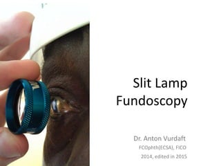
Slit Lamp
Fundoscopy
Dr. Anton Vurdaft
2014, edited in 2015
FCOphth(ECSA), FICO
Slit Lamp Fundoscopy
Biomicroscopy with simultaneous fundus
examination.
Using high-diopter lens (+60D, +78D, +90D,
+120D and others).
Ophthalmoscopy with slit lamp is the most
difficult skill in ophthalmic examination.
Ophthalmoscopy using slit lamp and the lens
gives the most clear picture of the fundus:
magnified, 3D, very bright).
Slit Lamp Fundoscopy
Slit lamp with
teaching piece
Slit Lamp Fundoscopy
It is possible to do fundoscopy with wide AND
with narrow pupil most of the times.
The latter is surely more difficult for
inexperienced examiner.
Moreover, it is difficult and sometimes not
possible to examine fundus periphery with
narrow pupils.
Slit Lamp Fundoscopy Principles
Use slit lamp as you do for anterior
segment.
The task is to focus the beam light on the
fundus.
The beam is narrow. Examine fundus
stepwise.
Everything what is not into the beam light
focus – is not seen or seen poorly.
Slit Lamp Fundoscopy Principles
With slit lamp fundoscopy we only see SLIT image, we do not see
the picture as a whole at one time. We have to move the slit
in certain directions. NOTICE the glare, which is moved out of
the picture with a mild tilting of the lens.
Slit Lamp Fundoscopy Technique
Prepare for the examination:
• Make the room as dark as possible
• Fixate your arms comfortably, using the
armrest
• Dilate the pupils of the patient (important if
you are not experienced) – use tropicamide or
combination of tropicamide with
phenylephrine
Slit Lamp Fundoscopy Technique
DILATE THE PUPILS!
Slit Lamp Fundoscopy Technique
The armrest you can
make yourself of any
suitable material or
involving carpenters.
This is especially
important for those who
have short hands.
Sometimes you can use
the case of high-diopter
lens
Slit Lamp Fundoscopy: Set Slit Lamp
First use magnification of x15 or less (i.e. x10). The
bigger the magnification, the harder it is for
inexperienced examiner to set the focus on the
fundus and to orient within the fundus.
Set the light beam as narrow as possible. The narrower
the pupil, the narrower the beam should be. The
height of the beam is not so important. The width
can be regulated during the examination.
Slit Lamp Fundoscopy: Set Slit Lamp
Set the brightness of the beam to the level most
comfortable for you (usually it is the brightest) and
for the patient (usually patients are more
comfortable with darker beam).
Height, width and brightness of the beam can be
regulated during the procedure, that is especially
important with uncooperative patients and patients
with photophobia.
The light-source and the pieces of the slit lamp should
be aligned into one axis (90 degree to the patient).
Slit Lamp Fundoscopy: Start Exam
Fixate a chin and a forehead of the patient (using
chinrest and headrest).
Ask patient to look straight or in other needed gauze
position.
Focus the beam on the center of the pupil.
Fix your hand with the lens on a cheek of a patient or
using a headrest.
It is more suitable to use left hand for a right eye of a
patient, and vice versa.
Slit Lamp Fundoscopy: Technique
DO NOT USE ONE HAND FOR BOTH EYES.
SWITH YOUR HANDS. IT IS EASY TO GET
USED TO ONE HAND, AND IT WILL SPOIL
YOUR EXAMINATIONS LATER.
Slit Lamp Fundoscopy: Technique
Grab the lens with big and index finger, the rest of the
fingers will help you to tilt the lens and pull the
upper eyelid up.
Set the center of the lens on the center of the pupil
(use the beam to check it).
The working distance of the lens is 5-10 mm.
It does not matter what side of the lens should be
faced to the patient, if the lens is double aspheric
(volk, ocular instruments). Otherwise – the letters on
the lens rim should be directed towards the patient
(see the foto).
Slit Lamp Fundoscopy: Technique
The picture is floating in the air actually.
Notice the direction of letters of the lens rim.
That is how you
focus a beam on
the center of a
pupil.
Notice how the
fingers are fixed.
Slit Lamp Fundoscopy: Technique
Patient should look in the needed direction.
Light source of the microscope moved closely
to the patient.
Slowly you pull the microscope back to you.
Focus of the beam is moving from the cornea
to the lens, than we see the image of the
fundus, it is formed somewhere in the air.
Stepwise examination follows.
Slit Lamp Fundoscopy: Technique
Practice makes perfect!
Do at least one dilated and one non-
dilated fundoscopy of the disc EVERY
DAY.
After 2-3 weeks you may add the
macula to your daily MUST DO list.
Slit Lamp Fundoscopy: The Image
The image which you see – is inverted. It means that
you see it upwards down and left-to-right. If you see
something in the upper part of your picture – it is
actually in the lower part of that part of the fundus,
which you are examining. The same goes to “left or
right” parameters.
The illustration of that fact is the relation of optic disc
and macula. The latter is always temporally (closer to
the temple), the disc is nasally (closer to the nose).
But with the slit lamp fundoscopy we see the picture
inverted – disc is always temporally there.
Slit Lamp Fundoscopy: The Image
Fundus of the left eye (egg), as if we see the eye in horizontal cross-
section. As it should be with the left eye – the optic disc is closer to the
nose than macula.
Slit Lamp Fundoscopy: The Image
Fundus of the same left eye (egg) with the using of high-diopter “+” lens.
Equally to the +90D or +78D, the image gets inverted. Macula and disc have
changed their places and got upside-down.
Slit Lamp Fundoscopy: The Image
When you are drawing the fundus picture in the file – there
are two ways you can do it. The first one implies mental
gymnastics – you may try to picture the findings immediately
in the way they are really located in the fundus (direct
picture).
The other way is to rotate your file on 180 degree and draw
the picture JUST exactly how you’ve seen it in inverted way. If
you’ll rotate this file back to 180 degree – the findings will be
in correct position and direct.
Use mental gymnastics! It is useful for a brain and will help
you to understand the topography of the fundus better.
Slit Lamp Fundoscopy: The Image
The area of fundus which you can see
depends on refractive power of the lens.
The more powerful is the lens, the bigger
part of fundus you see at a one time. For
example, with +60D you will struggle
trying to see the periphery with narrow
pupil, but +120D lens will easily allow you
to see it.
Slit Lamp Fundoscopy: The Image
The field of fundus which is seen – depends
not only on the lens, but also on the
patient’s cooperation. The more precisely
patient follows your instructions, the bigger
piece of fundus you will see.
It is wise to do the fundoscopy in the next
order: optic disc (patient fixates the
contralateral ear of the examiner), macula
(patient fixates straighth), periphery (patient
fixated every needed direction).
Slit Lamp Fundoscopy: Retinal Periphery
It is most comfortable for the beginner to
examine periphery of the retina starting
from 12 o’clock meridian going clockwise
and seeing the 1-30, 3, 4-30, 6, 7-30, 9 and
10-30 o’clock meridians.
Slit Lamp Fundoscopy: Retinal Periphery
For the beginner it is difficult to orient
within the fundus in a certain gauze
direction of a patient. It is quiet easy.
Patient looks at the direction of the piece
of fundus which you see. Patient looks
down – we see the lower periphery.
Patient looks up – we see the upper
periphery. Etc.
Slit Lamp Fundoscopy: Retinal Periphery
Patient fixated straight. We receive macula in the field of
view.
Slit Lamp Fundoscopy: Retinal Periphery
Patient fixated the contralateral ear of examiner. We receive
optic disc in the center of a fundus picture.
Slit Lamp Fundoscopy: Retinal Periphery
Patient looked at the ceiling, we see upper periphery (12
o’clock).
Slit Lamp Fundoscopy: Retinal Periphery
Patient looked at the floor, we see lower periphery (6 o’clock).
Slit Lamp Fundoscopy: Retinal Periphery
Patient looked left, we see temporal periphery (3 o’clock).
Slit Lamp Fundoscopy: Retinal Periphery
Patient looked right, we see nasal periphery (9 o’clock).
Slit Lamp Fundoscopy: Retinal Periphery
Inversion of the picture of upper periphery
Slit Lamp Fundoscopy: Retinal Periphery
Inversion of the picture of upper periphery
Slit Lamp Fundoscopy: Fundus Structure
Fundus Structure
Picturing The Fundus
Optic disc is always nasally, while macula is temporally
Optic Disc Structure
Measuring the Cup of the Disc
Measurement is not so precise, do not struggle
Measuring the Cup of the Disc
For our practice we need approximate cup/disc ratio
Measuring the Cup of the Disc
This cup/disc ratio is approximately 0,7. Notice the absence of the
neuroretinal rim from the temporal side
Measuring the Cup of the Disc
This cup/disc ratio is 0,9, almost total (1,0). Notice the absence of the
neuroretinal rim everywhere except nasal side of the disc.
Measuring the Cup of the Disc: Stereo
The color is not enough to assess the real cup-disc ratio. Use stereopsis!
Notice the absence of neuroretinal rim at the lower part of the disc
Measuring the Cup of the Disc: Stereo
Vessels bayoneting demarcates the borders of cupping
Measuring the Cup of the Disc: Stereo
Normal disc picture (10)
Lenses Care:
Lenses Care:
1)Do not touch the lens itself, touch only the
rim!
2)Occasionally you may clean the lens
surface gently with a smooth tissue
without fibers.
3)Put the lens into the case immediately
after use!
References:
(1) S.Barnard, A.Field, "Slit Lamp Indirect Ophthalmoscopy", American Academy of
Optometry, http://www.academy.org.uk/tutorials/volklens.htm
(2) "Practical Ophthalmology - A Manual for Beginning Residents", 5th ed., American
Academy of Ophthalmology, 2005
(3) James Benjamin, "Ophthalmology Investigation and Examination Techniques", 2006
(4) Контактные диагностические линзы и фиксация локтя:
http://terra-ophthalmica.blogspot.com/2012/06/blog-post_21.html
(5) Rosalind A. Stevens, Patrick J. Saine, Marshall E. Tyler, "Stereo atlas of fluorescein
and indocyanine green angiography", 1999
(6) William L. Jones, "Peripheral Ocular Fundus (Third Edition)", 2007
(7) Vortex Veins and Blonde Fundus,
http://www.optos.com/RecognizingPathology/pages/Visualization/VortexVeins-
8.html
(8) S.A. Rizvi, "A simple method to view stereo-images of the optic nerve head on
ordinary computer monitors", Eye (2009)
(9) My own fotos
Thank you!

More Related Content

What's hot

Refrective surgery ppt
Refrective surgery pptRefrective surgery ppt
Refrective surgery pptsubhadri manna
 
FUNDUS FLUORESCEIN ANGIOGRAPHY
FUNDUS FLUORESCEIN ANGIOGRAPHYFUNDUS FLUORESCEIN ANGIOGRAPHY
FUNDUS FLUORESCEIN ANGIOGRAPHYAnuraag Singh
 
Lenses of slit lamp biomicroscope & indirect ophthalmoscope.
Lenses of slit lamp biomicroscope & indirect ophthalmoscope.Lenses of slit lamp biomicroscope & indirect ophthalmoscope.
Lenses of slit lamp biomicroscope & indirect ophthalmoscope.Ayat AbuJazar
 
Indirect ophthalmoscopy
Indirect ophthalmoscopyIndirect ophthalmoscopy
Indirect ophthalmoscopyShahanaSherin9
 
Gonioscopy: gonioscopic lenses, principle and clinical aspects
Gonioscopy: gonioscopic lenses, principle and clinical aspectsGonioscopy: gonioscopic lenses, principle and clinical aspects
Gonioscopy: gonioscopic lenses, principle and clinical aspectsDr Samarth Mishra
 
Slit lamp biomicroscopy
Slit lamp biomicroscopySlit lamp biomicroscopy
Slit lamp biomicroscopyHira Dahal
 
Direct ophthalmoscopy final
Direct ophthalmoscopy finalDirect ophthalmoscopy final
Direct ophthalmoscopy finalshovon2026
 
Direct & indirect ophthalmoscopy
Direct & indirect ophthalmoscopyDirect & indirect ophthalmoscopy
Direct & indirect ophthalmoscopyDR. ROHIT AGRAWAL
 
Slit Lamp Illumination Techniques
Slit Lamp Illumination TechniquesSlit Lamp Illumination Techniques
Slit Lamp Illumination TechniquesIrina Kezik
 
Biometry & Iol calculations
Biometry & Iol calculationsBiometry & Iol calculations
Biometry & Iol calculationsrakesh jaiswal
 
Slit Lamp Biomicroscopy
Slit  Lamp BiomicroscopySlit  Lamp Biomicroscopy
Slit Lamp BiomicroscopyManoj Aryal
 
Tangent screen
Tangent screenTangent screen
Tangent screensana butt
 

What's hot (20)

Refrective surgery ppt
Refrective surgery pptRefrective surgery ppt
Refrective surgery ppt
 
FUNDUS FLUORESCEIN ANGIOGRAPHY
FUNDUS FLUORESCEIN ANGIOGRAPHYFUNDUS FLUORESCEIN ANGIOGRAPHY
FUNDUS FLUORESCEIN ANGIOGRAPHY
 
Lenses of slit lamp biomicroscope & indirect ophthalmoscope.
Lenses of slit lamp biomicroscope & indirect ophthalmoscope.Lenses of slit lamp biomicroscope & indirect ophthalmoscope.
Lenses of slit lamp biomicroscope & indirect ophthalmoscope.
 
Indirect ophthalmoscopy
Indirect ophthalmoscopyIndirect ophthalmoscopy
Indirect ophthalmoscopy
 
Gonioscopy: gonioscopic lenses, principle and clinical aspects
Gonioscopy: gonioscopic lenses, principle and clinical aspectsGonioscopy: gonioscopic lenses, principle and clinical aspects
Gonioscopy: gonioscopic lenses, principle and clinical aspects
 
Slit lamp biomicroscopy
Slit lamp biomicroscopySlit lamp biomicroscopy
Slit lamp biomicroscopy
 
Slit lamp biomicroscope
Slit lamp biomicroscopeSlit lamp biomicroscope
Slit lamp biomicroscope
 
Direct ophthalmoscopy final
Direct ophthalmoscopy finalDirect ophthalmoscopy final
Direct ophthalmoscopy final
 
Hess chart
Hess chartHess chart
Hess chart
 
Direct & indirect ophthalmoscopy
Direct & indirect ophthalmoscopyDirect & indirect ophthalmoscopy
Direct & indirect ophthalmoscopy
 
Synaptophore in ophthalmology
Synaptophore in ophthalmologySynaptophore in ophthalmology
Synaptophore in ophthalmology
 
Phacodynamics
PhacodynamicsPhacodynamics
Phacodynamics
 
Slit Lamp Illumination Techniques
Slit Lamp Illumination TechniquesSlit Lamp Illumination Techniques
Slit Lamp Illumination Techniques
 
Macular function tests
Macular function testsMacular function tests
Macular function tests
 
Biometry & Iol calculations
Biometry & Iol calculationsBiometry & Iol calculations
Biometry & Iol calculations
 
Slit Lamp Biomicroscopy
Slit  Lamp BiomicroscopySlit  Lamp Biomicroscopy
Slit Lamp Biomicroscopy
 
Gonioscopy
GonioscopyGonioscopy
Gonioscopy
 
Aphakia
AphakiaAphakia
Aphakia
 
Gonioscopy
GonioscopyGonioscopy
Gonioscopy
 
Tangent screen
Tangent screenTangent screen
Tangent screen
 

Viewers also liked

Looking deep into retina : indirect ophthalmoscopy and fundus drawing
Looking deep into retina : indirect ophthalmoscopy and fundus drawingLooking deep into retina : indirect ophthalmoscopy and fundus drawing
Looking deep into retina : indirect ophthalmoscopy and fundus drawingPrachir Agarwal
 
laser lenses for retinal diseases
 laser lenses for retinal diseases laser lenses for retinal diseases
laser lenses for retinal diseasesnalini2218
 
Diabetic retinopathy
Diabetic retinopathyDiabetic retinopathy
Diabetic retinopathyA V
 
Basic opthalmoscopy findings - presentation at www.eyenirvaan.com
Basic opthalmoscopy findings - presentation at www.eyenirvaan.comBasic opthalmoscopy findings - presentation at www.eyenirvaan.com
Basic opthalmoscopy findings - presentation at www.eyenirvaan.comEyenirvaan
 
Ophthalmoscopic slides
Ophthalmoscopic slidesOphthalmoscopic slides
Ophthalmoscopic slidesscorpio_80
 
Visual acuity, lens and fundus oculi
Visual acuity, lens and fundus oculiVisual acuity, lens and fundus oculi
Visual acuity, lens and fundus oculiFarhan Ali
 
Вскрытие абсцесса века: клинический случай и теоретическое обоснование
Вскрытие абсцесса века: клинический случай и теоретическое обоснованиеВскрытие абсцесса века: клинический случай и теоретическое обоснование
Вскрытие абсцесса века: клинический случай и теоретическое обоснованиеA V
 
копие от Varilux physio for attila 2
копие от Varilux physio for attila 2копие от Varilux physio for attila 2
копие от Varilux physio for attila 2pazoff
 
Volk Training Presentation Ns
Volk Training Presentation NsVolk Training Presentation Ns
Volk Training Presentation NsCaporex Oy
 
CHI'15 - WonderLens: Optical Lenses and Mirrors for Tangible Interactions on ...
CHI'15 - WonderLens: Optical Lenses and Mirrors for Tangible Interactions on ...CHI'15 - WonderLens: Optical Lenses and Mirrors for Tangible Interactions on ...
CHI'15 - WonderLens: Optical Lenses and Mirrors for Tangible Interactions on ...Rong-Hao Liang
 
Retina examination when to order
Retina examination when to orderRetina examination when to order
Retina examination when to orderinmyeyesslideshare
 
Ophthalmic Lens Design Prototype Facility – InVision Biomedical
Ophthalmic Lens Design Prototype Facility – InVision BiomedicalOphthalmic Lens Design Prototype Facility – InVision Biomedical
Ophthalmic Lens Design Prototype Facility – InVision BiomedicalInvisionBio
 
Slit lampbiomicroscopy-jinal & dhara
Slit lampbiomicroscopy-jinal & dharaSlit lampbiomicroscopy-jinal & dhara
Slit lampbiomicroscopy-jinal & dharadhara sorathiya
 

Viewers also liked (20)

Fundus examination
Fundus examinationFundus examination
Fundus examination
 
Funduscopy
Funduscopy Funduscopy
Funduscopy
 
Looking deep into retina : indirect ophthalmoscopy and fundus drawing
Looking deep into retina : indirect ophthalmoscopy and fundus drawingLooking deep into retina : indirect ophthalmoscopy and fundus drawing
Looking deep into retina : indirect ophthalmoscopy and fundus drawing
 
laser lenses for retinal diseases
 laser lenses for retinal diseases laser lenses for retinal diseases
laser lenses for retinal diseases
 
Slit lamp biomicroscopy.
Slit lamp biomicroscopy.Slit lamp biomicroscopy.
Slit lamp biomicroscopy.
 
Diabetic retinopathy
Diabetic retinopathyDiabetic retinopathy
Diabetic retinopathy
 
Basic opthalmoscopy findings - presentation at www.eyenirvaan.com
Basic opthalmoscopy findings - presentation at www.eyenirvaan.comBasic opthalmoscopy findings - presentation at www.eyenirvaan.com
Basic opthalmoscopy findings - presentation at www.eyenirvaan.com
 
Ophthalmoscopic slides
Ophthalmoscopic slidesOphthalmoscopic slides
Ophthalmoscopic slides
 
Complete Patient Work UP
Complete Patient Work UPComplete Patient Work UP
Complete Patient Work UP
 
Visual acuity, lens and fundus oculi
Visual acuity, lens and fundus oculiVisual acuity, lens and fundus oculi
Visual acuity, lens and fundus oculi
 
Вскрытие абсцесса века: клинический случай и теоретическое обоснование
Вскрытие абсцесса века: клинический случай и теоретическое обоснованиеВскрытие абсцесса века: клинический случай и теоретическое обоснование
Вскрытие абсцесса века: клинический случай и теоретическое обоснование
 
Poag case presentation
Poag case presentationPoag case presentation
Poag case presentation
 
копие от Varilux physio for attila 2
копие от Varilux physio for attila 2копие от Varilux physio for attila 2
копие от Varilux physio for attila 2
 
Primary Open Angle Glaucoma
Primary Open Angle GlaucomaPrimary Open Angle Glaucoma
Primary Open Angle Glaucoma
 
Volk Training Presentation Ns
Volk Training Presentation NsVolk Training Presentation Ns
Volk Training Presentation Ns
 
CHI'15 - WonderLens: Optical Lenses and Mirrors for Tangible Interactions on ...
CHI'15 - WonderLens: Optical Lenses and Mirrors for Tangible Interactions on ...CHI'15 - WonderLens: Optical Lenses and Mirrors for Tangible Interactions on ...
CHI'15 - WonderLens: Optical Lenses and Mirrors for Tangible Interactions on ...
 
Retina examination when to order
Retina examination when to orderRetina examination when to order
Retina examination when to order
 
Basic fundoscopy
Basic fundoscopyBasic fundoscopy
Basic fundoscopy
 
Ophthalmic Lens Design Prototype Facility – InVision Biomedical
Ophthalmic Lens Design Prototype Facility – InVision BiomedicalOphthalmic Lens Design Prototype Facility – InVision Biomedical
Ophthalmic Lens Design Prototype Facility – InVision Biomedical
 
Slit lampbiomicroscopy-jinal & dhara
Slit lampbiomicroscopy-jinal & dharaSlit lampbiomicroscopy-jinal & dhara
Slit lampbiomicroscopy-jinal & dhara
 

Similar to Slit lamp fundoscopy

Objective refraction
Objective refractionObjective refraction
Objective refractionsneha_thaps
 
Ophthalmoscope direct and indirect
Ophthalmoscope direct and indirectOphthalmoscope direct and indirect
Ophthalmoscope direct and indirectAnisur Rahman
 
Ophthalmoscopy
OphthalmoscopyOphthalmoscopy
OphthalmoscopySalalKhan5
 
Objective retinoscopy
Objective retinoscopyObjective retinoscopy
Objective retinoscopyBipin Koirala
 
ppt on direct opthalmoscopy
 ppt on direct opthalmoscopy ppt on direct opthalmoscopy
ppt on direct opthalmoscopymanisaikoduri
 
Fundoscopy ppt 2012
Fundoscopy ppt 2012Fundoscopy ppt 2012
Fundoscopy ppt 2012Reina Ramesh
 
2.ophthalmology workup.2
2.ophthalmology workup.22.ophthalmology workup.2
2.ophthalmology workup.2Reyad Yossif
 
Binocular Indirect Ophthalmoscopy
Binocular Indirect OphthalmoscopyBinocular Indirect Ophthalmoscopy
Binocular Indirect Ophthalmoscopyabhishek ghelani
 
07 ophthalmoscope direct and indirect
07 ophthalmoscope direct and indirect07 ophthalmoscope direct and indirect
07 ophthalmoscope direct and indirectAnisur Rahman
 
Direct ophthalmoscope.pptx
Direct ophthalmoscope.pptxDirect ophthalmoscope.pptx
Direct ophthalmoscope.pptxAnamSehreen
 
Direct ophthalmoscope Machine
Direct ophthalmoscope MachineDirect ophthalmoscope Machine
Direct ophthalmoscope MachineSunilAgrawal86
 
directophthalmoscope-180730142501 (2).pdf
directophthalmoscope-180730142501 (2).pdfdirectophthalmoscope-180730142501 (2).pdf
directophthalmoscope-180730142501 (2).pdfanju468752
 
ilovepdf_merged.pdf
ilovepdf_merged.pdfilovepdf_merged.pdf
ilovepdf_merged.pdfanju468752
 

Similar to Slit lamp fundoscopy (20)

Minggu Orientasi 2015. Year 1.
Minggu Orientasi 2015. Year 1.Minggu Orientasi 2015. Year 1.
Minggu Orientasi 2015. Year 1.
 
OPTHALMOSCOPY.pdf
OPTHALMOSCOPY.pdfOPTHALMOSCOPY.pdf
OPTHALMOSCOPY.pdf
 
Objective refraction
Objective refractionObjective refraction
Objective refraction
 
Ophthalmoscopy
OphthalmoscopyOphthalmoscopy
Ophthalmoscopy
 
Ophthalmoscopy
OphthalmoscopyOphthalmoscopy
Ophthalmoscopy
 
Ophthalmoscope direct and indirect
Ophthalmoscope direct and indirectOphthalmoscope direct and indirect
Ophthalmoscope direct and indirect
 
Ophthalmoscopy
OphthalmoscopyOphthalmoscopy
Ophthalmoscopy
 
Objective retinoscopy
Objective retinoscopyObjective retinoscopy
Objective retinoscopy
 
ppt on direct opthalmoscopy
 ppt on direct opthalmoscopy ppt on direct opthalmoscopy
ppt on direct opthalmoscopy
 
Fundoscopy ppt 2012
Fundoscopy ppt 2012Fundoscopy ppt 2012
Fundoscopy ppt 2012
 
2.ophthalmology workup.2
2.ophthalmology workup.22.ophthalmology workup.2
2.ophthalmology workup.2
 
Synaptophore
SynaptophoreSynaptophore
Synaptophore
 
Binocular Indirect Ophthalmoscopy
Binocular Indirect OphthalmoscopyBinocular Indirect Ophthalmoscopy
Binocular Indirect Ophthalmoscopy
 
07 ophthalmoscope direct and indirect
07 ophthalmoscope direct and indirect07 ophthalmoscope direct and indirect
07 ophthalmoscope direct and indirect
 
Slit lamp biomicroscope
Slit lamp biomicroscopeSlit lamp biomicroscope
Slit lamp biomicroscope
 
Direct ophthalmoscope.pptx
Direct ophthalmoscope.pptxDirect ophthalmoscope.pptx
Direct ophthalmoscope.pptx
 
Direct ophthalmoscope Machine
Direct ophthalmoscope MachineDirect ophthalmoscope Machine
Direct ophthalmoscope Machine
 
directophthalmoscope-180730142501 (2).pdf
directophthalmoscope-180730142501 (2).pdfdirectophthalmoscope-180730142501 (2).pdf
directophthalmoscope-180730142501 (2).pdf
 
Direct ophthalmoscope
Direct ophthalmoscopeDirect ophthalmoscope
Direct ophthalmoscope
 
ilovepdf_merged.pdf
ilovepdf_merged.pdfilovepdf_merged.pdf
ilovepdf_merged.pdf
 

More from A V

Intraokulärer Fremdkörper und Endophthalmitis
Intraokulärer Fremdkörper und EndophthalmitisIntraokulärer Fremdkörper und Endophthalmitis
Intraokulärer Fremdkörper und EndophthalmitisA V
 
Welches glaukom muss man nicht unbedingt behandeln?
Welches glaukom muss man nicht unbedingt behandeln?Welches glaukom muss man nicht unbedingt behandeln?
Welches glaukom muss man nicht unbedingt behandeln?A V
 
Severe chronic atopic keratoconjunctivitisSevere chronic atopic keratoconjunc...
Severe chronic atopic keratoconjunctivitisSevere chronic atopic keratoconjunc...Severe chronic atopic keratoconjunctivitisSevere chronic atopic keratoconjunc...
Severe chronic atopic keratoconjunctivitisSevere chronic atopic keratoconjunc...A V
 
Conjunctival Squamous Cell Carcinoma
Conjunctival Squamous Cell CarcinomaConjunctival Squamous Cell Carcinoma
Conjunctival Squamous Cell CarcinomaA V
 
Bilamellar tarsal plate rotation assistance
Bilamellar tarsal plate rotation assistanceBilamellar tarsal plate rotation assistance
Bilamellar tarsal plate rotation assistanceA V
 
Sind augenärztliche YAG-Laser gefährlich?
Sind augenärztliche YAG-Laser gefährlich?Sind augenärztliche YAG-Laser gefährlich?
Sind augenärztliche YAG-Laser gefährlich?A V
 
Emergencies in eye department
Emergencies in eye departmentEmergencies in eye department
Emergencies in eye departmentA V
 
On artificial eyes
On artificial eyesOn artificial eyes
On artificial eyesA V
 
Eyelid abscess drainage
Eyelid abscess drainageEyelid abscess drainage
Eyelid abscess drainageA V
 
Glaucoma for the students and general practitioners
Glaucoma for the students and general practitionersGlaucoma for the students and general practitioners
Glaucoma for the students and general practitionersA V
 
Poor vision healthy eyes
Poor vision healthy eyesPoor vision healthy eyes
Poor vision healthy eyesA V
 
Профилактика прогрессирования миопии с точки зрения доказательной медицины
Профилактика прогрессирования миопии с точки зрения доказательной медициныПрофилактика прогрессирования миопии с точки зрения доказательной медицины
Профилактика прогрессирования миопии с точки зрения доказательной медициныA V
 

More from A V (12)

Intraokulärer Fremdkörper und Endophthalmitis
Intraokulärer Fremdkörper und EndophthalmitisIntraokulärer Fremdkörper und Endophthalmitis
Intraokulärer Fremdkörper und Endophthalmitis
 
Welches glaukom muss man nicht unbedingt behandeln?
Welches glaukom muss man nicht unbedingt behandeln?Welches glaukom muss man nicht unbedingt behandeln?
Welches glaukom muss man nicht unbedingt behandeln?
 
Severe chronic atopic keratoconjunctivitisSevere chronic atopic keratoconjunc...
Severe chronic atopic keratoconjunctivitisSevere chronic atopic keratoconjunc...Severe chronic atopic keratoconjunctivitisSevere chronic atopic keratoconjunc...
Severe chronic atopic keratoconjunctivitisSevere chronic atopic keratoconjunc...
 
Conjunctival Squamous Cell Carcinoma
Conjunctival Squamous Cell CarcinomaConjunctival Squamous Cell Carcinoma
Conjunctival Squamous Cell Carcinoma
 
Bilamellar tarsal plate rotation assistance
Bilamellar tarsal plate rotation assistanceBilamellar tarsal plate rotation assistance
Bilamellar tarsal plate rotation assistance
 
Sind augenärztliche YAG-Laser gefährlich?
Sind augenärztliche YAG-Laser gefährlich?Sind augenärztliche YAG-Laser gefährlich?
Sind augenärztliche YAG-Laser gefährlich?
 
Emergencies in eye department
Emergencies in eye departmentEmergencies in eye department
Emergencies in eye department
 
On artificial eyes
On artificial eyesOn artificial eyes
On artificial eyes
 
Eyelid abscess drainage
Eyelid abscess drainageEyelid abscess drainage
Eyelid abscess drainage
 
Glaucoma for the students and general practitioners
Glaucoma for the students and general practitionersGlaucoma for the students and general practitioners
Glaucoma for the students and general practitioners
 
Poor vision healthy eyes
Poor vision healthy eyesPoor vision healthy eyes
Poor vision healthy eyes
 
Профилактика прогрессирования миопии с точки зрения доказательной медицины
Профилактика прогрессирования миопии с точки зрения доказательной медициныПрофилактика прогрессирования миопии с точки зрения доказательной медицины
Профилактика прогрессирования миопии с точки зрения доказательной медицины
 

Recently uploaded

Call Girl In Indore 📞9235973566📞 Just📲 Call Inaaya Indore Call Girls Service ...
Call Girl In Indore 📞9235973566📞 Just📲 Call Inaaya Indore Call Girls Service ...Call Girl In Indore 📞9235973566📞 Just📲 Call Inaaya Indore Call Girls Service ...
Call Girl In Indore 📞9235973566📞 Just📲 Call Inaaya Indore Call Girls Service ...Sheetaleventcompany
 
7 steps How to prevent Thalassemia : Dr Sharda Jain & Vandana Gupta
7 steps How to prevent Thalassemia : Dr Sharda Jain & Vandana Gupta7 steps How to prevent Thalassemia : Dr Sharda Jain & Vandana Gupta
7 steps How to prevent Thalassemia : Dr Sharda Jain & Vandana GuptaLifecare Centre
 
❤️Call Girl Service In Chandigarh☎️9814379184☎️ Call Girl in Chandigarh☎️ Cha...
❤️Call Girl Service In Chandigarh☎️9814379184☎️ Call Girl in Chandigarh☎️ Cha...❤️Call Girl Service In Chandigarh☎️9814379184☎️ Call Girl in Chandigarh☎️ Cha...
❤️Call Girl Service In Chandigarh☎️9814379184☎️ Call Girl in Chandigarh☎️ Cha...Sheetaleventcompany
 
Chennai ❣️ Call Girl 6378878445 Call Girls in Chennai Escort service book now
Chennai ❣️ Call Girl 6378878445 Call Girls in Chennai Escort service book nowChennai ❣️ Call Girl 6378878445 Call Girls in Chennai Escort service book now
Chennai ❣️ Call Girl 6378878445 Call Girls in Chennai Escort service book nowtanudubay92
 
Dehradun Call Girls Service {8854095900} ❤️VVIP ROCKY Call Girl in Dehradun U...
Dehradun Call Girls Service {8854095900} ❤️VVIP ROCKY Call Girl in Dehradun U...Dehradun Call Girls Service {8854095900} ❤️VVIP ROCKY Call Girl in Dehradun U...
Dehradun Call Girls Service {8854095900} ❤️VVIP ROCKY Call Girl in Dehradun U...Sheetaleventcompany
 
Bhawanipatna Call Girls 📞9332606886 Call Girls in Bhawanipatna Escorts servic...
Bhawanipatna Call Girls 📞9332606886 Call Girls in Bhawanipatna Escorts servic...Bhawanipatna Call Girls 📞9332606886 Call Girls in Bhawanipatna Escorts servic...
Bhawanipatna Call Girls 📞9332606886 Call Girls in Bhawanipatna Escorts servic...Dipal Arora
 
Call Girls Shahdol Just Call 8250077686 Top Class Call Girl Service Available
Call Girls Shahdol Just Call 8250077686 Top Class Call Girl Service AvailableCall Girls Shahdol Just Call 8250077686 Top Class Call Girl Service Available
Call Girls Shahdol Just Call 8250077686 Top Class Call Girl Service AvailableDipal Arora
 
Cardiac Output, Venous Return, and Their Regulation
Cardiac Output, Venous Return, and Their RegulationCardiac Output, Venous Return, and Their Regulation
Cardiac Output, Venous Return, and Their RegulationMedicoseAcademics
 
Difference Between Skeletal Smooth and Cardiac Muscles
Difference Between Skeletal Smooth and Cardiac MusclesDifference Between Skeletal Smooth and Cardiac Muscles
Difference Between Skeletal Smooth and Cardiac MusclesMedicoseAcademics
 
Kolkata Call Girls Naktala 💯Call Us 🔝 8005736733 🔝 💃 Top Class Call Girl Se...
Kolkata Call Girls Naktala  💯Call Us 🔝 8005736733 🔝 💃  Top Class Call Girl Se...Kolkata Call Girls Naktala  💯Call Us 🔝 8005736733 🔝 💃  Top Class Call Girl Se...
Kolkata Call Girls Naktala 💯Call Us 🔝 8005736733 🔝 💃 Top Class Call Girl Se...Namrata Singh
 
Pune Call Girl Service 📞9xx000xx09📞Just Call Divya📲 Call Girl In Pune No💰Adva...
Pune Call Girl Service 📞9xx000xx09📞Just Call Divya📲 Call Girl In Pune No💰Adva...Pune Call Girl Service 📞9xx000xx09📞Just Call Divya📲 Call Girl In Pune No💰Adva...
Pune Call Girl Service 📞9xx000xx09📞Just Call Divya📲 Call Girl In Pune No💰Adva...Sheetaleventcompany
 
💰Call Girl In Bangalore☎️63788-78445💰 Call Girl service in Bangalore☎️Bangalo...
💰Call Girl In Bangalore☎️63788-78445💰 Call Girl service in Bangalore☎️Bangalo...💰Call Girl In Bangalore☎️63788-78445💰 Call Girl service in Bangalore☎️Bangalo...
💰Call Girl In Bangalore☎️63788-78445💰 Call Girl service in Bangalore☎️Bangalo...gragneelam30
 
❤️Chandigarh Escorts Service☎️9814379184☎️ Call Girl service in Chandigarh☎️ ...
❤️Chandigarh Escorts Service☎️9814379184☎️ Call Girl service in Chandigarh☎️ ...❤️Chandigarh Escorts Service☎️9814379184☎️ Call Girl service in Chandigarh☎️ ...
❤️Chandigarh Escorts Service☎️9814379184☎️ Call Girl service in Chandigarh☎️ ...Sheetaleventcompany
 
Cara Menggugurkan Kandungan Dengan Cepat Selesai Dalam 24 Jam Secara Alami Bu...
Cara Menggugurkan Kandungan Dengan Cepat Selesai Dalam 24 Jam Secara Alami Bu...Cara Menggugurkan Kandungan Dengan Cepat Selesai Dalam 24 Jam Secara Alami Bu...
Cara Menggugurkan Kandungan Dengan Cepat Selesai Dalam 24 Jam Secara Alami Bu...Cara Menggugurkan Kandungan 087776558899
 
Call Girl in Chennai | Whatsapp No 📞 7427069034 📞 VIP Escorts Service Availab...
Call Girl in Chennai | Whatsapp No 📞 7427069034 📞 VIP Escorts Service Availab...Call Girl in Chennai | Whatsapp No 📞 7427069034 📞 VIP Escorts Service Availab...
Call Girl in Chennai | Whatsapp No 📞 7427069034 📞 VIP Escorts Service Availab...amritaverma53
 
Cheap Rate Call Girls Bangalore {9179660964} ❤️VVIP BEBO Call Girls in Bangal...
Cheap Rate Call Girls Bangalore {9179660964} ❤️VVIP BEBO Call Girls in Bangal...Cheap Rate Call Girls Bangalore {9179660964} ❤️VVIP BEBO Call Girls in Bangal...
Cheap Rate Call Girls Bangalore {9179660964} ❤️VVIP BEBO Call Girls in Bangal...Sheetaleventcompany
 
Circulatory Shock, types and stages, compensatory mechanisms
Circulatory Shock, types and stages, compensatory mechanismsCirculatory Shock, types and stages, compensatory mechanisms
Circulatory Shock, types and stages, compensatory mechanismsMedicoseAcademics
 
👉 Chennai Sexy Aunty’s WhatsApp Number 👉📞 7427069034 👉📞 Just📲 Call Ruhi Colle...
👉 Chennai Sexy Aunty’s WhatsApp Number 👉📞 7427069034 👉📞 Just📲 Call Ruhi Colle...👉 Chennai Sexy Aunty’s WhatsApp Number 👉📞 7427069034 👉📞 Just📲 Call Ruhi Colle...
👉 Chennai Sexy Aunty’s WhatsApp Number 👉📞 7427069034 👉📞 Just📲 Call Ruhi Colle...rajnisinghkjn
 
Race Course Road } Book Call Girls in Bangalore | Whatsapp No 6378878445 VIP ...
Race Course Road } Book Call Girls in Bangalore | Whatsapp No 6378878445 VIP ...Race Course Road } Book Call Girls in Bangalore | Whatsapp No 6378878445 VIP ...
Race Course Road } Book Call Girls in Bangalore | Whatsapp No 6378878445 VIP ...dishamehta3332
 
(RIYA)🎄Airhostess Call Girl Jaipur Call Now 8445551418 Premium Collection Of ...
(RIYA)🎄Airhostess Call Girl Jaipur Call Now 8445551418 Premium Collection Of ...(RIYA)🎄Airhostess Call Girl Jaipur Call Now 8445551418 Premium Collection Of ...
(RIYA)🎄Airhostess Call Girl Jaipur Call Now 8445551418 Premium Collection Of ...TanyaAhuja34
 

Recently uploaded (20)

Call Girl In Indore 📞9235973566📞 Just📲 Call Inaaya Indore Call Girls Service ...
Call Girl In Indore 📞9235973566📞 Just📲 Call Inaaya Indore Call Girls Service ...Call Girl In Indore 📞9235973566📞 Just📲 Call Inaaya Indore Call Girls Service ...
Call Girl In Indore 📞9235973566📞 Just📲 Call Inaaya Indore Call Girls Service ...
 
7 steps How to prevent Thalassemia : Dr Sharda Jain & Vandana Gupta
7 steps How to prevent Thalassemia : Dr Sharda Jain & Vandana Gupta7 steps How to prevent Thalassemia : Dr Sharda Jain & Vandana Gupta
7 steps How to prevent Thalassemia : Dr Sharda Jain & Vandana Gupta
 
❤️Call Girl Service In Chandigarh☎️9814379184☎️ Call Girl in Chandigarh☎️ Cha...
❤️Call Girl Service In Chandigarh☎️9814379184☎️ Call Girl in Chandigarh☎️ Cha...❤️Call Girl Service In Chandigarh☎️9814379184☎️ Call Girl in Chandigarh☎️ Cha...
❤️Call Girl Service In Chandigarh☎️9814379184☎️ Call Girl in Chandigarh☎️ Cha...
 
Chennai ❣️ Call Girl 6378878445 Call Girls in Chennai Escort service book now
Chennai ❣️ Call Girl 6378878445 Call Girls in Chennai Escort service book nowChennai ❣️ Call Girl 6378878445 Call Girls in Chennai Escort service book now
Chennai ❣️ Call Girl 6378878445 Call Girls in Chennai Escort service book now
 
Dehradun Call Girls Service {8854095900} ❤️VVIP ROCKY Call Girl in Dehradun U...
Dehradun Call Girls Service {8854095900} ❤️VVIP ROCKY Call Girl in Dehradun U...Dehradun Call Girls Service {8854095900} ❤️VVIP ROCKY Call Girl in Dehradun U...
Dehradun Call Girls Service {8854095900} ❤️VVIP ROCKY Call Girl in Dehradun U...
 
Bhawanipatna Call Girls 📞9332606886 Call Girls in Bhawanipatna Escorts servic...
Bhawanipatna Call Girls 📞9332606886 Call Girls in Bhawanipatna Escorts servic...Bhawanipatna Call Girls 📞9332606886 Call Girls in Bhawanipatna Escorts servic...
Bhawanipatna Call Girls 📞9332606886 Call Girls in Bhawanipatna Escorts servic...
 
Call Girls Shahdol Just Call 8250077686 Top Class Call Girl Service Available
Call Girls Shahdol Just Call 8250077686 Top Class Call Girl Service AvailableCall Girls Shahdol Just Call 8250077686 Top Class Call Girl Service Available
Call Girls Shahdol Just Call 8250077686 Top Class Call Girl Service Available
 
Cardiac Output, Venous Return, and Their Regulation
Cardiac Output, Venous Return, and Their RegulationCardiac Output, Venous Return, and Their Regulation
Cardiac Output, Venous Return, and Their Regulation
 
Difference Between Skeletal Smooth and Cardiac Muscles
Difference Between Skeletal Smooth and Cardiac MusclesDifference Between Skeletal Smooth and Cardiac Muscles
Difference Between Skeletal Smooth and Cardiac Muscles
 
Kolkata Call Girls Naktala 💯Call Us 🔝 8005736733 🔝 💃 Top Class Call Girl Se...
Kolkata Call Girls Naktala  💯Call Us 🔝 8005736733 🔝 💃  Top Class Call Girl Se...Kolkata Call Girls Naktala  💯Call Us 🔝 8005736733 🔝 💃  Top Class Call Girl Se...
Kolkata Call Girls Naktala 💯Call Us 🔝 8005736733 🔝 💃 Top Class Call Girl Se...
 
Pune Call Girl Service 📞9xx000xx09📞Just Call Divya📲 Call Girl In Pune No💰Adva...
Pune Call Girl Service 📞9xx000xx09📞Just Call Divya📲 Call Girl In Pune No💰Adva...Pune Call Girl Service 📞9xx000xx09📞Just Call Divya📲 Call Girl In Pune No💰Adva...
Pune Call Girl Service 📞9xx000xx09📞Just Call Divya📲 Call Girl In Pune No💰Adva...
 
💰Call Girl In Bangalore☎️63788-78445💰 Call Girl service in Bangalore☎️Bangalo...
💰Call Girl In Bangalore☎️63788-78445💰 Call Girl service in Bangalore☎️Bangalo...💰Call Girl In Bangalore☎️63788-78445💰 Call Girl service in Bangalore☎️Bangalo...
💰Call Girl In Bangalore☎️63788-78445💰 Call Girl service in Bangalore☎️Bangalo...
 
❤️Chandigarh Escorts Service☎️9814379184☎️ Call Girl service in Chandigarh☎️ ...
❤️Chandigarh Escorts Service☎️9814379184☎️ Call Girl service in Chandigarh☎️ ...❤️Chandigarh Escorts Service☎️9814379184☎️ Call Girl service in Chandigarh☎️ ...
❤️Chandigarh Escorts Service☎️9814379184☎️ Call Girl service in Chandigarh☎️ ...
 
Cara Menggugurkan Kandungan Dengan Cepat Selesai Dalam 24 Jam Secara Alami Bu...
Cara Menggugurkan Kandungan Dengan Cepat Selesai Dalam 24 Jam Secara Alami Bu...Cara Menggugurkan Kandungan Dengan Cepat Selesai Dalam 24 Jam Secara Alami Bu...
Cara Menggugurkan Kandungan Dengan Cepat Selesai Dalam 24 Jam Secara Alami Bu...
 
Call Girl in Chennai | Whatsapp No 📞 7427069034 📞 VIP Escorts Service Availab...
Call Girl in Chennai | Whatsapp No 📞 7427069034 📞 VIP Escorts Service Availab...Call Girl in Chennai | Whatsapp No 📞 7427069034 📞 VIP Escorts Service Availab...
Call Girl in Chennai | Whatsapp No 📞 7427069034 📞 VIP Escorts Service Availab...
 
Cheap Rate Call Girls Bangalore {9179660964} ❤️VVIP BEBO Call Girls in Bangal...
Cheap Rate Call Girls Bangalore {9179660964} ❤️VVIP BEBO Call Girls in Bangal...Cheap Rate Call Girls Bangalore {9179660964} ❤️VVIP BEBO Call Girls in Bangal...
Cheap Rate Call Girls Bangalore {9179660964} ❤️VVIP BEBO Call Girls in Bangal...
 
Circulatory Shock, types and stages, compensatory mechanisms
Circulatory Shock, types and stages, compensatory mechanismsCirculatory Shock, types and stages, compensatory mechanisms
Circulatory Shock, types and stages, compensatory mechanisms
 
👉 Chennai Sexy Aunty’s WhatsApp Number 👉📞 7427069034 👉📞 Just📲 Call Ruhi Colle...
👉 Chennai Sexy Aunty’s WhatsApp Number 👉📞 7427069034 👉📞 Just📲 Call Ruhi Colle...👉 Chennai Sexy Aunty’s WhatsApp Number 👉📞 7427069034 👉📞 Just📲 Call Ruhi Colle...
👉 Chennai Sexy Aunty’s WhatsApp Number 👉📞 7427069034 👉📞 Just📲 Call Ruhi Colle...
 
Race Course Road } Book Call Girls in Bangalore | Whatsapp No 6378878445 VIP ...
Race Course Road } Book Call Girls in Bangalore | Whatsapp No 6378878445 VIP ...Race Course Road } Book Call Girls in Bangalore | Whatsapp No 6378878445 VIP ...
Race Course Road } Book Call Girls in Bangalore | Whatsapp No 6378878445 VIP ...
 
(RIYA)🎄Airhostess Call Girl Jaipur Call Now 8445551418 Premium Collection Of ...
(RIYA)🎄Airhostess Call Girl Jaipur Call Now 8445551418 Premium Collection Of ...(RIYA)🎄Airhostess Call Girl Jaipur Call Now 8445551418 Premium Collection Of ...
(RIYA)🎄Airhostess Call Girl Jaipur Call Now 8445551418 Premium Collection Of ...
 

Slit lamp fundoscopy

  • 1. Slit Lamp Fundoscopy Dr. Anton Vurdaft 2014, edited in 2015 FCOphth(ECSA), FICO
  • 2. Slit Lamp Fundoscopy Biomicroscopy with simultaneous fundus examination. Using high-diopter lens (+60D, +78D, +90D, +120D and others). Ophthalmoscopy with slit lamp is the most difficult skill in ophthalmic examination. Ophthalmoscopy using slit lamp and the lens gives the most clear picture of the fundus: magnified, 3D, very bright).
  • 3. Slit Lamp Fundoscopy Slit lamp with teaching piece
  • 4. Slit Lamp Fundoscopy It is possible to do fundoscopy with wide AND with narrow pupil most of the times. The latter is surely more difficult for inexperienced examiner. Moreover, it is difficult and sometimes not possible to examine fundus periphery with narrow pupils.
  • 5. Slit Lamp Fundoscopy Principles Use slit lamp as you do for anterior segment. The task is to focus the beam light on the fundus. The beam is narrow. Examine fundus stepwise. Everything what is not into the beam light focus – is not seen or seen poorly.
  • 6. Slit Lamp Fundoscopy Principles With slit lamp fundoscopy we only see SLIT image, we do not see the picture as a whole at one time. We have to move the slit in certain directions. NOTICE the glare, which is moved out of the picture with a mild tilting of the lens.
  • 7. Slit Lamp Fundoscopy Technique Prepare for the examination: • Make the room as dark as possible • Fixate your arms comfortably, using the armrest • Dilate the pupils of the patient (important if you are not experienced) – use tropicamide or combination of tropicamide with phenylephrine
  • 8. Slit Lamp Fundoscopy Technique DILATE THE PUPILS!
  • 9. Slit Lamp Fundoscopy Technique The armrest you can make yourself of any suitable material or involving carpenters. This is especially important for those who have short hands. Sometimes you can use the case of high-diopter lens
  • 10. Slit Lamp Fundoscopy: Set Slit Lamp First use magnification of x15 or less (i.e. x10). The bigger the magnification, the harder it is for inexperienced examiner to set the focus on the fundus and to orient within the fundus. Set the light beam as narrow as possible. The narrower the pupil, the narrower the beam should be. The height of the beam is not so important. The width can be regulated during the examination.
  • 11. Slit Lamp Fundoscopy: Set Slit Lamp Set the brightness of the beam to the level most comfortable for you (usually it is the brightest) and for the patient (usually patients are more comfortable with darker beam). Height, width and brightness of the beam can be regulated during the procedure, that is especially important with uncooperative patients and patients with photophobia. The light-source and the pieces of the slit lamp should be aligned into one axis (90 degree to the patient).
  • 12. Slit Lamp Fundoscopy: Start Exam Fixate a chin and a forehead of the patient (using chinrest and headrest). Ask patient to look straight or in other needed gauze position. Focus the beam on the center of the pupil. Fix your hand with the lens on a cheek of a patient or using a headrest. It is more suitable to use left hand for a right eye of a patient, and vice versa.
  • 13. Slit Lamp Fundoscopy: Technique DO NOT USE ONE HAND FOR BOTH EYES. SWITH YOUR HANDS. IT IS EASY TO GET USED TO ONE HAND, AND IT WILL SPOIL YOUR EXAMINATIONS LATER.
  • 14. Slit Lamp Fundoscopy: Technique Grab the lens with big and index finger, the rest of the fingers will help you to tilt the lens and pull the upper eyelid up. Set the center of the lens on the center of the pupil (use the beam to check it). The working distance of the lens is 5-10 mm. It does not matter what side of the lens should be faced to the patient, if the lens is double aspheric (volk, ocular instruments). Otherwise – the letters on the lens rim should be directed towards the patient (see the foto).
  • 15. Slit Lamp Fundoscopy: Technique The picture is floating in the air actually. Notice the direction of letters of the lens rim.
  • 16. That is how you focus a beam on the center of a pupil. Notice how the fingers are fixed. Slit Lamp Fundoscopy: Technique
  • 17. Slit Lamp Fundoscopy: Technique Patient should look in the needed direction. Light source of the microscope moved closely to the patient. Slowly you pull the microscope back to you. Focus of the beam is moving from the cornea to the lens, than we see the image of the fundus, it is formed somewhere in the air. Stepwise examination follows.
  • 18. Slit Lamp Fundoscopy: Technique Practice makes perfect! Do at least one dilated and one non- dilated fundoscopy of the disc EVERY DAY. After 2-3 weeks you may add the macula to your daily MUST DO list.
  • 19. Slit Lamp Fundoscopy: The Image The image which you see – is inverted. It means that you see it upwards down and left-to-right. If you see something in the upper part of your picture – it is actually in the lower part of that part of the fundus, which you are examining. The same goes to “left or right” parameters. The illustration of that fact is the relation of optic disc and macula. The latter is always temporally (closer to the temple), the disc is nasally (closer to the nose). But with the slit lamp fundoscopy we see the picture inverted – disc is always temporally there.
  • 20. Slit Lamp Fundoscopy: The Image Fundus of the left eye (egg), as if we see the eye in horizontal cross- section. As it should be with the left eye – the optic disc is closer to the nose than macula.
  • 21. Slit Lamp Fundoscopy: The Image Fundus of the same left eye (egg) with the using of high-diopter “+” lens. Equally to the +90D or +78D, the image gets inverted. Macula and disc have changed their places and got upside-down.
  • 22. Slit Lamp Fundoscopy: The Image When you are drawing the fundus picture in the file – there are two ways you can do it. The first one implies mental gymnastics – you may try to picture the findings immediately in the way they are really located in the fundus (direct picture). The other way is to rotate your file on 180 degree and draw the picture JUST exactly how you’ve seen it in inverted way. If you’ll rotate this file back to 180 degree – the findings will be in correct position and direct. Use mental gymnastics! It is useful for a brain and will help you to understand the topography of the fundus better.
  • 23. Slit Lamp Fundoscopy: The Image The area of fundus which you can see depends on refractive power of the lens. The more powerful is the lens, the bigger part of fundus you see at a one time. For example, with +60D you will struggle trying to see the periphery with narrow pupil, but +120D lens will easily allow you to see it.
  • 24. Slit Lamp Fundoscopy: The Image The field of fundus which is seen – depends not only on the lens, but also on the patient’s cooperation. The more precisely patient follows your instructions, the bigger piece of fundus you will see. It is wise to do the fundoscopy in the next order: optic disc (patient fixates the contralateral ear of the examiner), macula (patient fixates straighth), periphery (patient fixated every needed direction).
  • 25. Slit Lamp Fundoscopy: Retinal Periphery It is most comfortable for the beginner to examine periphery of the retina starting from 12 o’clock meridian going clockwise and seeing the 1-30, 3, 4-30, 6, 7-30, 9 and 10-30 o’clock meridians.
  • 26. Slit Lamp Fundoscopy: Retinal Periphery For the beginner it is difficult to orient within the fundus in a certain gauze direction of a patient. It is quiet easy. Patient looks at the direction of the piece of fundus which you see. Patient looks down – we see the lower periphery. Patient looks up – we see the upper periphery. Etc.
  • 27. Slit Lamp Fundoscopy: Retinal Periphery Patient fixated straight. We receive macula in the field of view.
  • 28. Slit Lamp Fundoscopy: Retinal Periphery Patient fixated the contralateral ear of examiner. We receive optic disc in the center of a fundus picture.
  • 29. Slit Lamp Fundoscopy: Retinal Periphery Patient looked at the ceiling, we see upper periphery (12 o’clock).
  • 30. Slit Lamp Fundoscopy: Retinal Periphery Patient looked at the floor, we see lower periphery (6 o’clock).
  • 31. Slit Lamp Fundoscopy: Retinal Periphery Patient looked left, we see temporal periphery (3 o’clock).
  • 32. Slit Lamp Fundoscopy: Retinal Periphery Patient looked right, we see nasal periphery (9 o’clock).
  • 33. Slit Lamp Fundoscopy: Retinal Periphery Inversion of the picture of upper periphery
  • 34. Slit Lamp Fundoscopy: Retinal Periphery Inversion of the picture of upper periphery
  • 35. Slit Lamp Fundoscopy: Fundus Structure
  • 36. Slit Lamp Fundoscopy: Fundus Structure
  • 38. Picturing The Fundus Optic disc is always nasally, while macula is temporally
  • 40. Measuring the Cup of the Disc Measurement is not so precise, do not struggle
  • 41. Measuring the Cup of the Disc For our practice we need approximate cup/disc ratio
  • 42. Measuring the Cup of the Disc This cup/disc ratio is approximately 0,7. Notice the absence of the neuroretinal rim from the temporal side
  • 43. Measuring the Cup of the Disc This cup/disc ratio is 0,9, almost total (1,0). Notice the absence of the neuroretinal rim everywhere except nasal side of the disc.
  • 44. Measuring the Cup of the Disc: Stereo The color is not enough to assess the real cup-disc ratio. Use stereopsis!
  • 45. Notice the absence of neuroretinal rim at the lower part of the disc Measuring the Cup of the Disc: Stereo
  • 46. Measuring the Cup of the Disc: Stereo Vessels bayoneting demarcates the borders of cupping
  • 47. Measuring the Cup of the Disc: Stereo Normal disc picture (10)
  • 49. Lenses Care: 1)Do not touch the lens itself, touch only the rim! 2)Occasionally you may clean the lens surface gently with a smooth tissue without fibers. 3)Put the lens into the case immediately after use!
  • 50. References: (1) S.Barnard, A.Field, "Slit Lamp Indirect Ophthalmoscopy", American Academy of Optometry, http://www.academy.org.uk/tutorials/volklens.htm (2) "Practical Ophthalmology - A Manual for Beginning Residents", 5th ed., American Academy of Ophthalmology, 2005 (3) James Benjamin, "Ophthalmology Investigation and Examination Techniques", 2006 (4) Контактные диагностические линзы и фиксация локтя: http://terra-ophthalmica.blogspot.com/2012/06/blog-post_21.html (5) Rosalind A. Stevens, Patrick J. Saine, Marshall E. Tyler, "Stereo atlas of fluorescein and indocyanine green angiography", 1999 (6) William L. Jones, "Peripheral Ocular Fundus (Third Edition)", 2007 (7) Vortex Veins and Blonde Fundus, http://www.optos.com/RecognizingPathology/pages/Visualization/VortexVeins- 8.html (8) S.A. Rizvi, "A simple method to view stereo-images of the optic nerve head on ordinary computer monitors", Eye (2009) (9) My own fotos