
Recommended
More Related Content
What's hot
What's hot (20)
Similar to Dr. r.subramaniyan, 08 3-17 tear film
Similar to Dr. r.subramaniyan, 08 3-17 tear film (20)
More from ophthalmgmcri
More from ophthalmgmcri (20)
Recently uploaded
Recently uploaded (20)
Dr. r.subramaniyan, 08 3-17 tear film
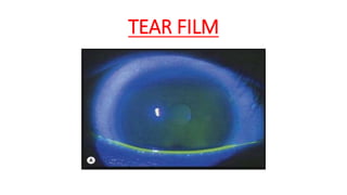
- 1. TEAR FILM
- 2. LAYERS OF TEAR FILM • Outer Lipid layer • Middle Aqueous layer • Inner Mucin layer
- 5. STRUCTURE • LIPID LAYER • Derived from secretions from Meibomian, Zeiss and Moll glands. • Cholesterol esters , phospholipids and wax • 0.1 µm in thickness • Prevents the overflow of tears • Retards their evaporation
- 6. • AQUEOUS LAYER • Secreted by the lacrimal gland and the accessory glands of Krause and Wolfring. • Responsible for reflex and basal secretion. • 10 µm in thickness • Main bulk of the tear film. • Aqueous solution of inorganic salts, glucose, urea, proteins and glycoproteins. • Lysozyme, lactoferrin, albumin immunoglobulin A are the main constituents of protein fraction. • Functions: • 1. provides atmospheric oxygen to the corneal epithelium • 2. washes away debris • 3. Contains antibacterial substances like lysozyme.
- 7. • MUCOUS LAYER • Innermost layer • Secreted by the conjunctival goblet cells. • The superficial epithelial cells of the cornea and conjunctiva produce transmembrane mucins that form their glycocalyx. • Plays a vital role in the stability of the tear film. • Clear corneal epithelium is hydrophobic surface. The mucin gets adsorbed on the cell membrane of epithelial cells and anchored by their microvilli forming a new hydrophilic surface. • Provides a slippery coating over the foreign bodies, thereby protecting the cornea and conjunctiva against abrasive effects of particles.
- 11. TEAR SECRETION BASAL SECRETION • Tears are continuously secreted by the accessory glands. • Keeps the cornea moist, lubricates the eye and help to keep it clear of dust. REFLEX SECRETION • Due to irritation of the eye by foreign particles. • Evaporation of tear film. • Bright light • These reflex tears attempt to wash out irritants that have come in contact with the eye. • By main lacrimal gland.
- 12. FORMATION OF PRE-OCULAR TEAR FILM
- 13. MECHANISM OF TEAR FILM BREAKUP
- 16. • On eye closure: • Contraction of pretarsal orbicularis oculi shortens the ampulla and canaliculi and propels the tear fluid present into lacrimal sac. • Contraction of preseptal fibres of orbicularis pulls the lacrimal fascia and lacrimal sac, thereby opening the normally closed lacrimal sac. This produces negative pressure and draws tears from canaliculi into the lacrimal sac.
- 17. • ON EYE OPENING:
- 20. TEAR FILM ABNORMALITIES • AQUEOUS DEFICIENCY (Keratoconjunctivitis sicca) Decreased secretion by the lacrimal gland Obstruction of the lacrimal gland Burns, inflammation. Sjogren’s syndrome, idiopathic hyposecretion.
- 21. • MUCIN DEFICIENCY • Destruction of the normal conjunctival architecture (goblet cells). • Xerophthalmia, Stevens Johnsons syndrome, Trachoma, Burns
- 22. • LIPID ABNORMALITIES • Absence of Meibomian gland openings • Congenital anhidrotic ectodermal dysplasia • Destruction of Meibomian glands • Burns, chronic blepharitis
- 23. • LID SURFACE ABNORMALITIES • Exposure keratitis due to eyelid surgeries, trauma, Bell’s palsy, incomplete lid closure
- 24. TESTS FOR TEAR FILM ADEQUACY
- 25. • TEAR FILM BREAK UP TIME • Defined as the interval between a complete blink and appearance of the first randomly distributed dry spot on the cornea. • Noted after instilling a drop of fluorescein and examining in the cobalt blue light. • 10 seconds is the cut off point for normal individuals. • Mucin deficiency
- 26. FLUORESCEIN STAINING • The intact corneal epithelium because of its high lipid content resists penetration of water soluble fluorescein and is not stained by it. • Any break in the epithelium barrier permits fluorescein penetration and staining.
- 27. ROSE BENGAL STAINING • Stains mucous, debris and devitalized cells of corneal and conjunctival epithelium.
- 28. LISSAMINE GREEN STAINING • Vital staining properties almost identical with those of rose Bengal. • Stains degenerated and dead cells and mucus. • Less irritating as compared to rose Bengal.
- 29. MARGINAL TEAR STRIP CHARACTERISTICS • Marginal tear meniscus is a continuous, full and slightly concave meniscus formed by tears between the eyelid margin and the inferior bulbar conjunctiva where the lid touches the globe. • 0.5- 1 mm is considered as normal. • Less than 1 suggests tear deficiency.
- 30. CONJUNCTIVAL IMPRESSION CYTOLOGY • Non- invasive technique of impression cytology, where conjunctival impressions are taken to examine cellular structure. • Cells are obtained by using cellulose acetate filter material to make impression.
- 31. HYPERLACRIMATION PRIMARY • direct stimulation • Lacrimal gland tumours SECONDARY (REFLEX) Stimulation of sensory branches of 5th nerve due to irritation of conjunctiva and cornea. Trichiasis, corneal ulcers, trauma, allergic conjunctivitis.
- 32. EPIPHORA • Inadequate drainage of tears. • PHYSIOLOGICAL: • Lacrimal pump failure, weakness of the orbicularis muscle • MECHANICAL : • Obstruction at the level of punctum, canaliculus, lacrimal sac, or nasolacrimal sac.
- 33. QUESTIONS • Name 2 investigations for dry eye • Draw the layers of Tear film • Functions of tear film