
Recommended
Recommended
More Related Content
Similar to AFM.pptx
Similar to AFM.pptx (20)
Recently uploaded
Recently uploaded (20)
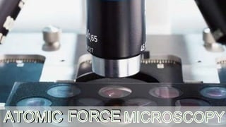
AFM.pptx
- 3. HISTORY • In 1929 Shmalz described Stylus Profiler. • In 1950 Becker suggested oscillating the probe that approach contact with surface. • In 1971 Young described non contact type stylys. • In 1982 Binning and Rohrer described STM(Scanning Tunnelling Microscope). • AFM was invented in 1986 by Binning.
- 4. ATOMIC FORCE MICROSCOPY Atomic force microscopy or scanning force microscopy is a very-high-resolution type of scanning probe microscopy, with demonstrated resolution on the order of fractions of a nanometer, more than 1000 times better than the optical diffraction limit. It is an invaluable tool not only to obtain high-resolution topographical images, but also to determine certain physical properties of specimens, such as their mechanical properties and composition. The atomic force microscope (AFM) was developed to overcome a basic drawback with STM – it can only image conducting or semiconducting surfaces. The AFM has the advantage of imaging almost any type of surface, including polymers, ceramics, composites, glass, and biological samples. Binnig, Quate, and Gerber invented the AFM in 1985. Their original AFM consisted of a diamond shard attached to a strip of gold foil.
- 5. THE FIRST ORIGINAL ATOMIC FORCE MICROSCOPE
- 6. BASIC COMPONENTS • The basic components of an Atomic force microscope include : • Photo-detector and feedback control • Probe • Scanner • Cantilever and tip • Laser
- 7. PROBE
- 8. CANTILEVER
- 9. SCANNER
- 10. PHOTO DETECTOR AND FEEDBACK CONTROL
- 11. OVERVIEW OF COMPONENTS AND FUNCTIONS
- 12. PATHWAY
- 15. TAPPING MODE
- 16. NON CONTACT MODE
- 17. ADVANTAGES DISADVANTAGES ADVANTAGES AND DISADVANTAGES OF AFM ‣ Easy sample preparation. ‣ Non-destructive imaging. ‣ Accurate height information. ‣ Works in vacuum , air and liquids. ‣ Living systems can be studied. ‣ Sample not required to be conductive. ‣ Polymers , ceramics , glass , metals , ‣ Limited vertical range. ‣ Limited magnification range. ‣ Highly dependent on AFM probes. ‣ Tip or sample can be damaged.
- 18. APPLICATIONS A. Digitally images a topographical surface. B. Determines the roughness of a surface sample or to measure the thickness of a crystal growth layer. C. Any sample like ceramic material , human cells or individual molecules of DNA. D. In biological applications: 1.Study of unfolding particles 2.Imagining of biomolecules 3.Force measurements in real solvent environments 4.Antibody-Antigen Binding studies 5.Ligand-Receptor Binding studies 6.Binding Forces of complimentary DNA strands
- 20. SUMMARY • AFM stands for Atomic Force Microscopy. • It works by scanning a probe over the sample surface, building up a map of the height or topography of the surface as it goes along. • AFM microscopy is different from other microscopes as it physically feels the sample’s surface with a sharp probe , building up a map of the height of sample’s surface. • It provides single atomic level structure so provides high resolution. • It doesn’t need focusing , illumination , depth of field.
- 21. • Main components of the AFM instrument include: • Microscope stage : Moving AFM tip , Sample holder , Force sensor • Control electronics : Optical microscope , Vibration controller • Computer : The control electronics usually takes the form of a large box interfaced to both the microscope stage and the computer.
- 22. AFM has 3 basic modes of operation: 1. Contact mode : Strong , Repulsive • High Resolution Images • Fastest of all the topographic modes. • Sensitive to the nature of sample.
- 23. 2. Non-Contact Mode : Weak , Attractive • Oscillating modes can measure images with a small probe-sample force. 3. Tapping Mode : Strong , Repulsive • No Capillary effect. • Amplitude signals are used in feedback. • Used for imaging in air.