SlideShare a Scribd company logo
1 of 1
Download to read offline
MRI In Vivo Assessment of Optic Nerve
                                                                                Degeneration in Experimental Glaucoma
                          Miller J. Ogidigben1, Alex Coimbra2, Lynn O’Neill-Davis1, M. Holahan2, Cathy Thut1, J. Cook2, D. Williams2
                                                      1
                                                       Ophthalmics Research, and 2Imaging Department, Merck Research Laboratories, West Point, PA, United States


                              Purpose                                                                                       Methods (continued)                                                                                                           Results
Glaucoma is a multifactorial neurodegenerative disease of the optic nerve        • Reconstructed images and parametric maps reoriented to standard position.            In normotensive eyes, IOP was 15.0±1.7 mmHg (mean±S.D.) and ON area was                     Figure 4. Correlation between diffusivity and IOP. Scatter
(ON) with pathophysiological consequences of unilateral or bilateral             • The chiasms were identified and cross-sectional areas (CAs) were estimated on the     6.4±0.8 mm2. In contrast, IOP in hypertensive eyes was 34.1±6.6 mmHg and                              plots showing relationship between intraocular
blindness. The clinical management is currently limited to intraocular             chiasm central coronal planes.                                                       ON area was 4.5±4.7 mm2 (single-sided t-test, P=0.027). ON CAs correlated with                        pressure and diffusivity parameters, axial and
pressure (IOP) reduction strategy. Current methods for examining ON                                                                                                     IOPs (Figure 2, Pearson’s r=0.66, P=0.008). DTI diffusivity parameters in ON                          radial diffusivity, and ADC (top panel) and
                                                                                 • CAs of left and right ONs were computed 4 mm anterior to the chiasm plane
degeneration in glaucoma are ex vivo preclinical studies and in vivo imaging                                                                                            of normotensive eyes were 1.16±0.2 and 0.71±0.23 s/mm2 for axial and radial                           fractional anisotropy (bottom panel) of the
                                                                                   (Figure 1).
of nerve fiber layer in the periphery of the ON head. The present study                                                                                                  diffusivity, respectively. In the ON of hypertensive eyes, those parameters were                      optic nerve. All diffusivity parameters of the ON
assessed the ability of MRI to quantify degeneration of the ON in the retro-     • Four DTI parameters were analyzed: axial and radial diffusivity; fractional          0.95±0.19 and 0.47±0.12 s/mm2, respectively (P=0.034 for axial and P=0.013 for
                                                                                   anisotropy (FA); and apparent diffusion coefficient (ADC).
                                                                                                                                                                                                                                                                              increased as a linear function of IOP. Fractional
orbital space of laser-induced glaucoma monkeys, and shows that MRI                                                                                                     radial diffusivity). Fractional anisotropy was also lower in the ON of the hypertensive
                                                                                                                                                                                                                                                                              anisotropy decreased with increasing IOP.
renders quantitative indices that correlate with atrophy and IOP and may         • Intraocular pressure (IOP) was measured in all 16 eyes.                              eye (0.35±0.09) when compared with the normotensive eye (0.45±0.07, single-
be used to study IOP-induced ON degeneration (radial with respect to the                                                                                                sided t-test, P=0.0167). Finally, the overall ADC in the ON of the hypertensive eye
                                                                                 • However, data from one eye was dropped because of failure to obtain a reliable                                                                                                                                  1.6
nerve’s main axis).                                                                                                                                                     was larger (0.86±0.23 s/mm2) than in the normotensive eye (0.63±0.14 s/mm2,
                                                                                   IOP measurement.
                                                                                                                                                                        P=0.017). Pearson correlation coefficients between IOP and axial and radial                                                 1.4
                                                                                 • Comparative statistical analysis was performed between ON MRI parameters of
                         Introduction                                              normotensive and hypertensive eyes. Correlation between ON parameters and
                                                                                                                                                                        diffusivity, FA, and ADC were 0.58 (P=0.02), 0.71 (P=0.003), -0.59 (P=0.02), and
                                                                                                                                                                        0.67 (P=0.006), respectively (Figure 3).


                                                                                                                                                                                                                                                                      Diffusivity (s/mm2)
                                                                                                                                                                                                                                                                                                   1.2
                                                                                   IOPs was also assessed.
The animal model of glaucoma that is close to the human disease and
most frequently used in glaucoma drug discovery studies is the monkey            Atrophy                                                                                Diffusivity                                                                                                                1.0
laser photocoagulation model. This model, unlike human glaucoma, is an                                                                                                  Figure 3. Typical diffusivity parametric maps and respective
                                                                                 Figure 1. Typical horizontal (top) and coronal (bottom) views of                                                                                                                                                  0.8
artificial model that induces intraocular pressure (IOP) elevation in two                                                                                                          anatomical images.
                                                                                           the chiasm and ON delineated by yellow contours.
to three months. However, like human glaucoma, the monkey model of                                                                                                                                                                                                                                 0.6
elevated IOP induced by repeated circumferential laser photocoagulation
of the trabecular meshwork is considered by many to be the best choice of                                                                                                                                                                                                                                                                                          axial diff
                                                                                                                                                                                                           chiasm                                                                                  0.4
animal models to study human glaucoma. The anatomy of the iridocorneal                                                                                                                                       eyes                                                                                                                                                  radial diff
angle and physiology in the monkey eye are similar to the human eye. The
                                                                                                                                                                                     Anatomy                                                                                                       0.2
                                                                                                                                                                                                                                                                                                                                                                   ADC
sustained elevated IOP in the monkey glaucoma model is associated with
a reduction of outflow facility, nerve fiber layer defects, and progressive                                                                                                                                                                                                                          0.0
enlargement of the cup-to-disc ratio, similar to human chronic open-angle                                                                                                            Fractional                                                                                                          5              15               25               35               45
glaucoma. The IOP of the monkey glaucoma model responds to single and                                                                                                                                                                                                                                                                 IOP (mmHg)
multiple dosing of antiglaucoma drugs in a manner similar to humans.The
                                                                                                                                                                                     Anisotropy
effects of laser treatment on uveoscleral outflow and aqueous flow, two
parameters that are important in the control of IOP, showed that aqueous                                                                                                                                                                                                                           0.6


                                                                                                                                                                                                                                                                      Fractional Anisotropy (FA)
humor dynamics could be understood in this model. The model, therefore,                                                                                                              Trace = 3x


                                                                                                                                                                          DTI Data
                                                                                                                                                                                                                                                                                                   0.6
provides for a better prediction of the human response to a drug. The optic                                                                                                          Mean Diff
nerves of laser-induced glaucoma monkeys show similar characteristics of                                                                                                                                                                                                                           0.5                                                           FA
cupping as humans, and visual field changes have been demonstrated in
clinical studies. However, it has been assumed that degenerating ganglion                                                                                                                                                                                                                          0.5
cell axons could result in atrophic or degenerative optic nerve. Since ocular                                                                                                        Axial                                                                                                         0.4
imaging technology is primarily restricted to use in viewing inside the eye,                                                                                                         Diffusivity
we employed magnetic resonance imaging technology to explore regions                                                                                                                                                                                                                               0.4
of the optic nerve beyond the globe in a monkey model of elevated IOP.
The objective of this study was to assess diffusion tensor imaging (DTI)                                                                                                                                                                                                                           0.3
parameters as a biomarker for optic nerve degeneration in monkeys with
                                                                                                                                                                                     Radial
                                                                                                                                                                                                                                                                                                   0.3
chronically elevated IOP.                                                                                                                                                            Diffusivity
                                                                                                                                                                                                                                                                                                   0.2
                              Methods                                                                                                                                                              right        left                   right       left
                                                                                                                                                                                                                                                                                                         5              15              25
                                                                                                                                                                                                                                                                                                                                      IOP (mmHg)
                                                                                                                                                                                                                                                                                                                                                 35                        45
                                                                                 Figure 2. ON atrophy and IOP. Scatter plot showing relationship
Animal:                                                                                    between intraocular pressure and cross-sectional area
Female cynomolgus monkeys (Macaca fascicularis) (3-5 kg) were maintained                   of the optic nerve of corresponding eye in monkeys with
on a 12-hour light/dark cycle during these experiments. Animal care and                    experimental glaucoma. Cross-sectional area of the ON
treatment in this investigation complied with the ARVO resolution in the
                                                                                           is reduced as a linear function of IOP.
use of animals for research, and all procedures had prior approval by the
Institutional Animal Care and Use Committee (IACUC) of Merck & Co., Inc.
• Experiments were conducted in 8 cynomolgus monkeys (10-11 yrs)
  scanned ~2 years after photocoagulation of the right eye trabecular
                                                                                                                8.0                                                                                                                                   Conclusion
                                                                                      ON Cross-sectional Area


  meshwork to induce increase of intraocular pressure.
                                                                                                                7.0
                                                                                                                                                                           This study shows that MRI may be used to study ON degeneration induced by increased IOP in vivo. Longitudinal studies will be
Imaging:                                                                                                        6.0
                                                                                                                                                                           required to determine the onset and rate of optic nerve degeneration to confirm the suitability of this MRI methodology to delineate
• Siemens Trio 3T magnet.
                                                                                                                5.0                                                        surrogate markers for disease progression and address potential benefit of novel therapies to glaucoma patients.
                                                                                              (mm2)


• T1 weighted MPRAGE pulse sequence (TR/TE/TI/FA=1.47/4.38/870/12)
  was used to obtain high-resolution images (0.5x0.5x0.8 mm3) of the                                            4.0
  animals’ whole heads.                                                                                                          y = -0.1122x + 8.2041
                                                                                                                3.0
• Diffusion tensor imaging (DTI) scans to assess integrity of the ON                                                                 R2 = 0.4321
  (TR/TE/TI=11.4 s/118ms/2.2 s; beta=0, 1000 s/mm2, 30 diffusion-                                               2.0
  sensitizing gradient directions, image resolution 1.5x1.5x1.5 mm3).                                           1.0
Analysis:
                                                                                                                0.0
• Parametric maps of diffusivity and diffusion anisotropy extracted from the
                                                                                                                      0.0     10.0      20.0       30.0   40.0   50.0
                                                                                                                                                                                                                                                                    Reference
  DTI data (DTI Studio).
                                                                                                                                                                                                                                                                    1. Radius and Pederson. Arch Ophthalmol. 1984.
                                                                                                                                         IOP (mmHg)

                                                                                                                                                                                                                                                                                                   Copyright ©2009 Merck & Co., Inc., Whitehouse Station, NJ, USA. All Rights Reserved.

                                                                                                                                                                                                                    2009-0359ogidigbe_ARVO.indd 04/22/09 ARVO, 5/3/2009 Final Size: 66” x 44”, Scale 200%

More Related Content

What's hot

anomalous retinal correspondence
anomalous retinal correspondenceanomalous retinal correspondence
anomalous retinal correspondenceRajeshwori
 
Accommodation of eye
Accommodation of eye Accommodation of eye
Accommodation of eye Rohit Rao
 
Corneal Astigmatism
Corneal AstigmatismCorneal Astigmatism
Corneal Astigmatismkaushyk
 
Diplopia charting
Diplopia charting Diplopia charting
Diplopia charting ANUJA DHAKAL
 
Anomalies of accomodation ‫‬
Anomalies of accomodation ‫‬Anomalies of accomodation ‫‬
Anomalies of accomodation ‫‬Ayat AbuJazar
 
Accommodation insufficiency treatment
Accommodation insufficiency treatment Accommodation insufficiency treatment
Accommodation insufficiency treatment Amrit Pokharel
 
Vergence and its anomalies
Vergence and its anomaliesVergence and its anomalies
Vergence and its anomaliesNandhu Krishna
 
Sensory & motor evaluation of strabismus
Sensory & motor evaluation of strabismusSensory & motor evaluation of strabismus
Sensory & motor evaluation of strabismusDevdutta Nayak
 
Perimetry by Pandian M, Dept of Physiology, DYPMC, KOP, MH
Perimetry by Pandian M, Dept of Physiology, DYPMC, KOP, MHPerimetry by Pandian M, Dept of Physiology, DYPMC, KOP, MH
Perimetry by Pandian M, Dept of Physiology, DYPMC, KOP, MHPandian M
 
Sensory Adaptation to Strabismus
Sensory Adaptation to StrabismusSensory Adaptation to Strabismus
Sensory Adaptation to StrabismusHossein Mirzaie
 

What's hot (20)

Synoptophore 2
Synoptophore 2Synoptophore 2
Synoptophore 2
 
anomalous retinal correspondence
anomalous retinal correspondenceanomalous retinal correspondence
anomalous retinal correspondence
 
Aniseikonia
Aniseikonia Aniseikonia
Aniseikonia
 
Accommodation of eye
Accommodation of eye Accommodation of eye
Accommodation of eye
 
Vergence
VergenceVergence
Vergence
 
Corneal Astigmatism
Corneal AstigmatismCorneal Astigmatism
Corneal Astigmatism
 
Diplopia charting
Diplopia charting Diplopia charting
Diplopia charting
 
Anomalies of accomodation ‫‬
Anomalies of accomodation ‫‬Anomalies of accomodation ‫‬
Anomalies of accomodation ‫‬
 
Accommodation insufficiency treatment
Accommodation insufficiency treatment Accommodation insufficiency treatment
Accommodation insufficiency treatment
 
Vergence and its anomalies
Vergence and its anomaliesVergence and its anomalies
Vergence and its anomalies
 
Aniso saiful
Aniso saifulAniso saiful
Aniso saiful
 
fixation disparity
fixation disparityfixation disparity
fixation disparity
 
Sensory & motor evaluation of strabismus
Sensory & motor evaluation of strabismusSensory & motor evaluation of strabismus
Sensory & motor evaluation of strabismus
 
Synoptophore
Synoptophore   Synoptophore
Synoptophore
 
Presbyopia
PresbyopiaPresbyopia
Presbyopia
 
Perimetry by Pandian M, Dept of Physiology, DYPMC, KOP, MH
Perimetry by Pandian M, Dept of Physiology, DYPMC, KOP, MHPerimetry by Pandian M, Dept of Physiology, DYPMC, KOP, MH
Perimetry by Pandian M, Dept of Physiology, DYPMC, KOP, MH
 
Sensory Adaptation to Strabismus
Sensory Adaptation to StrabismusSensory Adaptation to Strabismus
Sensory Adaptation to Strabismus
 
spectacle refraction versus ocular refraction
spectacle refraction versus ocular refractionspectacle refraction versus ocular refraction
spectacle refraction versus ocular refraction
 
Anisometropia
AnisometropiaAnisometropia
Anisometropia
 
Accommodation
AccommodationAccommodation
Accommodation
 

Viewers also liked

Ministry of National planning
Ministry of National planningMinistry of National planning
Ministry of National planningTransformNG
 
Training and development # 4
Training and development # 4Training and development # 4
Training and development # 4Sorab Sadri
 
Maycol And Lilian
Maycol And LilianMaycol And Lilian
Maycol And Lilianlilianesca
 
The Arab Economic Forum - History Highlights
The Arab Economic Forum - History HighlightsThe Arab Economic Forum - History Highlights
The Arab Economic Forum - History HighlightsAl-Iktissad Wal-Aamal
 
Changing paradigms of corporate social responsibility
Changing paradigms of corporate social responsibilityChanging paradigms of corporate social responsibility
Changing paradigms of corporate social responsibilitySorab Sadri
 
Verd Fort9
Verd Fort9Verd Fort9
Verd Fort9allaura2
 
Organisational culture
Organisational cultureOrganisational culture
Organisational cultureSorab Sadri
 
key elements to make the most out of enterprise mobility efforts
key elements to make the most out of enterprise mobility effortskey elements to make the most out of enterprise mobility efforts
key elements to make the most out of enterprise mobility effortsSpace-O Technologies
 
Anuncios Interior Contra
Anuncios Interior ContraAnuncios Interior Contra
Anuncios Interior Contraammnessia
 

Viewers also liked (12)

Arvo 2013 presentation
Arvo 2013 presentation Arvo 2013 presentation
Arvo 2013 presentation
 
AMPK
AMPKAMPK
AMPK
 
Grana5
Grana5Grana5
Grana5
 
Ministry of National planning
Ministry of National planningMinistry of National planning
Ministry of National planning
 
Training and development # 4
Training and development # 4Training and development # 4
Training and development # 4
 
Maycol And Lilian
Maycol And LilianMaycol And Lilian
Maycol And Lilian
 
The Arab Economic Forum - History Highlights
The Arab Economic Forum - History HighlightsThe Arab Economic Forum - History Highlights
The Arab Economic Forum - History Highlights
 
Changing paradigms of corporate social responsibility
Changing paradigms of corporate social responsibilityChanging paradigms of corporate social responsibility
Changing paradigms of corporate social responsibility
 
Verd Fort9
Verd Fort9Verd Fort9
Verd Fort9
 
Organisational culture
Organisational cultureOrganisational culture
Organisational culture
 
key elements to make the most out of enterprise mobility efforts
key elements to make the most out of enterprise mobility effortskey elements to make the most out of enterprise mobility efforts
key elements to make the most out of enterprise mobility efforts
 
Anuncios Interior Contra
Anuncios Interior ContraAnuncios Interior Contra
Anuncios Interior Contra
 

Similar to 2009 0359ogidigbe Arvo Final

Vision therapy in_adults_with_convergence.11
Vision therapy in_adults_with_convergence.11Vision therapy in_adults_with_convergence.11
Vision therapy in_adults_with_convergence.11Yesenia Castillo Salinas
 
Vision therapy in_adults_with_convergence_insufficiency_clinical_and_function...
Vision therapy in_adults_with_convergence_insufficiency_clinical_and_function...Vision therapy in_adults_with_convergence_insufficiency_clinical_and_function...
Vision therapy in_adults_with_convergence_insufficiency_clinical_and_function...Yesenia Castillo Salinas
 
Vision therapy in_adults_with_convergence.11
Vision therapy in_adults_with_convergence.11Vision therapy in_adults_with_convergence.11
Vision therapy in_adults_with_convergence.11Yesenia Castillo Salinas
 
Electrophysiological assessment of optic neuritis: is there still a role
Electrophysiological assessment of optic neuritis: is there still a roleElectrophysiological assessment of optic neuritis: is there still a role
Electrophysiological assessment of optic neuritis: is there still a roleClare Fraser
 
The Baby and the Bathwater: Signal and Noise in Psychiatric Neuroimaging
The Baby and the Bathwater: Signal and Noise in Psychiatric NeuroimagingThe Baby and the Bathwater: Signal and Noise in Psychiatric Neuroimaging
The Baby and the Bathwater: Signal and Noise in Psychiatric Neuroimagingwef
 
Positron-emission tomography studies of cross-modality inhibition in selectiv...
Positron-emission tomography studies of cross-modality inhibition in selectiv...Positron-emission tomography studies of cross-modality inhibition in selectiv...
Positron-emission tomography studies of cross-modality inhibition in selectiv...Dr Brendan O'Sullivan
 
Assink Lockwood Duffy poster 7-26-2015
Assink Lockwood Duffy poster 7-26-2015Assink Lockwood Duffy poster 7-26-2015
Assink Lockwood Duffy poster 7-26-2015Cormac Assink
 
[KHBM] Application of network analysis based on cortical thickness to obsessi...
[KHBM] Application of network analysis based on cortical thickness to obsessi...[KHBM] Application of network analysis based on cortical thickness to obsessi...
[KHBM] Application of network analysis based on cortical thickness to obsessi...Seung-Goo Kim
 
Laser Blended Vision for Presbyopia:
Laser Blended Vision for Presbyopia: Laser Blended Vision for Presbyopia:
Laser Blended Vision for Presbyopia: London Vision Clinic
 
Tomographic fundus features in Pseudoxanthoma Elasticum
Tomographic fundus features in Pseudoxanthoma Elasticum Tomographic fundus features in Pseudoxanthoma Elasticum
Tomographic fundus features in Pseudoxanthoma Elasticum Abdallah Ellabban
 
Binocular balance in_normal_vision_and_its.9
Binocular balance in_normal_vision_and_its.9Binocular balance in_normal_vision_and_its.9
Binocular balance in_normal_vision_and_its.9Yesenia Castillo Salinas
 
Optical Coherence Tomography in Multiple Sclerosis: a Structural & Functional...
Optical Coherence Tomography in Multiple Sclerosis: a Structural & Functional...Optical Coherence Tomography in Multiple Sclerosis: a Structural & Functional...
Optical Coherence Tomography in Multiple Sclerosis: a Structural & Functional...Manal AlRomeih
 
Srk2 Formula in Sri Lanka
Srk2 Formula in Sri LankaSrk2 Formula in Sri Lanka
Srk2 Formula in Sri Lankaaanilanga
 
HRT and GDx VCC
HRT and GDx VCCHRT and GDx VCC
HRT and GDx VCCSSSIHMS-PG
 
Optic Neuritis and OCT in Multiple Sclerosis
Optic Neuritis and OCT in Multiple Sclerosis Optic Neuritis and OCT in Multiple Sclerosis
Optic Neuritis and OCT in Multiple Sclerosis neurophq8
 

Similar to 2009 0359ogidigbe Arvo Final (20)

GDx
GDxGDx
GDx
 
Vision therapy in_adults_with_convergence.11
Vision therapy in_adults_with_convergence.11Vision therapy in_adults_with_convergence.11
Vision therapy in_adults_with_convergence.11
 
Vision therapy in_adults_with_convergence_insufficiency_clinical_and_function...
Vision therapy in_adults_with_convergence_insufficiency_clinical_and_function...Vision therapy in_adults_with_convergence_insufficiency_clinical_and_function...
Vision therapy in_adults_with_convergence_insufficiency_clinical_and_function...
 
Vision therapy in_adults_with_convergence_insufficiency_clinical_and_function...
Vision therapy in_adults_with_convergence_insufficiency_clinical_and_function...Vision therapy in_adults_with_convergence_insufficiency_clinical_and_function...
Vision therapy in_adults_with_convergence_insufficiency_clinical_and_function...
 
Vision therapy in_adults_with_convergence.11
Vision therapy in_adults_with_convergence.11Vision therapy in_adults_with_convergence.11
Vision therapy in_adults_with_convergence.11
 
Electrophysiological assessment of optic neuritis: is there still a role
Electrophysiological assessment of optic neuritis: is there still a roleElectrophysiological assessment of optic neuritis: is there still a role
Electrophysiological assessment of optic neuritis: is there still a role
 
The Baby and the Bathwater: Signal and Noise in Psychiatric Neuroimaging
The Baby and the Bathwater: Signal and Noise in Psychiatric NeuroimagingThe Baby and the Bathwater: Signal and Noise in Psychiatric Neuroimaging
The Baby and the Bathwater: Signal and Noise in Psychiatric Neuroimaging
 
Positron-emission tomography studies of cross-modality inhibition in selectiv...
Positron-emission tomography studies of cross-modality inhibition in selectiv...Positron-emission tomography studies of cross-modality inhibition in selectiv...
Positron-emission tomography studies of cross-modality inhibition in selectiv...
 
pòster-anglès-ok2
pòster-anglès-ok2pòster-anglès-ok2
pòster-anglès-ok2
 
Aniseikonia
AniseikoniaAniseikonia
Aniseikonia
 
Assink Lockwood Duffy poster 7-26-2015
Assink Lockwood Duffy poster 7-26-2015Assink Lockwood Duffy poster 7-26-2015
Assink Lockwood Duffy poster 7-26-2015
 
[KHBM] Application of network analysis based on cortical thickness to obsessi...
[KHBM] Application of network analysis based on cortical thickness to obsessi...[KHBM] Application of network analysis based on cortical thickness to obsessi...
[KHBM] Application of network analysis based on cortical thickness to obsessi...
 
Laser Blended Vision for Presbyopia:
Laser Blended Vision for Presbyopia: Laser Blended Vision for Presbyopia:
Laser Blended Vision for Presbyopia:
 
Tomographic fundus features in Pseudoxanthoma Elasticum
Tomographic fundus features in Pseudoxanthoma Elasticum Tomographic fundus features in Pseudoxanthoma Elasticum
Tomographic fundus features in Pseudoxanthoma Elasticum
 
Binocular balance in_normal_vision_and_its.9
Binocular balance in_normal_vision_and_its.9Binocular balance in_normal_vision_and_its.9
Binocular balance in_normal_vision_and_its.9
 
Optical Coherence Tomography in Multiple Sclerosis: a Structural & Functional...
Optical Coherence Tomography in Multiple Sclerosis: a Structural & Functional...Optical Coherence Tomography in Multiple Sclerosis: a Structural & Functional...
Optical Coherence Tomography in Multiple Sclerosis: a Structural & Functional...
 
Srk2 Formula in Sri Lanka
Srk2 Formula in Sri LankaSrk2 Formula in Sri Lanka
Srk2 Formula in Sri Lanka
 
HRT and GDx VCC
HRT and GDx VCCHRT and GDx VCC
HRT and GDx VCC
 
Optic Neuritis and OCT in Multiple Sclerosis
Optic Neuritis and OCT in Multiple Sclerosis Optic Neuritis and OCT in Multiple Sclerosis
Optic Neuritis and OCT in Multiple Sclerosis
 
Visual acuity by Optom Ankit Varshney
Visual acuity by Optom Ankit VarshneyVisual acuity by Optom Ankit Varshney
Visual acuity by Optom Ankit Varshney
 

2009 0359ogidigbe Arvo Final

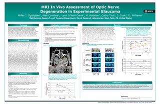
  • 1. MRI In Vivo Assessment of Optic Nerve Degeneration in Experimental Glaucoma Miller J. Ogidigben1, Alex Coimbra2, Lynn O’Neill-Davis1, M. Holahan2, Cathy Thut1, J. Cook2, D. Williams2 1 Ophthalmics Research, and 2Imaging Department, Merck Research Laboratories, West Point, PA, United States Purpose Methods (continued) Results Glaucoma is a multifactorial neurodegenerative disease of the optic nerve • Reconstructed images and parametric maps reoriented to standard position. In normotensive eyes, IOP was 15.0±1.7 mmHg (mean±S.D.) and ON area was Figure 4. Correlation between diffusivity and IOP. Scatter (ON) with pathophysiological consequences of unilateral or bilateral • The chiasms were identified and cross-sectional areas (CAs) were estimated on the 6.4±0.8 mm2. In contrast, IOP in hypertensive eyes was 34.1±6.6 mmHg and plots showing relationship between intraocular blindness. The clinical management is currently limited to intraocular chiasm central coronal planes. ON area was 4.5±4.7 mm2 (single-sided t-test, P=0.027). ON CAs correlated with pressure and diffusivity parameters, axial and pressure (IOP) reduction strategy. Current methods for examining ON IOPs (Figure 2, Pearson’s r=0.66, P=0.008). DTI diffusivity parameters in ON radial diffusivity, and ADC (top panel) and • CAs of left and right ONs were computed 4 mm anterior to the chiasm plane degeneration in glaucoma are ex vivo preclinical studies and in vivo imaging of normotensive eyes were 1.16±0.2 and 0.71±0.23 s/mm2 for axial and radial fractional anisotropy (bottom panel) of the (Figure 1). of nerve fiber layer in the periphery of the ON head. The present study diffusivity, respectively. In the ON of hypertensive eyes, those parameters were optic nerve. All diffusivity parameters of the ON assessed the ability of MRI to quantify degeneration of the ON in the retro- • Four DTI parameters were analyzed: axial and radial diffusivity; fractional 0.95±0.19 and 0.47±0.12 s/mm2, respectively (P=0.034 for axial and P=0.013 for anisotropy (FA); and apparent diffusion coefficient (ADC). increased as a linear function of IOP. Fractional orbital space of laser-induced glaucoma monkeys, and shows that MRI radial diffusivity). Fractional anisotropy was also lower in the ON of the hypertensive anisotropy decreased with increasing IOP. renders quantitative indices that correlate with atrophy and IOP and may • Intraocular pressure (IOP) was measured in all 16 eyes. eye (0.35±0.09) when compared with the normotensive eye (0.45±0.07, single- be used to study IOP-induced ON degeneration (radial with respect to the sided t-test, P=0.0167). Finally, the overall ADC in the ON of the hypertensive eye • However, data from one eye was dropped because of failure to obtain a reliable 1.6 nerve’s main axis). was larger (0.86±0.23 s/mm2) than in the normotensive eye (0.63±0.14 s/mm2, IOP measurement. P=0.017). Pearson correlation coefficients between IOP and axial and radial 1.4 • Comparative statistical analysis was performed between ON MRI parameters of Introduction normotensive and hypertensive eyes. Correlation between ON parameters and diffusivity, FA, and ADC were 0.58 (P=0.02), 0.71 (P=0.003), -0.59 (P=0.02), and 0.67 (P=0.006), respectively (Figure 3). Diffusivity (s/mm2) 1.2 IOPs was also assessed. The animal model of glaucoma that is close to the human disease and most frequently used in glaucoma drug discovery studies is the monkey Atrophy Diffusivity 1.0 laser photocoagulation model. This model, unlike human glaucoma, is an Figure 3. Typical diffusivity parametric maps and respective Figure 1. Typical horizontal (top) and coronal (bottom) views of 0.8 artificial model that induces intraocular pressure (IOP) elevation in two anatomical images. the chiasm and ON delineated by yellow contours. to three months. However, like human glaucoma, the monkey model of 0.6 elevated IOP induced by repeated circumferential laser photocoagulation of the trabecular meshwork is considered by many to be the best choice of axial diff chiasm 0.4 animal models to study human glaucoma. The anatomy of the iridocorneal eyes radial diff angle and physiology in the monkey eye are similar to the human eye. The Anatomy 0.2 ADC sustained elevated IOP in the monkey glaucoma model is associated with a reduction of outflow facility, nerve fiber layer defects, and progressive 0.0 enlargement of the cup-to-disc ratio, similar to human chronic open-angle Fractional 5 15 25 35 45 glaucoma. The IOP of the monkey glaucoma model responds to single and IOP (mmHg) multiple dosing of antiglaucoma drugs in a manner similar to humans.The Anisotropy effects of laser treatment on uveoscleral outflow and aqueous flow, two parameters that are important in the control of IOP, showed that aqueous 0.6 Fractional Anisotropy (FA) humor dynamics could be understood in this model. The model, therefore, Trace = 3x DTI Data 0.6 provides for a better prediction of the human response to a drug. The optic Mean Diff nerves of laser-induced glaucoma monkeys show similar characteristics of 0.5 FA cupping as humans, and visual field changes have been demonstrated in clinical studies. However, it has been assumed that degenerating ganglion 0.5 cell axons could result in atrophic or degenerative optic nerve. Since ocular Axial 0.4 imaging technology is primarily restricted to use in viewing inside the eye, Diffusivity we employed magnetic resonance imaging technology to explore regions 0.4 of the optic nerve beyond the globe in a monkey model of elevated IOP. The objective of this study was to assess diffusion tensor imaging (DTI) 0.3 parameters as a biomarker for optic nerve degeneration in monkeys with Radial 0.3 chronically elevated IOP. Diffusivity 0.2 Methods right left right left 5 15 25 IOP (mmHg) 35 45 Figure 2. ON atrophy and IOP. Scatter plot showing relationship Animal: between intraocular pressure and cross-sectional area Female cynomolgus monkeys (Macaca fascicularis) (3-5 kg) were maintained of the optic nerve of corresponding eye in monkeys with on a 12-hour light/dark cycle during these experiments. Animal care and experimental glaucoma. Cross-sectional area of the ON treatment in this investigation complied with the ARVO resolution in the is reduced as a linear function of IOP. use of animals for research, and all procedures had prior approval by the Institutional Animal Care and Use Committee (IACUC) of Merck & Co., Inc. • Experiments were conducted in 8 cynomolgus monkeys (10-11 yrs) scanned ~2 years after photocoagulation of the right eye trabecular 8.0 Conclusion ON Cross-sectional Area meshwork to induce increase of intraocular pressure. 7.0 This study shows that MRI may be used to study ON degeneration induced by increased IOP in vivo. Longitudinal studies will be Imaging: 6.0 required to determine the onset and rate of optic nerve degeneration to confirm the suitability of this MRI methodology to delineate • Siemens Trio 3T magnet. 5.0 surrogate markers for disease progression and address potential benefit of novel therapies to glaucoma patients. (mm2) • T1 weighted MPRAGE pulse sequence (TR/TE/TI/FA=1.47/4.38/870/12) was used to obtain high-resolution images (0.5x0.5x0.8 mm3) of the 4.0 animals’ whole heads. y = -0.1122x + 8.2041 3.0 • Diffusion tensor imaging (DTI) scans to assess integrity of the ON R2 = 0.4321 (TR/TE/TI=11.4 s/118ms/2.2 s; beta=0, 1000 s/mm2, 30 diffusion- 2.0 sensitizing gradient directions, image resolution 1.5x1.5x1.5 mm3). 1.0 Analysis: 0.0 • Parametric maps of diffusivity and diffusion anisotropy extracted from the 0.0 10.0 20.0 30.0 40.0 50.0 Reference DTI data (DTI Studio). 1. Radius and Pederson. Arch Ophthalmol. 1984. IOP (mmHg) Copyright ©2009 Merck & Co., Inc., Whitehouse Station, NJ, USA. All Rights Reserved. 2009-0359ogidigbe_ARVO.indd 04/22/09 ARVO, 5/3/2009 Final Size: 66” x 44”, Scale 200%