
Recommended
More Related Content
What's hot
What's hot (20)
Similar to Red eye syndrome
Similar to Red eye syndrome (20)
Recently uploaded
Recently uploaded (20)
Red eye syndrome
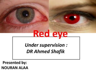
- 1. Under supervision : DR Ahmed Shafik Red eye Presented by: NOURAN ALAA
- 2. Red eye Red eye“ is sign of pathology of anterior or posterior ocular segment, of orbit or of ocular adnexa
- 3. Red Eye Etiologies Infection Inflammation Irritation Allergy(Air pollution , Contact lenses ,Smoke , Fume) Trauma Chemicals Tumor Systemic conditions
- 4. dilation of blood vessels in the eye. differentiation Ciliary injection branches of the anterior ciliary arteries and indicates inflammation of the cornea, iris, or ciliary body. Conjunctival injection the posterior conjunctival blood vessels are more superficial than the ciliary arteries, they produce more redness, Pathophysiology
- 7. Red Eye Disorders: Non-Vision Threatening Blepharitis Hordeolum Chalazion Conjunctivitis Dry eyes Subconjunctival hemorrhage
- 8. Blepharitis Staph blepharitis may occur with seborrhea. associated problems. Marginal infiltrates. Hordeolum. Chalazion. Meibomitis. Marginal infiltrates.
- 9. Acute Hordeolum Acute staph infection of lid External-glands of Zeiss, moll or lash follicle Internal- Meibomian Warm compresses Systemic antibiotics if preseptal cellulitis develops
- 10. Chalazion Obstruction of Meibomian gland with extrusion of lipid into surrounding tissue Lipogranulomatous reaction, not infectious May cause astigmatism secondary to pressure on the cornea
- 12. MEIBOMITIS Meibomian orifice shows erythema and edema with secretions thick and tenacious Often diffusely inflamed lid margins Oral teracycline helpful (doxy 100 BID)
- 13. Phlyctenulosis •Round elevated infiltrate which moves centrally from limbus with “leash of vessels” •Sterile type IV hypersensitivity immune rxn , usually to Staph but may be secondary to T.B., or fungal infections
- 15. Allergic Conjunctivitis Usually allergy to air born allergen. Mediated by IgE. May occur with hay fever, asthma or rhinitis. Associated with itching, hyperemia, chemosis, watery ,mucoid discharge. Topical vasoconstrictors and mast cell stabilizers helpful.
- 16. VERNAL CONJUNCTIVITIS •Seasonally recurring •History of atopy common •Occurs in children and young adults •Hyperemia and chemosis progress to diffuse papillary hypertrophy on upper tarsus
- 17. VIRAL CONJUNCTIVITIS Adenoviral conjunctivitis presents with acute onset of red, watery eyes. Follicular response worse inferiorly. Hemorrhagic or pseudomembranous response can occur.
- 18. Bacterial Conjunctivitis Mucopurulent discharge. Must consider gonococcus since it can cause a perforation-hyperacute, needs systemic antibiotics. And has a preauricular node like Adeno.
- 19. Traumatic Subconjunctival Hemorrhage Bright blood red eye. Normal vision. No pain. May occur in cases of trauma, or in cases of coughing, vomiting, or straining. If traumatic must do thorough exam to other pathology.
- 20. DRY EYE Symptoms of tear deficiency include; FB sensation Tearing Ropy mucus Burning Scratchiness ALL WORSE LATER IN THE DAY or in HEAT< WIND OR LOW HUMIDITY Rose Bengal staining.. Sjogrens syndome xerostomia,and arthritis usually in middle aged women. Tear replacement, plugs, rarely lateral tarsorraphy.
- 21. Pinguecula Benign pathologic change in the bulbar conjunctiva at the palpebral fissure Associated with sun and wind exposure Red secondary to increased vascularity of the lesion Can be intermittently inflamed
- 22. Pterygium
- 23. Red Eye Disorders: Vision Threatening Orbital Cellulitis Scleritis Uveitis Trauma Hyphema Acute glaucoma Corneal infections
- 24. ORBITAL CELLULITIS Lid swelling and erythema +/- Proptosis +/- Conjunctival chemosis and/or injection Reduced motility Pain Fever +/- Optic nerve: decreased vision, APD, disc edema
- 25. Episcleritis May be benign or signify underlying disease Red eye usually localized, but may be diffuse, or nodular Dilated episcleral vessels Mild tenderness and irritation
- 27. Scleromalacia Perforans Usually associated with long standing rheumatoid arthritis. Progressive scleral thinning without signs of inflammation. Large abnormal vessels cross the devitalized area.
- 28. Corneal & Conjunctival Foreign Body Presents with c/o pain, tearing, photophobia and foreign body sensation Foreign body (FB) may be flushed out if superficial, cotton tip after anesthetic If not easily dislodged – can be removed with 25 gauge needle, rust ring with Alger brush Subsequent defect to be treated with antibiotics Flip lid if no FB seen and linear abrasion
- 29. Chemical Injury
- 30. Contact Lens Wear Associated Red Eye Prolonged contact lens wear or poorly fitting lenses may cause a red eye. Severe pain. Tearing. If opacity is noted or corneal infection is suspected,treat as if infected. Bacterial, parasite, fungus are possible pathogens.
- 31. Bacterial Corneal Ulcer Predisposing factors usually include trauma. All may contribute: Immunosuppression. Alcoholism. Aging. Dry eye. Exposed sutures.Contact lens wear. Bullous Keratopathy. Topical steroid use.
- 32. Fungal Corneal Ulcer Can mimic bacterial or viral keratitis. Often occur after trauma with plant or vegetable matter. Aspergillus, and Penicillium occur in otherwise normal eyes wheras Candida occurs in immunocompromised anterior segments. Natamycin5% is available. Bad prognosis ,may need PK.
- 33. Viral Keratitis (HSV) Replicates along the corneal nerves. Decreased corneal sensation. Heals spontaneously in 21 days but Trifluridine 8x/day hastens the process. Avoid steroids unless DISCIFORM or KERATOUVEITIS occurs and then with 1:1antivirals.
- 34. Uveitis Limbal (circumcorneal) flush (redness) Pain Photophobia Decreased vision Pupillary abnormalities AC Rxn possibly hypopyon
- 35. Acute Angle Closure Glaucoma Sudden rise in intraocular High pressure can lead to optic nerve &/or retinal damage, including, but not limited to vascular occlusions pressure ( IOP) Mid-dilated pupil Halos, decrease in vision Pain Red eye Cloudy cornea (corneal edema) Nausea and vomiting Headache
- 36. Red Eye Management Short-term solutions for red eyes Warm compress Cool compress Artificial tear Long-term solutions for red eyes Switch contacts Pay attention todiet Be aware of your surroundings
- 38. References 1- American Academy of ophthalmology http://www.allaboutvision.com/conditions/conjunctivitis.htm https://www.aao.org/eye-health/diseases 2-Health http://www.health.com/eye-health 3- Kansaki Clinical ophthalmology and kansaki signs in ophthalmology 4-Wikipedia https://en.wikipedia.org/wiki American optometric associations https://www.aoa.org/patients-and-public/eye-and-vision- problems/glossary-of-eye-and-vision-
- 39. Thank