SlideShare a Scribd company logo
1 of 7
Download to read offline
NeuroQuantology | January 2022 | Volume 20 | Issue 1 | Page 34-40 | doi: 10.14704/nq.2022.20.1.NQ22005
Mohammed Kadhom Awajel et al / Assessment of Carpal Tunnel Syndrome Severity by Ultrasound and Electrodiagnosis Study
eISSN 1303-5150 www.neuroquantology.com
34
Assessment of Carpal Tunnel Syndrome Severity
by Ultrasound and Electrodiagnosis Study
Mohammed Kadhom Awajel1*, Abdullnasir Hussin2, Mohammed Ab-Khudhur3
Abstract
Carpal tunnel syndrome (CTS) is the upper limb neuropathic entrapment. Electrodiagnostic study (EDX) is the gold
standard for diagnosis. However, EDX is not always abnormal in CTS beside its relatively high cost which requires
the search for alternative modalities.
Objectives: To investigate ultrasound (US) has a function in the evalution of CTS as well as determination disease
severity.
Subjects and Methods: A case-control experiment which involve 50 CTS patients (65 wrists) and 30 healthy
subjects (30 wrists). Electrodiagnostic study involve nerve coduction test (NCS) were performed for all included
wrists, with ultrasound measure the cross-sectional area (CSA) of the median nerve.
Results: The result of 65 affected wrists with CTS is classified for 40 (61.15%) wrist mild disease, 19 wrists
(29.23%) demonstrated a moderate disease and 6 wrists (9.23%) had severe disease. The mean in mild, moderate,
and severe cases, median nerve CSA was determined 11.21±1.22 mm2, 15.09±0.1.14 mm2 and 17.88±0.2.1 mm2,
respectively with highly significant differences between the three categories. Furthermore, all three categories had
higher CSA than control (8.73±0.45 mm2) with highly significant differences. In patients, CSA demonstrated a
significant linked with median distal sensory latency (r = 0.71 8, p <0.001), median motor distal latency (r = 0.818,
p < 0.001) and age (r = 0.316, p = 0.0 1) and a negative significant with median nerve conduction velocity (r= -0.837,
p < 0.001). US demonstrated excellent diagnostic values. The sensitivity and specificity of US in the context of
discriminate patients and controls at cut off value of CSA = 10 mm2 were 92% and 100%, respectively. Furthermore,
US can effectively discriminate between mild, moderate and severe cases CTS with sensitivity levels ranging from
83%- 100% and 94%-100% specificity. The optimal cut off values ranges between 13.2-16.2 mm2.
Conclusions: The CSA of the median nerve is increased and the degree of enlargement significantly correlates with
the parameters of disease severity. However, the CSA of the nerve does not associate with the gender of patients or
the side of the affected wrist. Ultrasound of CST of median nerve has an excellent diagnostic value in the disinguish
between patients and controls, with 92 percent sensitivity and 99 percent specificity, respectively. The optimum cut
off value is 10 mm2. Also, US can effectively distinguish mild, moderate and severe cases CTS with Sensitivity levels
ranging from 83%- 100% and 94%-100% Specificity. The optimal values cut off ranges between 13.2-16.2 mm2.
Key Words: Carpal Tunnel Syndrome, Ultrasound, Median Cross–sectional Area, Nerve Conduction Study,
Electrodiagnostic Study.
DOI Number: 10.14704/nq.2022.20.1.NQ22005 NeuroQuantology 2022; 20(1):34-40
Introduction
Is the neuropathic lesion of the nerve at the region
of wrist that travels during canal along with nine
flexor tendons. It is also is known as the most
prevalent neuropathy of entrapment affecting
upper extremities described by numbness and
tingling, and discomfort or pain in the hand.
Corresponding author: Mohammed Kadhom Awajel
Address: 1*,2,3College of Medicine, University of Baghdad, Iraq.
1*E-mail: awjel_87@yahoo.com
Relevant conflicts of interest/financial disclosures: The authors declare that the research was conducted in the absence of
any commercial or financial relationships that could be construed as a potential conflict of interest.
Received: 02 November 2021 Accepted: 05 December 2021
NeuroQuantology | January 2022 | Volume 20 | Issue 1 | Page 34-40 | doi: 10.14704/nq.2022.20.1.NQ22005
Mohammed Kadhom Awajel et al / Assessment of Carpal Tunnel Syndrome Severity by Ultrasound and Electrodiagnosis Study
eISSN 1303-5150 www.neuroquantology.com
35
It is consider and is remembered as one of the most
significant causes of morbidity in the workplace
(Coraci., 2014). Trauma, pressure increase in the
canal, ischemia lesion to median nerve in the carpal
tunnel are all factors lead to CTS, the normal
pressure ranged from 2 and 10 mm Hg. Carpal
tunnel pressure changes during wrist extension
and flexion leading to an increase 8 to 10 folds.
Therefore, recurrent hand movement is the main
risk factor of carpal tunnel syndrome (Ibrahim et al,
2012). It can be detected by taking a medical
history and performing physical examinations
depend on American Academy of Neurology
practice.
1. Discomfort, pain, paresthesia, espicialy at
night.
2. Sensory fiber involvement.
3. Motor involvement, weakness of the thumb
abduction and hypotrophy of abductor pollicis
brevis.
4. Positive Phalen’s maneuver and/or Tinel’s
sign (Chen 2011).
Electrodiagnosis is the most useful tool for
diagnosing CTS and determining its severity,
however it is considered intrusive, another
diagnostic noninvasive, feasible, simple like high
resolution ultrasonography. (Kang 2012) The
etiology of carpal tunnel is demyelination and
accompanied by axonal loss due to severity In a
nerve electrodiagnosis, a demyelination lesion in
the median nerve reflected in a slowing of the
sensory median nerve's distal latency, If very tiny
discrepancies between the median and ulnar
latencies (usually 0.4–0.5 ms) are discovered in
these sensitive median-versus-ulnar comparison
investigations, they are regarded abnormal.
(Preston, D., C. & Shapiro, B., E. 2021).
Over the last few years, neuromuscular
ultrasonography has become more widely
employed in conjunction with EDX in the
evaluation of patients with neuromuscular
illnesses. (Preston, D., C. & Shapiro, B., E. (2021).
used of ultrasound growing for many factors
simple, accurate, rapid, non-invasive method low
cost, fesible, (coraci 2014). The prevalent criterion
in CTS is increase diameter of nerve
(cross–sectional area >10 mm2 which has a high
sensitivity as 97.9%. (Cara McDonagh, 2015).
When examining nerve, there are a number of
factors to consider. The cross-sectional area (CSA)
above 10mm is significant, and it is the most
important and well-validated parameter. The
ultrasound image of median compression at the
wrist is an enlarged and hypoechoic nerve next to
the entrapment site with loss of the usual fascicular
architecture, which is evaluated using a tracing
method immediately inside the hyperechoic
epineurium. (Descatha, A., L. Huard 2012).
Materials and Methods
A case - control experiment which conducted in
Al-Shaheed Ghazi Al-Hariri teaching hospital, Iraq,
Baghdad in from 2021/1/20 to 2021/7/23 Which
included a total of 50 patients with carpal tunnel
syndrome and 30 normal subjects As control, they
referred from neurosurgery department,
orthopaedic department, electrodiagnosis study
cover NCS (nerve conduction test) perform for all
hands and ultrasound measure median cross-
sectional area, Data were collected via direct
interface with the patients and examination using a
data collecting sheet including; age, gender, date of
examination, RT or LT hand affected or Both.
Exclusion Criteria Were
Patient with diabetic mellitus, Patient with wrist
trauma, Patient with cervical radiculopathy, Patient
connective tissue diseases, Patient with rheumatoid
arthritis, Thyroid disease Natus
electroneuromyography was used, which involved
assessing bilateral motor with sensory fibers of the
ulnar and median nerves and recording abnormal
parameters in the median nerve. The results
classified into mild (sensory fibers affected),
moderate (sensory and motor fibers affected) and
sever (sever motor fiber affected) according to the
neurophysiological reference values' local severity
scale (J.D. Bland, 2000). During examination during
the test procedures, the room temperature was
kept between (25C°-28C°) and the skin
temperature was kept between (36C°-37C°) with a
mean of 36.7C° using a skin thermometer.
The following parameters were studied in sensory
nerve: Amplitude, conduction velocity, distal
latency. When stimulating sensory fiber the current
of Supramaximal stimulation was used to elicit the
maximum potential of the sensory nerve. The
following parameters were studied in motor nerve:
Amplitude, velocity, latency, duration the motor
nerve is stimulated at two sites along its path
(proximal and distal), and the muscle supplied by
the same nerve is recorded. The stimulus intensity
must be high enough to activate all motor nerve
fibers (Preston and Shapiro, 2013). The HD11XE
Philips 2009 was used for the US evaluation, and
NeuroQuantology | January 2022 | Volume 20 | Issue 1 | Page 34-40 | doi: 10.14704/nq.2022.20.1.NQ22005
Mohammed Kadhom Awajel et al / Assessment of Carpal Tunnel Syndrome Severity by Ultrasound and Electrodiagnosis Study
eISSN 1303-5150 www.neuroquantology.com
36
the US model was equipped with linear probe of
(7.5 MHz frequency) at the same time with nerve
conduction study, the setting position is apply to
examine the patient, with his hand open and flat on
the coach, examination done with transverse
direction. We used the upper normal CS area limit
for the median nerve (<10 mm²) (Preston, D., C. &
Shapiro, B., E. (2021) (Cara McDonagh, M ichael
Alexander, David Kane, 2015).
Statistic analyse conducted by S P S S software (S P
S S, Chicago). Data were offered as mean and root
mean square deviation, and analysed with unpaired
Student t - test. Relative operating characteristic
curve (ROC) estimated value diagnostic of cross -
sectional area (CSA) of the median nerve in the
context of discrimination between patients and
controls. Pearson’s r to explore possible correlation
CSA with other variable in patients and controls. P -
value < 0. 0 5 significant difference.
Result
Demographic data of population study, mean
patients age 46.1±11.27 years significantly
different from that of controls group (35.35±8.49
years), no significant distinction in gender
distribution between the two groups. Likewise,
patient group and control group had comparable
weight, height and BMI with no significant
differences There were 65 affected wrists in
patients, of which 38 (58.46%) were right wrist
and 27(41.54%) were left wrists. On the other
wrist, there were 13 right wrists (56.52%) and 10
left wrists (43.4 8 %) in control group with no
significant distinction (Table 1).
Table 1. Demographic data of population study
variables Patients (N= 50) Controls (N=30) P value
Age, years
Mean±SD
Range
46.1±11.27
21-64
35.35±8.49
20-52
<0.001
Gender
Male
Female
20(40%)
30(60%)
12(52.17%)
11(47.83%)
0.330
Height, cm
Mean±SD
Range
75.82±10.17
57-110
75.39±8.13
59-90
0.860
Weight, kg
Mean±SD
Range
164.97±9.23
150-179
167.35±7.44
157-180
0.281
BMI, kg/m2
Mean±SD
Range
27.85±2.88
20.76-38.06
26.89±2.05
23.89-23.25
0.156
Side
Right
Left
38(58.46%)
27(41.54%)
13(56.52%)
10(43.48%)
0.871
Electrophysiological Parameters and
Ultrasonography
The mean time for median sensory latency, median
motor latency and conduction velocity in CTS
patients was 4.18±0.63 ms, 4.0±1.41 ms and
49.62±9.79 m/s, respectively compared with
2.8±0.3 ms, 2.92±0.34 ms and 61.32±4.75 m/s,
respectively in controls with highly significant
differences. However, there was no significant
differentiation between them patients group and
controls group in ulnar nerve sensory distal latency
(2.61±0.29 ms versus 2.57±3.66 ms). According to
the result of ultrasound, mean median CSA in CTS
patients 13.11±2.57 mm2 (range= 8.5-19.9 mm2)
which was much higher than that of controls (mean
= 8.73±0.45 mm2, range= 7.5-9.0 mm2) as shown in
table 2.
Table 2. Electrophysiological parameters and sonography
Variables
CTS (65
wrists)
Controls
(30 wrists)
P-
value
Median N. sensory
distal latency, ms
Mean±SD
Range
4.18±0.63
3.5-6.0
2.8±0.3
2.3-3.3
<0.00
Median N. motor distal
latency, ms
Mean±SD
Range
4.0±1.41
2.0-8.09
2.92±0.34
2.2-3.5
0.001
Median N. conduction
velocity, m/s
Mean±SD
Range
49.62±9.79
25.0-60.0
61.32±4.75
52.0-68.0
<0.001
Ulna N. sensory distal
latency, ms
Mean±SD
Range
2.61±0.29
2.06-3.34
2.57±3.66
2.1-3.32
0.644
Median N. cross
sectional area, mm2
Mean±SD
Range
13.11±2.57
8.5-19.9
8.73±0.45
7.5-9.0
<0.001
Disease Severity
The result of 65 affected wrists with CTS is
classified for, 40 (61.15%) had mild disease, 19
wrists (29.23%) demonstrated a moderate disease
and 6 wrist (9.23%) had severe disease (Figure 1).
NeuroQuantology | January 2022 | Volume 20 | Issue 1 | Page 34-40 | doi: 10.14704/nq.2022.20.1.NQ22005
Mohammed Kadhom Awajel et al / Assessment of Carpal Tunnel Syndrome Severity by Ultrasound and Electrodiagnosis Study
eISSN 1303-5150 www.neuroquantology.com
37
Figure 1. CTS disease severity
Association of MN CSA (median nerve
cross-sectional area) with Disease Severity: The
mean CSA of median nerve in mild, moderate,
severe cases was 11.21±1.22 mm2, 15.09±0.1.14
mm2 and 17.88±0.2.1 mm2, respectively with highly
significant differences between the three
categories. Furthermore, all three categories had
higher CSA than control (8.73±0.45 mm2) with
highly significant differences (Figure 2).
Figure 2. Association of CSA of median Nerve with Disease Severity
**Highly significant difference. Analysis of test
variance was to analyse data
Diagnostic Performance of Ultrasound
Value diagnostic investigated by a Relative
Operating Characteristic (ROC) curve median CSA
(calculated through US) in detection and
differentiation of CTS. Generally, US demonstrated
excellent diagnostic values. In the context
distinguish between CTS patients groub and control
group, area under the curve (AUC) was 0.950 and
95% CI = 0.903 - 0.996, p < 0.001. Sensitivity,
Specificity of US at value cut off of CSA = 10 mm2
were 92% and 100 %, respectively (Figure 3).
Figure 3. Relative operating characteristic curve for Ultrasound
context of discriminate between CTS patients and controls
In the context of discriminate between mild and
moderate CTS, the AUC was 0 .9 74, 95 % CI = 0.
925-1.00, < 0.00 1. Sensitivity, Specificity of the
Ultrasound at value cut-off of CSA = 13.1 mm2 were
95% and 97.5%, respectively (Figure 4).
Figure 4. Relative operating characteristic curve for US in context of
discrimination between mild and moderate CTS
In the context of discrimination mild and severe
CTS, AUC was 1 .0 0, 95 % CI = 1 .0 0 - 1 .0 0,
 < 0 .001. Sensitivity and Specificity of US at cut off
value of CSA = 14.0 mm2 100% for both (Figure 5).
NeuroQuantology | January 2022 | Volume 20 | Issue 1 | Page 34-40 | doi: 10.14704/nq.2022.20.1.NQ22005
Mohammed Kadhom Awajel et al / Assessment of Carpal Tunnel Syndrome Severity by Ultrasound and Electrodiagnosis Study
eISSN 1303-5150 www.neuroquantology.com
38
Figure 5. Relative operating characteristic curve for US in context of
discriminate between mild and severe CTS
In the context of discriminate between moderate
and severe CTS, AUC 0. 8 68, 95 % CI = 0. 645-1.00,
p = 0.008. Sensitivity, Specificity of US at cut off
value of CSA = 16.2 mm2 83% and 94%,
respectively (Figure 6).
Figure 6. Relative operating characteristic curve for US in context of
discriminate between moderate and severe CTS
Discussion
In the present study, patients were older than
controls with a significant difference. This result
corroborates with many previous studies. In A
Turkish study including 547 patients with CTS,
wrists were categorized into four groups normal,
mild, moderate, sever.
After adjusting for BMI, the CTS development risk
raised by a factor of 1.86 for ages thirty–sixty years
and by 4.17 for ages sixty when the age group
eighteen–thirty-five years was used as the control
group. Almost similar result regarding the risk of
older age in development of CTS were reported by
two other studies (Kouyoumdjian, 2002;
Kouyoumdjian, 1999). The precise reasons beyond
this association are not precisely known. However,
it was assumed axon loss, development of nerve
conduction, vessels abnormalities increase with age
(Kommalage, 2011). In the present study, motor
and sensory latency of the median nerve increased
significantly in CTS, while the conduction velocity
of this nerve significantly decreased compared with
the controls. These electrophysiological changes
are the basic for gold standard diagnosis of CTS.
The median nerve is always affected with different
degree of damage. Several mechanisms have been
proposed that results in median nerve injury with
an eventual increase sensory and motor latency
and decrease conduction velocity (Arrori and
Spence, 2008). An interesting finding in this study
was median cross-sectional area was significantly
different on CTS patients than controls, and the CSA
of median was significantly linked with severity
disease. Such results were frequently reported by a
plethora of previous studies (Mohammadi 2012;
Ajeena, 2013; El-Shintenawy 2019). (Tsai 2013)
and (Chan 2011) identified a strong link between
median nerve conduction anomalies detected by
electro diagnostic testing and CSA measured by
ultrasound. In the same context, there was
correlating between CSA medain at the tunnel inlet,
median motor distal latency (DML). As prolonged
median DML is a marker of focal nerve
demyelination across the carpal tunnel, this
provides a biological basis for the US finding in CTS
(Chan 2011). The most important finding in the
present study were that the US had excellent
diagnostic values either discrimination between
control and patients with CTS in the context of CTS
or in the context of discrimination between
different severities of the CTS. In accordance with
these results are many studies worldwide. In many
of these studies the CSA of the median nerve
yielded sensitivities range from sixty-seven to
ninety four percent, and from fifty-seven to ninety
seven percent to specificities, with value cut off 8 .5
to 9.5 percent 15mm2 (Wang 2008; Mondelli 2008;
Padua 2008; Polykandriotis, 2007; Hobson-Webb,
2008). Also, in agreement with the present result is
the result by (Karadag 2010) who claimed that
NeuroQuantology | January 2022 | Volume 20 | Issue 1 | Page 34-40 | doi: 10.14704/nq.2022.20.1.NQ22005
Mohammed Kadhom Awajel et al / Assessment of Carpal Tunnel Syndrome Severity by Ultrasound and Electrodiagnosis Study
eISSN 1303-5150 www.neuroquantology.com
39
ultrasound was helpful in determining the severity
of CTS They came to the conclusion that ultrasound
measurements of CSA could provide information
regarding the severity of median involvement, it
established value cutoff in the US to differentiate
between different levels of CTS severity as follows:
>thirteen mm2 for mild, thirteen to fifteen mm2 for
moderate, and < fifteen mm2 for severe symptoms.
In a Turkish study, (Kaymak 2008) conducted a
cross-sectional study to clarification whether US or
electrophysiologic testing is a preferable foreteller
of grading severity and to estimated value
diagnostic US in CTS patients. The median CSA at
the tunnel entrance and proximal tunnel were 12. 5
± 2.6 and 10.6 ±2.6, respectively in patients versus
15.6 ±4.2 and 11.5 ±3.2, respectively in control
group, with highly significant differences. The
optimum value cut off for median CSA was
11.2mm2 at the entrance tunnel and 11.9mm2 at the
proximal carpal tunnel. Sensitivity and specificity at
the proximal tunnel (eighty-eight%, sixty-six%,
respectively) were higher than those at the carpal
tunnel entrance (sixty-eight% and sixty-two%,
respectively).
The differences cut off value is mainly related to the
severity of disease. However, these values are
variable and not universally applicable as there are
differences in study population, demographic
differences and the “gold standard” used in the
diagnosis of CTS.
NCS is not always abnormal in CTS (Chan 2011),
and in two studies, US revealed abnormal findings
in CTS patients who had normal NCS (Altinok 2004;
Koyuncuoglu 2005). On the other hand, US appear
to be relatively quite specific for CTS (Pastare
2009). Furthermore, US may be useful in severe
CTS, where NCS may be unrecordable (Chan 2011).
Lower cost, non-invasive - test and shorter time for
test are additional advantages of sonography over
Electrodiagnostic for appraisal of CTS (Mohammadi
2013).
Conclusions
1. The majority of CTS patients in the study had
a mild to moderate disease.
2. The CSA median is largest in patients with CTS
and degree of enlargement significantly
correlates with the parameters of disease
severity. However, the CSA of the nerve not
associated with the gender of patients or the
side of the affected wrist.
3. Ultrasound of CST of median nerve has an
excellent diagnostic value in the
discrimination between patients and controls,
with Sensitivity, Specificity of 92%, 99%. The
optimum value cut off is ten mm2.
4. Also, US can effectively discriminate between
mild, moderate and severe cases of CTS with
sensitivity ranges 83%- 100% and 94%-100%
specificity. The optimal cut off values ranges
between 13.2-16.2 mm2.
Refrecnses
Altinok T, Baysal O, Karakas HM. Ultrasonographic assessment
of mild and moderate idiopathic carpal tunnel syndrome.
Clinical radiology 2004; 59(10): 916-925.
Aroori S, Spence RA. Carpal tunnel syndrome. Ulster Medical
Journal 2008; 77(1): 6-17.
Bland JD. A neurophysiological grading scale for carpal tunnel
syndrome. Muscle & Nerve: Official Journal of the American
Association of Electrodiagnostic Medicine 2000; 23(8):
1280-1283.
McDonagh C, Alexander M, Kane D. The role of ultrasound in
the diagnosis and management of carpal tunnel syndrome:
a new paradigm. Rheumatology 2015; 54(1): 9-19.
Chan KY, George J, Goh KJ, Ahmad TS. Ultrasonography in the
evaluation of carpal tunnel syndrome: diagnostic criteria
and comparison with nerve conduction studies. Neurology
Asia 2011; 16(1): 57–64.
Chen SF. Ultrasonographic median nerve cross-section areas
measured by 8-point “inching test” for idiopathic carpal
tunnel syndrome: A correlation of nerve conduction study
severity and duration of clinical symptoms. BMC Medical
Imaging 2011; 11: 1–9.
http://doi.org/10.1186/1471-2342-11-22
Coraci D, Santilli V, De Franco P, Padua L. Comment to
“ultrasonic assessment of females with carpal tunnel
syndrome proved by nerve conduction study. Neural
plasticity 2014. http://doi.org/10.1155/2014/893963
El Miedany YM, Aty SA, Ashour S. Ultrasonography versus
nerve conduction study in patients with carpal tunnel
syndrome: substantive or complementary tests.
Rheumatology 2004; 43(7): 887–889.
El-Shintenawy AA. Diagnostic potential of high resolution
ultrasound and nerve conduction study in patients with
idiopathic carpal tunnel syndrome. Egyptian
Rheumatologist 2019; 41(1): 71–75.
http://doi.org/10.1016/j.ejr.2018.04.001
Hobson-Webb LD, Massey JM, Juel VC, Sanders DB. The
ultrasonographic wrist-to-forearm median nerve area ratio
in carpal tunnel syndrome. Clinical neurophysiology 2008;
119(6): 1353-1357.
Ibrahim I, Khan WS, Goddard N, Smitham P. Suppl 1: carpal
tunnel syndrome: a review of the recent literature. The
open orthopaedics journal 2012; 6: 69-70.
Kang S. Ultrasonography of median nerve and
electrophysiologic severity in carpal tunnel syndrome.
Annals of Rehabilitation Medicine 2012; 36(1): 72–79.
http://doi.org/10.5535/arm.2012.36.1.72
Karadag YS, Karadag O, Cicekli E. Severity of carpal tunnel
syndrome assessed with high frequency ultrasonography.
Rheumatology international 2010; 30: 761-765.
NeuroQuantology | January 2022 | Volume 20 | Issue 1 | Page 34-40 | doi: 10.14704/nq.2022.20.1.NQ22005
Mohammed Kadhom Awajel et al / Assessment of Carpal Tunnel Syndrome Severity by Ultrasound and Electrodiagnosis Study
eISSN 1303-5150 www.neuroquantology.com
40
Kaymak B, Özçakar L, Çetin A. A comparison of the benefits of
sonography and electrophysiologic measurements as
predictors of symptom severity and functional status in
patients with carpal tunnel syndrome. Archives of physical
medicine and rehabilitation 2008; 89(4): 743-748.
Kommalage M, Pathirana KD. Influence of age and the severity
of median nerve compression on forearm median motor
conduction velocity in carpal tunnel syndrome. Journal of
Clinical Neurophysiology 2011; 28(6): 642-646.
Kouyoumdjian JA, Zanetta DM, Morita MP: Evaluation of age,
body mass index, and wrist index as risk factors for carpal
tunnel syndrome severity. Muscle & Nerve: Official Journal
of the American Association of Electrodiagnostic Medicine
2002; 25(1): 93– 97.
Kouyoumdjian JA. Carpal tunnel syndrome. Age, nerve
conduction severity and duration of symptomatology.
Arquivos de neuro-psiquiatria 1999; 57(2B): 382–386.
Koyuncuoglu HR, Kutluhan S, Yesildag A, Oyar O, Guler K,
Ozden A. The value of ultrasonographic measurement in
carpal tunnel syndrome in patients with negative
electrodiagnostic tests. European journal of radiology 2005;
56(3): 365-369.
Mohammadi A, Ghasemi-Rad M, Mladkova-Suchy N, Ansari S.
Correlation between the severity of carpal tunnel
syndrome and color Doppler sonography findings.
American Journal of Roentgenology 2012; 198(2):
W181-W184.
Mondelli M, Filippou G, Gallo A, Frediani B. Diagnostic utility of
ultrasonography versus nerve conduction studies in mild
carpal tunnel syndrome. Arthritis Care & Research 2008;
59(3): 357-366.
Padua L, Pazzaglia C, Caliandro P, Granata G, Foschini M, Briani
C, Martinoli, C. Carpal tunnel syndrome: ultrasound,
neurophysiology, clinical and patient-oriented assessment.
Clinical Neurophysiology 2008; 119(9):2064-2069.
Pastare D, Therimadasamy AK, Lee E, Wilder‐Smith EP.
Sonography versus nerve conduction studies in patients
referred with a clinical diagnosis of carpal tunnel
syndrome. Journal of Clinical Ultrasound 2009; 37(7):
380-393.
Polykandriotis E, Premm W, Horch RE. Carpal tunnel syndrome
in young adults-an ultrasonographic and
neurophysiological study. Min-Minimally Invasive
Neurosurgery 2007; 50(6): 328-334.
Preston DC, Shapiro BE. Approach to pediatric
electromyography. Electromyograghy and neuromuscular
disorders, chapter 38, 3rd edition. China: Elsevier Inc,
2011: 597-600.
Preston DC, Shapiro BE. Electromyograghy and neuromuscular
disorders clinical – electrophysiological - ultrasound
correlation, chapter 20, 4rd edition. China: Elsevier Inc,
2021: 342-328.
Tsai NW, Lee LH, Huang CR, Chang WN, Wang HC, Lin YJ, Lu CH.
The diagnostic value of ultrasonography in carpal tunnel
syndrome: a comparison between diabetic and
non-diabetic patients. BMC neurology 2013; 13(1):1-8.
Wang LY, Leong CP, Huang YC, Hung JW, Cheung SM, Pong YP.
Best diagnostic criterion in high-resolution
ultrasonography for car-pal tunnel syndrome. Chang Gung
Medical Journal 2008; 31(5): 469-476.
Al-Mamoori MHK, Alshrefi SM, Jader MJ, Kodeary AK.
Structural characteristics, synthesis of novel TiO2/VO (II)
composites thin films decorated with chlorophyllvia
solvothermal-laser dual technique. NeuroQuantology 2020;
18(3): 6-15.

More Related Content

Similar to scientific report journal.pdf

CROSS CORRELATION ANALYSIS OF MULTI-CHANNEL NEAR INFRARED SPECTROSCOPY
CROSS CORRELATION ANALYSIS OF MULTI-CHANNEL NEAR INFRARED SPECTROSCOPYCROSS CORRELATION ANALYSIS OF MULTI-CHANNEL NEAR INFRARED SPECTROSCOPY
CROSS CORRELATION ANALYSIS OF MULTI-CHANNEL NEAR INFRARED SPECTROSCOPYcscpconf
 
Peripheral nerve ultrasonography in patients with transthyretin amyloidosis
Peripheral nerve ultrasonography in patients with transthyretin amyloidosis Peripheral nerve ultrasonography in patients with transthyretin amyloidosis
Peripheral nerve ultrasonography in patients with transthyretin amyloidosis MIDEAS
 
Micro-Neuro-Sensor Recording of STN Neurons of the Human Brain
Micro-Neuro-Sensor Recording of STN Neurons of the Human BrainMicro-Neuro-Sensor Recording of STN Neurons of the Human Brain
Micro-Neuro-Sensor Recording of STN Neurons of the Human BrainMangaiK4
 
Different Approaches for the Detection of Epilepsy and Schizophrenia Using EE...
Different Approaches for the Detection of Epilepsy and Schizophrenia Using EE...Different Approaches for the Detection of Epilepsy and Schizophrenia Using EE...
Different Approaches for the Detection of Epilepsy and Schizophrenia Using EE...IRJET Journal
 
Comparison of resting electroencephalogram coherence in patients with mild co...
Comparison of resting electroencephalogram coherence in patients with mild co...Comparison of resting electroencephalogram coherence in patients with mild co...
Comparison of resting electroencephalogram coherence in patients with mild co...IJECEIAES
 
EMG PRESENTATION RAGHU KIMS CME 2023 .pptx
EMG PRESENTATION RAGHU KIMS CME 2023 .pptxEMG PRESENTATION RAGHU KIMS CME 2023 .pptx
EMG PRESENTATION RAGHU KIMS CME 2023 .pptxRaghu Nadh
 
UW Undergrad Research Poster 2015 ATW
UW Undergrad Research Poster 2015 ATWUW Undergrad Research Poster 2015 ATW
UW Undergrad Research Poster 2015 ATWAlexander Wickstrom
 
2014 CAN J NEUROL SCI - AD Initial clinical implementation of automated volum...
2014 CAN J NEUROL SCI - AD Initial clinical implementation of automated volum...2014 CAN J NEUROL SCI - AD Initial clinical implementation of automated volum...
2014 CAN J NEUROL SCI - AD Initial clinical implementation of automated volum...Julio Vargas
 
Focal Cortical Dysplasia Lesion Analysis with Complex Diffusion Approach
Focal Cortical Dysplasia Lesion Analysis with Complex Diffusion ApproachFocal Cortical Dysplasia Lesion Analysis with Complex Diffusion Approach
Focal Cortical Dysplasia Lesion Analysis with Complex Diffusion ApproachQuEST Global (erstwhile NeST Software)
 
Deep Brain Stimulation surgery experience at Apollo Hospital, New Delhi
Deep Brain Stimulation surgery experience at Apollo Hospital, New DelhiDeep Brain Stimulation surgery experience at Apollo Hospital, New Delhi
Deep Brain Stimulation surgery experience at Apollo Hospital, New DelhiApollo Hospitals
 

Similar to scientific report journal.pdf (20)

CROSS CORRELATION ANALYSIS OF MULTI-CHANNEL NEAR INFRARED SPECTROSCOPY
CROSS CORRELATION ANALYSIS OF MULTI-CHANNEL NEAR INFRARED SPECTROSCOPYCROSS CORRELATION ANALYSIS OF MULTI-CHANNEL NEAR INFRARED SPECTROSCOPY
CROSS CORRELATION ANALYSIS OF MULTI-CHANNEL NEAR INFRARED SPECTROSCOPY
 
Peripheral nerve ultrasonography in patients with transthyretin amyloidosis
Peripheral nerve ultrasonography in patients with transthyretin amyloidosis Peripheral nerve ultrasonography in patients with transthyretin amyloidosis
Peripheral nerve ultrasonography in patients with transthyretin amyloidosis
 
signals ppt.pptx
signals ppt.pptxsignals ppt.pptx
signals ppt.pptx
 
Micro-Neuro-Sensor Recording of STN Neurons of the Human Brain
Micro-Neuro-Sensor Recording of STN Neurons of the Human BrainMicro-Neuro-Sensor Recording of STN Neurons of the Human Brain
Micro-Neuro-Sensor Recording of STN Neurons of the Human Brain
 
RVS CONFERENCE.pptx
RVS CONFERENCE.pptxRVS CONFERENCE.pptx
RVS CONFERENCE.pptx
 
Fneur 12-601153
Fneur 12-601153Fneur 12-601153
Fneur 12-601153
 
Neuroimagen estructural demencias
Neuroimagen estructural demenciasNeuroimagen estructural demencias
Neuroimagen estructural demencias
 
Different Approaches for the Detection of Epilepsy and Schizophrenia Using EE...
Different Approaches for the Detection of Epilepsy and Schizophrenia Using EE...Different Approaches for the Detection of Epilepsy and Schizophrenia Using EE...
Different Approaches for the Detection of Epilepsy and Schizophrenia Using EE...
 
Comparison of resting electroencephalogram coherence in patients with mild co...
Comparison of resting electroencephalogram coherence in patients with mild co...Comparison of resting electroencephalogram coherence in patients with mild co...
Comparison of resting electroencephalogram coherence in patients with mild co...
 
Clinical dx of carpal tunel
Clinical dx of carpal tunelClinical dx of carpal tunel
Clinical dx of carpal tunel
 
NM
NMNM
NM
 
EMG PRESENTATION RAGHU KIMS CME 2023 .pptx
EMG PRESENTATION RAGHU KIMS CME 2023 .pptxEMG PRESENTATION RAGHU KIMS CME 2023 .pptx
EMG PRESENTATION RAGHU KIMS CME 2023 .pptx
 
ESSKA Poster: Risk of nerve injury with sub pectoral biceps tenodesis
ESSKA Poster: Risk of nerve injury with sub pectoral biceps tenodesisESSKA Poster: Risk of nerve injury with sub pectoral biceps tenodesis
ESSKA Poster: Risk of nerve injury with sub pectoral biceps tenodesis
 
UW Undergrad Research Poster 2015 ATW
UW Undergrad Research Poster 2015 ATWUW Undergrad Research Poster 2015 ATW
UW Undergrad Research Poster 2015 ATW
 
2014 CAN J NEUROL SCI - AD Initial clinical implementation of automated volum...
2014 CAN J NEUROL SCI - AD Initial clinical implementation of automated volum...2014 CAN J NEUROL SCI - AD Initial clinical implementation of automated volum...
2014 CAN J NEUROL SCI - AD Initial clinical implementation of automated volum...
 
Focal Cortical Dysplasia Lesion Analysis with Complex Diffusion Approach
Focal Cortical Dysplasia Lesion Analysis with Complex Diffusion ApproachFocal Cortical Dysplasia Lesion Analysis with Complex Diffusion Approach
Focal Cortical Dysplasia Lesion Analysis with Complex Diffusion Approach
 
Imaging of brain tumours
Imaging of brain tumoursImaging of brain tumours
Imaging of brain tumours
 
Int journal 04
Int journal 04Int journal 04
Int journal 04
 
Deep Brain Stimulation surgery experience at Apollo Hospital, New Delhi
Deep Brain Stimulation surgery experience at Apollo Hospital, New DelhiDeep Brain Stimulation surgery experience at Apollo Hospital, New Delhi
Deep Brain Stimulation surgery experience at Apollo Hospital, New Delhi
 
Cr31618621
Cr31618621Cr31618621
Cr31618621
 

More from nareshkotra

ugc carelist journals 22nov.pdf
ugc carelist journals 22nov.pdfugc carelist journals 22nov.pdf
ugc carelist journals 22nov.pdfnareshkotra
 
materials science journal 21 nov.pdf
materials science journal 21 nov.pdfmaterials science journal 21 nov.pdf
materials science journal 21 nov.pdfnareshkotra
 
indian journal of pharmaceutical science 21 pdf.pdf
indian journal of pharmaceutical science 21 pdf.pdfindian journal of pharmaceutical science 21 pdf.pdf
indian journal of pharmaceutical science 21 pdf.pdfnareshkotra
 
ugc carelist 17 nov.pdf
ugc carelist 17 nov.pdfugc carelist 17 nov.pdf
ugc carelist 17 nov.pdfnareshkotra
 
science research journal 15 nov.pdf
science research journal 15 nov.pdfscience research journal 15 nov.pdf
science research journal 15 nov.pdfnareshkotra
 
scopus database journal 11 n.pdf
scopus database journal 11 n.pdfscopus database journal 11 n.pdf
scopus database journal 11 n.pdfnareshkotra
 
scientific report journal 09.pdf
scientific report journal     09.pdfscientific report journal     09.pdf
scientific report journal 09.pdfnareshkotra
 
ugc carelist 07 nov.pdf
ugc carelist 07 nov.pdfugc carelist 07 nov.pdf
ugc carelist 07 nov.pdfnareshkotra
 
scopus database journal.pdf
scopus database journal.pdfscopus database journal.pdf
scopus database journal.pdfnareshkotra
 
scientific report journal 04 nov.pdf
scientific report journal 04 nov.pdfscientific report journal 04 nov.pdf
scientific report journal 04 nov.pdfnareshkotra
 
medical scopus journals pdf 03.pdf
medical scopus journals pdf 03.pdfmedical scopus journals pdf 03.pdf
medical scopus journals pdf 03.pdfnareshkotra
 
international research journal of engineering and technology 3 nov.pdf
international research journal of engineering and technology 3 nov.pdfinternational research journal of engineering and technology 3 nov.pdf
international research journal of engineering and technology 3 nov.pdfnareshkotra
 
scopus database journal 02 nov.pdf
scopus database journal  02 nov.pdfscopus database journal  02 nov.pdf
scopus database journal 02 nov.pdfnareshkotra
 
ugc list of approved journals 02 nov.pdf
ugc list of approved journals 02 nov.pdfugc list of approved journals 02 nov.pdf
ugc list of approved journals 02 nov.pdfnareshkotra
 
best publications28.pdf
best publications28.pdfbest publications28.pdf
best publications28.pdfnareshkotra
 
science research journal 27.pdf
science research journal    27.pdfscience research journal    27.pdf
science research journal 27.pdfnareshkotra
 
PHD research publications 26.pdf
PHD research publications 26.pdfPHD research publications 26.pdf
PHD research publications 26.pdfnareshkotra
 
PHD research publications 26.pdf
PHD research publications 26.pdfPHD research publications 26.pdf
PHD research publications 26.pdfnareshkotra
 
scientific report journal 25.pdf
scientific report journal 25.pdfscientific report journal 25.pdf
scientific report journal 25.pdfnareshkotra
 

More from nareshkotra (20)

ugc carelist journals 22nov.pdf
ugc carelist journals 22nov.pdfugc carelist journals 22nov.pdf
ugc carelist journals 22nov.pdf
 
materials science journal 21 nov.pdf
materials science journal 21 nov.pdfmaterials science journal 21 nov.pdf
materials science journal 21 nov.pdf
 
indian journal of pharmaceutical science 21 pdf.pdf
indian journal of pharmaceutical science 21 pdf.pdfindian journal of pharmaceutical science 21 pdf.pdf
indian journal of pharmaceutical science 21 pdf.pdf
 
ugc carelist 17 nov.pdf
ugc carelist 17 nov.pdfugc carelist 17 nov.pdf
ugc carelist 17 nov.pdf
 
science research journal 15 nov.pdf
science research journal 15 nov.pdfscience research journal 15 nov.pdf
science research journal 15 nov.pdf
 
scopus database journal 11 n.pdf
scopus database journal 11 n.pdfscopus database journal 11 n.pdf
scopus database journal 11 n.pdf
 
scientific report journal 09.pdf
scientific report journal     09.pdfscientific report journal     09.pdf
scientific report journal 09.pdf
 
scopu s 07.pdf
scopu s 07.pdfscopu s 07.pdf
scopu s 07.pdf
 
ugc carelist 07 nov.pdf
ugc carelist 07 nov.pdfugc carelist 07 nov.pdf
ugc carelist 07 nov.pdf
 
scopus database journal.pdf
scopus database journal.pdfscopus database journal.pdf
scopus database journal.pdf
 
scientific report journal 04 nov.pdf
scientific report journal 04 nov.pdfscientific report journal 04 nov.pdf
scientific report journal 04 nov.pdf
 
medical scopus journals pdf 03.pdf
medical scopus journals pdf 03.pdfmedical scopus journals pdf 03.pdf
medical scopus journals pdf 03.pdf
 
international research journal of engineering and technology 3 nov.pdf
international research journal of engineering and technology 3 nov.pdfinternational research journal of engineering and technology 3 nov.pdf
international research journal of engineering and technology 3 nov.pdf
 
scopus database journal 02 nov.pdf
scopus database journal  02 nov.pdfscopus database journal  02 nov.pdf
scopus database journal 02 nov.pdf
 
ugc list of approved journals 02 nov.pdf
ugc list of approved journals 02 nov.pdfugc list of approved journals 02 nov.pdf
ugc list of approved journals 02 nov.pdf
 
best publications28.pdf
best publications28.pdfbest publications28.pdf
best publications28.pdf
 
science research journal 27.pdf
science research journal    27.pdfscience research journal    27.pdf
science research journal 27.pdf
 
PHD research publications 26.pdf
PHD research publications 26.pdfPHD research publications 26.pdf
PHD research publications 26.pdf
 
PHD research publications 26.pdf
PHD research publications 26.pdfPHD research publications 26.pdf
PHD research publications 26.pdf
 
scientific report journal 25.pdf
scientific report journal 25.pdfscientific report journal 25.pdf
scientific report journal 25.pdf
 

Recently uploaded

KAKINADA CALL GIRL 92628/71154 KAKINADA C
KAKINADA CALL GIRL 92628/71154 KAKINADA CKAKINADA CALL GIRL 92628/71154 KAKINADA C
KAKINADA CALL GIRL 92628/71154 KAKINADA CNiteshKumar82226
 
Call Now ☎9870417354|| Call Girls in Noida Sector 12 Escort Service Noida N.C.R.
Call Now ☎9870417354|| Call Girls in Noida Sector 12 Escort Service Noida N.C.R.Call Now ☎9870417354|| Call Girls in Noida Sector 12 Escort Service Noida N.C.R.
Call Now ☎9870417354|| Call Girls in Noida Sector 12 Escort Service Noida N.C.R.riyadelhic riyadelhic
 
BHOPAL CALL GIRL 92628*71154 BHOPAL CALL
BHOPAL CALL GIRL 92628*71154 BHOPAL CALLBHOPAL CALL GIRL 92628*71154 BHOPAL CALL
BHOPAL CALL GIRL 92628*71154 BHOPAL CALLNiteshKumar82226
 
Best VIP Call Girls Noida Sector 24 Call Me: 8700611579
Best VIP Call Girls Noida Sector 24 Call Me: 8700611579Best VIP Call Girls Noida Sector 24 Call Me: 8700611579
Best VIP Call Girls Noida Sector 24 Call Me: 8700611579diyaspanoida
 
Call Girls in Majnu ka Tilla Delhi 💯 Call Us 🔝9711014705🔝
Call Girls in Majnu ka Tilla Delhi 💯 Call Us 🔝9711014705🔝Call Girls in Majnu ka Tilla Delhi 💯 Call Us 🔝9711014705🔝
Call Girls in Majnu ka Tilla Delhi 💯 Call Us 🔝9711014705🔝thapagita
 
Call Girls In {Laxmi Nagar Delhi} 9667938988 Indian Russian High Profile Girl...
Call Girls In {Laxmi Nagar Delhi} 9667938988 Indian Russian High Profile Girl...Call Girls In {Laxmi Nagar Delhi} 9667938988 Indian Russian High Profile Girl...
Call Girls In {Laxmi Nagar Delhi} 9667938988 Indian Russian High Profile Girl...aakahthapa70
 
JABALPUR CALL GIRL 92628/71154 JABALPUR K
JABALPUR CALL GIRL 92628/71154 JABALPUR KJABALPUR CALL GIRL 92628/71154 JABALPUR K
JABALPUR CALL GIRL 92628/71154 JABALPUR KNiteshKumar82226
 
Call US Pooja📞 9892124323 ✅Call Girls In Mira Road ( Mumbai ) secure service...
Call US  Pooja📞 9892124323 ✅Call Girls In Mira Road ( Mumbai ) secure service...Call US  Pooja📞 9892124323 ✅Call Girls In Mira Road ( Mumbai ) secure service...
Call US Pooja📞 9892124323 ✅Call Girls In Mira Road ( Mumbai ) secure service...Pooja Nehwal
 
Call Girls | 😏💦 03274100048 | Call Girls Near Me
Call Girls | 😏💦 03274100048 | Call Girls Near MeCall Girls | 😏💦 03274100048 | Call Girls Near Me
Call Girls | 😏💦 03274100048 | Call Girls Near MeIfra Zohaib
 
Low Rate Russian Call Girls In Lajpat Nagar ➡️ 7836950116 Call Girls Service ...
Low Rate Russian Call Girls In Lajpat Nagar ➡️ 7836950116 Call Girls Service ...Low Rate Russian Call Girls In Lajpat Nagar ➡️ 7836950116 Call Girls Service ...
Low Rate Russian Call Girls In Lajpat Nagar ➡️ 7836950116 Call Girls Service ...riyasharma00119
 
VAPI CALL GIRL 92628/71154 VAPI CALL GIR
VAPI CALL GIRL 92628/71154 VAPI CALL GIRVAPI CALL GIRL 92628/71154 VAPI CALL GIR
VAPI CALL GIRL 92628/71154 VAPI CALL GIRNiteshKumar82226
 
Hot Vip Call Girls Service In Sector 149,9818099198 Young Female Escorts Serv...
Hot Vip Call Girls Service In Sector 149,9818099198 Young Female Escorts Serv...Hot Vip Call Girls Service In Sector 149,9818099198 Young Female Escorts Serv...
Hot Vip Call Girls Service In Sector 149,9818099198 Young Female Escorts Serv...riyaescorts54
 
💚😋Bangalore Escort Service Call Girls, ₹5000 To 25K With AC💚😋
💚😋Bangalore Escort Service Call Girls, ₹5000 To 25K With AC💚😋💚😋Bangalore Escort Service Call Girls, ₹5000 To 25K With AC💚😋
💚😋Bangalore Escort Service Call Girls, ₹5000 To 25K With AC💚😋Sheetaleventcompany
 
9811611494,Low Rate Call Girls In Connaught Place Delhi 24hrs Available
9811611494,Low Rate Call Girls In Connaught Place Delhi 24hrs Available9811611494,Low Rate Call Girls In Connaught Place Delhi 24hrs Available
9811611494,Low Rate Call Girls In Connaught Place Delhi 24hrs Availablenitugupta1209
 
Call Girls In Sector 85 Noida 9711911712 Escorts ServiCe Noida
Call Girls In Sector 85 Noida 9711911712 Escorts ServiCe NoidaCall Girls In Sector 85 Noida 9711911712 Escorts ServiCe Noida
Call Girls In Sector 85 Noida 9711911712 Escorts ServiCe NoidaDelhi Escorts Service
 
RAJKOT CALL GIRLS 92628/71154 RAJKOT CAL
RAJKOT CALL GIRLS 92628/71154 RAJKOT CALRAJKOT CALL GIRLS 92628/71154 RAJKOT CAL
RAJKOT CALL GIRLS 92628/71154 RAJKOT CALNiteshKumar82226
 
Best VIP Call Girl Noida Sector 48 Call Me: 8700611579
Best VIP Call Girl Noida Sector 48 Call Me: 8700611579Best VIP Call Girl Noida Sector 48 Call Me: 8700611579
Best VIP Call Girl Noida Sector 48 Call Me: 8700611579diyaspanoida
 
Call Girls In {Laxmi Nagar Delhi} 9667938988 Indian Russian High Profile Girl...
Call Girls In {Laxmi Nagar Delhi} 9667938988 Indian Russian High Profile Girl...Call Girls In {Laxmi Nagar Delhi} 9667938988 Indian Russian High Profile Girl...
Call Girls In {Laxmi Nagar Delhi} 9667938988 Indian Russian High Profile Girl...aakahthapa70
 
Call Girl Rohini ❤️7065000506 Pooja@ Rohini Call Girls Near Me ❤️♀️@ Sexy Cal...
Call Girl Rohini ❤️7065000506 Pooja@ Rohini Call Girls Near Me ❤️♀️@ Sexy Cal...Call Girl Rohini ❤️7065000506 Pooja@ Rohini Call Girls Near Me ❤️♀️@ Sexy Cal...
Call Girl Rohini ❤️7065000506 Pooja@ Rohini Call Girls Near Me ❤️♀️@ Sexy Cal...Sheetaleventcompany
 

Recently uploaded (20)

KAKINADA CALL GIRL 92628/71154 KAKINADA C
KAKINADA CALL GIRL 92628/71154 KAKINADA CKAKINADA CALL GIRL 92628/71154 KAKINADA C
KAKINADA CALL GIRL 92628/71154 KAKINADA C
 
Call Now ☎9870417354|| Call Girls in Noida Sector 12 Escort Service Noida N.C.R.
Call Now ☎9870417354|| Call Girls in Noida Sector 12 Escort Service Noida N.C.R.Call Now ☎9870417354|| Call Girls in Noida Sector 12 Escort Service Noida N.C.R.
Call Now ☎9870417354|| Call Girls in Noida Sector 12 Escort Service Noida N.C.R.
 
BHOPAL CALL GIRL 92628*71154 BHOPAL CALL
BHOPAL CALL GIRL 92628*71154 BHOPAL CALLBHOPAL CALL GIRL 92628*71154 BHOPAL CALL
BHOPAL CALL GIRL 92628*71154 BHOPAL CALL
 
Best VIP Call Girls Noida Sector 24 Call Me: 8700611579
Best VIP Call Girls Noida Sector 24 Call Me: 8700611579Best VIP Call Girls Noida Sector 24 Call Me: 8700611579
Best VIP Call Girls Noida Sector 24 Call Me: 8700611579
 
Call Girls in Majnu ka Tilla Delhi 💯 Call Us 🔝9711014705🔝
Call Girls in Majnu ka Tilla Delhi 💯 Call Us 🔝9711014705🔝Call Girls in Majnu ka Tilla Delhi 💯 Call Us 🔝9711014705🔝
Call Girls in Majnu ka Tilla Delhi 💯 Call Us 🔝9711014705🔝
 
Goa Call Girls 🥰 +91 9540619990 📍Service Girls In Goa
Goa Call Girls 🥰 +91 9540619990 📍Service Girls In GoaGoa Call Girls 🥰 +91 9540619990 📍Service Girls In Goa
Goa Call Girls 🥰 +91 9540619990 📍Service Girls In Goa
 
Call Girls In {Laxmi Nagar Delhi} 9667938988 Indian Russian High Profile Girl...
Call Girls In {Laxmi Nagar Delhi} 9667938988 Indian Russian High Profile Girl...Call Girls In {Laxmi Nagar Delhi} 9667938988 Indian Russian High Profile Girl...
Call Girls In {Laxmi Nagar Delhi} 9667938988 Indian Russian High Profile Girl...
 
JABALPUR CALL GIRL 92628/71154 JABALPUR K
JABALPUR CALL GIRL 92628/71154 JABALPUR KJABALPUR CALL GIRL 92628/71154 JABALPUR K
JABALPUR CALL GIRL 92628/71154 JABALPUR K
 
Call US Pooja📞 9892124323 ✅Call Girls In Mira Road ( Mumbai ) secure service...
Call US  Pooja📞 9892124323 ✅Call Girls In Mira Road ( Mumbai ) secure service...Call US  Pooja📞 9892124323 ✅Call Girls In Mira Road ( Mumbai ) secure service...
Call US Pooja📞 9892124323 ✅Call Girls In Mira Road ( Mumbai ) secure service...
 
Call Girls | 😏💦 03274100048 | Call Girls Near Me
Call Girls | 😏💦 03274100048 | Call Girls Near MeCall Girls | 😏💦 03274100048 | Call Girls Near Me
Call Girls | 😏💦 03274100048 | Call Girls Near Me
 
Low Rate Russian Call Girls In Lajpat Nagar ➡️ 7836950116 Call Girls Service ...
Low Rate Russian Call Girls In Lajpat Nagar ➡️ 7836950116 Call Girls Service ...Low Rate Russian Call Girls In Lajpat Nagar ➡️ 7836950116 Call Girls Service ...
Low Rate Russian Call Girls In Lajpat Nagar ➡️ 7836950116 Call Girls Service ...
 
VAPI CALL GIRL 92628/71154 VAPI CALL GIR
VAPI CALL GIRL 92628/71154 VAPI CALL GIRVAPI CALL GIRL 92628/71154 VAPI CALL GIR
VAPI CALL GIRL 92628/71154 VAPI CALL GIR
 
Hot Vip Call Girls Service In Sector 149,9818099198 Young Female Escorts Serv...
Hot Vip Call Girls Service In Sector 149,9818099198 Young Female Escorts Serv...Hot Vip Call Girls Service In Sector 149,9818099198 Young Female Escorts Serv...
Hot Vip Call Girls Service In Sector 149,9818099198 Young Female Escorts Serv...
 
💚😋Bangalore Escort Service Call Girls, ₹5000 To 25K With AC💚😋
💚😋Bangalore Escort Service Call Girls, ₹5000 To 25K With AC💚😋💚😋Bangalore Escort Service Call Girls, ₹5000 To 25K With AC💚😋
💚😋Bangalore Escort Service Call Girls, ₹5000 To 25K With AC💚😋
 
9811611494,Low Rate Call Girls In Connaught Place Delhi 24hrs Available
9811611494,Low Rate Call Girls In Connaught Place Delhi 24hrs Available9811611494,Low Rate Call Girls In Connaught Place Delhi 24hrs Available
9811611494,Low Rate Call Girls In Connaught Place Delhi 24hrs Available
 
Call Girls In Sector 85 Noida 9711911712 Escorts ServiCe Noida
Call Girls In Sector 85 Noida 9711911712 Escorts ServiCe NoidaCall Girls In Sector 85 Noida 9711911712 Escorts ServiCe Noida
Call Girls In Sector 85 Noida 9711911712 Escorts ServiCe Noida
 
RAJKOT CALL GIRLS 92628/71154 RAJKOT CAL
RAJKOT CALL GIRLS 92628/71154 RAJKOT CALRAJKOT CALL GIRLS 92628/71154 RAJKOT CAL
RAJKOT CALL GIRLS 92628/71154 RAJKOT CAL
 
Best VIP Call Girl Noida Sector 48 Call Me: 8700611579
Best VIP Call Girl Noida Sector 48 Call Me: 8700611579Best VIP Call Girl Noida Sector 48 Call Me: 8700611579
Best VIP Call Girl Noida Sector 48 Call Me: 8700611579
 
Call Girls In {Laxmi Nagar Delhi} 9667938988 Indian Russian High Profile Girl...
Call Girls In {Laxmi Nagar Delhi} 9667938988 Indian Russian High Profile Girl...Call Girls In {Laxmi Nagar Delhi} 9667938988 Indian Russian High Profile Girl...
Call Girls In {Laxmi Nagar Delhi} 9667938988 Indian Russian High Profile Girl...
 
Call Girl Rohini ❤️7065000506 Pooja@ Rohini Call Girls Near Me ❤️♀️@ Sexy Cal...
Call Girl Rohini ❤️7065000506 Pooja@ Rohini Call Girls Near Me ❤️♀️@ Sexy Cal...Call Girl Rohini ❤️7065000506 Pooja@ Rohini Call Girls Near Me ❤️♀️@ Sexy Cal...
Call Girl Rohini ❤️7065000506 Pooja@ Rohini Call Girls Near Me ❤️♀️@ Sexy Cal...
 

scientific report journal.pdf

  • 1. NeuroQuantology | January 2022 | Volume 20 | Issue 1 | Page 34-40 | doi: 10.14704/nq.2022.20.1.NQ22005 Mohammed Kadhom Awajel et al / Assessment of Carpal Tunnel Syndrome Severity by Ultrasound and Electrodiagnosis Study eISSN 1303-5150 www.neuroquantology.com 34 Assessment of Carpal Tunnel Syndrome Severity by Ultrasound and Electrodiagnosis Study Mohammed Kadhom Awajel1*, Abdullnasir Hussin2, Mohammed Ab-Khudhur3 Abstract Carpal tunnel syndrome (CTS) is the upper limb neuropathic entrapment. Electrodiagnostic study (EDX) is the gold standard for diagnosis. However, EDX is not always abnormal in CTS beside its relatively high cost which requires the search for alternative modalities. Objectives: To investigate ultrasound (US) has a function in the evalution of CTS as well as determination disease severity. Subjects and Methods: A case-control experiment which involve 50 CTS patients (65 wrists) and 30 healthy subjects (30 wrists). Electrodiagnostic study involve nerve coduction test (NCS) were performed for all included wrists, with ultrasound measure the cross-sectional area (CSA) of the median nerve. Results: The result of 65 affected wrists with CTS is classified for 40 (61.15%) wrist mild disease, 19 wrists (29.23%) demonstrated a moderate disease and 6 wrists (9.23%) had severe disease. The mean in mild, moderate, and severe cases, median nerve CSA was determined 11.21±1.22 mm2, 15.09±0.1.14 mm2 and 17.88±0.2.1 mm2, respectively with highly significant differences between the three categories. Furthermore, all three categories had higher CSA than control (8.73±0.45 mm2) with highly significant differences. In patients, CSA demonstrated a significant linked with median distal sensory latency (r = 0.71 8, p <0.001), median motor distal latency (r = 0.818, p < 0.001) and age (r = 0.316, p = 0.0 1) and a negative significant with median nerve conduction velocity (r= -0.837, p < 0.001). US demonstrated excellent diagnostic values. The sensitivity and specificity of US in the context of discriminate patients and controls at cut off value of CSA = 10 mm2 were 92% and 100%, respectively. Furthermore, US can effectively discriminate between mild, moderate and severe cases CTS with sensitivity levels ranging from 83%- 100% and 94%-100% specificity. The optimal cut off values ranges between 13.2-16.2 mm2. Conclusions: The CSA of the median nerve is increased and the degree of enlargement significantly correlates with the parameters of disease severity. However, the CSA of the nerve does not associate with the gender of patients or the side of the affected wrist. Ultrasound of CST of median nerve has an excellent diagnostic value in the disinguish between patients and controls, with 92 percent sensitivity and 99 percent specificity, respectively. The optimum cut off value is 10 mm2. Also, US can effectively distinguish mild, moderate and severe cases CTS with Sensitivity levels ranging from 83%- 100% and 94%-100% Specificity. The optimal values cut off ranges between 13.2-16.2 mm2. Key Words: Carpal Tunnel Syndrome, Ultrasound, Median Cross–sectional Area, Nerve Conduction Study, Electrodiagnostic Study. DOI Number: 10.14704/nq.2022.20.1.NQ22005 NeuroQuantology 2022; 20(1):34-40 Introduction Is the neuropathic lesion of the nerve at the region of wrist that travels during canal along with nine flexor tendons. It is also is known as the most prevalent neuropathy of entrapment affecting upper extremities described by numbness and tingling, and discomfort or pain in the hand. Corresponding author: Mohammed Kadhom Awajel Address: 1*,2,3College of Medicine, University of Baghdad, Iraq. 1*E-mail: awjel_87@yahoo.com Relevant conflicts of interest/financial disclosures: The authors declare that the research was conducted in the absence of any commercial or financial relationships that could be construed as a potential conflict of interest. Received: 02 November 2021 Accepted: 05 December 2021
  • 2. NeuroQuantology | January 2022 | Volume 20 | Issue 1 | Page 34-40 | doi: 10.14704/nq.2022.20.1.NQ22005 Mohammed Kadhom Awajel et al / Assessment of Carpal Tunnel Syndrome Severity by Ultrasound and Electrodiagnosis Study eISSN 1303-5150 www.neuroquantology.com 35 It is consider and is remembered as one of the most significant causes of morbidity in the workplace (Coraci., 2014). Trauma, pressure increase in the canal, ischemia lesion to median nerve in the carpal tunnel are all factors lead to CTS, the normal pressure ranged from 2 and 10 mm Hg. Carpal tunnel pressure changes during wrist extension and flexion leading to an increase 8 to 10 folds. Therefore, recurrent hand movement is the main risk factor of carpal tunnel syndrome (Ibrahim et al, 2012). It can be detected by taking a medical history and performing physical examinations depend on American Academy of Neurology practice. 1. Discomfort, pain, paresthesia, espicialy at night. 2. Sensory fiber involvement. 3. Motor involvement, weakness of the thumb abduction and hypotrophy of abductor pollicis brevis. 4. Positive Phalen’s maneuver and/or Tinel’s sign (Chen 2011). Electrodiagnosis is the most useful tool for diagnosing CTS and determining its severity, however it is considered intrusive, another diagnostic noninvasive, feasible, simple like high resolution ultrasonography. (Kang 2012) The etiology of carpal tunnel is demyelination and accompanied by axonal loss due to severity In a nerve electrodiagnosis, a demyelination lesion in the median nerve reflected in a slowing of the sensory median nerve's distal latency, If very tiny discrepancies between the median and ulnar latencies (usually 0.4–0.5 ms) are discovered in these sensitive median-versus-ulnar comparison investigations, they are regarded abnormal. (Preston, D., C. & Shapiro, B., E. 2021). Over the last few years, neuromuscular ultrasonography has become more widely employed in conjunction with EDX in the evaluation of patients with neuromuscular illnesses. (Preston, D., C. & Shapiro, B., E. (2021). used of ultrasound growing for many factors simple, accurate, rapid, non-invasive method low cost, fesible, (coraci 2014). The prevalent criterion in CTS is increase diameter of nerve (cross–sectional area >10 mm2 which has a high sensitivity as 97.9%. (Cara McDonagh, 2015). When examining nerve, there are a number of factors to consider. The cross-sectional area (CSA) above 10mm is significant, and it is the most important and well-validated parameter. The ultrasound image of median compression at the wrist is an enlarged and hypoechoic nerve next to the entrapment site with loss of the usual fascicular architecture, which is evaluated using a tracing method immediately inside the hyperechoic epineurium. (Descatha, A., L. Huard 2012). Materials and Methods A case - control experiment which conducted in Al-Shaheed Ghazi Al-Hariri teaching hospital, Iraq, Baghdad in from 2021/1/20 to 2021/7/23 Which included a total of 50 patients with carpal tunnel syndrome and 30 normal subjects As control, they referred from neurosurgery department, orthopaedic department, electrodiagnosis study cover NCS (nerve conduction test) perform for all hands and ultrasound measure median cross- sectional area, Data were collected via direct interface with the patients and examination using a data collecting sheet including; age, gender, date of examination, RT or LT hand affected or Both. Exclusion Criteria Were Patient with diabetic mellitus, Patient with wrist trauma, Patient with cervical radiculopathy, Patient connective tissue diseases, Patient with rheumatoid arthritis, Thyroid disease Natus electroneuromyography was used, which involved assessing bilateral motor with sensory fibers of the ulnar and median nerves and recording abnormal parameters in the median nerve. The results classified into mild (sensory fibers affected), moderate (sensory and motor fibers affected) and sever (sever motor fiber affected) according to the neurophysiological reference values' local severity scale (J.D. Bland, 2000). During examination during the test procedures, the room temperature was kept between (25C°-28C°) and the skin temperature was kept between (36C°-37C°) with a mean of 36.7C° using a skin thermometer. The following parameters were studied in sensory nerve: Amplitude, conduction velocity, distal latency. When stimulating sensory fiber the current of Supramaximal stimulation was used to elicit the maximum potential of the sensory nerve. The following parameters were studied in motor nerve: Amplitude, velocity, latency, duration the motor nerve is stimulated at two sites along its path (proximal and distal), and the muscle supplied by the same nerve is recorded. The stimulus intensity must be high enough to activate all motor nerve fibers (Preston and Shapiro, 2013). The HD11XE Philips 2009 was used for the US evaluation, and
  • 3. NeuroQuantology | January 2022 | Volume 20 | Issue 1 | Page 34-40 | doi: 10.14704/nq.2022.20.1.NQ22005 Mohammed Kadhom Awajel et al / Assessment of Carpal Tunnel Syndrome Severity by Ultrasound and Electrodiagnosis Study eISSN 1303-5150 www.neuroquantology.com 36 the US model was equipped with linear probe of (7.5 MHz frequency) at the same time with nerve conduction study, the setting position is apply to examine the patient, with his hand open and flat on the coach, examination done with transverse direction. We used the upper normal CS area limit for the median nerve (<10 mm²) (Preston, D., C. & Shapiro, B., E. (2021) (Cara McDonagh, M ichael Alexander, David Kane, 2015). Statistic analyse conducted by S P S S software (S P S S, Chicago). Data were offered as mean and root mean square deviation, and analysed with unpaired Student t - test. Relative operating characteristic curve (ROC) estimated value diagnostic of cross - sectional area (CSA) of the median nerve in the context of discrimination between patients and controls. Pearson’s r to explore possible correlation CSA with other variable in patients and controls. P - value < 0. 0 5 significant difference. Result Demographic data of population study, mean patients age 46.1±11.27 years significantly different from that of controls group (35.35±8.49 years), no significant distinction in gender distribution between the two groups. Likewise, patient group and control group had comparable weight, height and BMI with no significant differences There were 65 affected wrists in patients, of which 38 (58.46%) were right wrist and 27(41.54%) were left wrists. On the other wrist, there were 13 right wrists (56.52%) and 10 left wrists (43.4 8 %) in control group with no significant distinction (Table 1). Table 1. Demographic data of population study variables Patients (N= 50) Controls (N=30) P value Age, years Mean±SD Range 46.1±11.27 21-64 35.35±8.49 20-52 <0.001 Gender Male Female 20(40%) 30(60%) 12(52.17%) 11(47.83%) 0.330 Height, cm Mean±SD Range 75.82±10.17 57-110 75.39±8.13 59-90 0.860 Weight, kg Mean±SD Range 164.97±9.23 150-179 167.35±7.44 157-180 0.281 BMI, kg/m2 Mean±SD Range 27.85±2.88 20.76-38.06 26.89±2.05 23.89-23.25 0.156 Side Right Left 38(58.46%) 27(41.54%) 13(56.52%) 10(43.48%) 0.871 Electrophysiological Parameters and Ultrasonography The mean time for median sensory latency, median motor latency and conduction velocity in CTS patients was 4.18±0.63 ms, 4.0±1.41 ms and 49.62±9.79 m/s, respectively compared with 2.8±0.3 ms, 2.92±0.34 ms and 61.32±4.75 m/s, respectively in controls with highly significant differences. However, there was no significant differentiation between them patients group and controls group in ulnar nerve sensory distal latency (2.61±0.29 ms versus 2.57±3.66 ms). According to the result of ultrasound, mean median CSA in CTS patients 13.11±2.57 mm2 (range= 8.5-19.9 mm2) which was much higher than that of controls (mean = 8.73±0.45 mm2, range= 7.5-9.0 mm2) as shown in table 2. Table 2. Electrophysiological parameters and sonography Variables CTS (65 wrists) Controls (30 wrists) P- value Median N. sensory distal latency, ms Mean±SD Range 4.18±0.63 3.5-6.0 2.8±0.3 2.3-3.3 <0.00 Median N. motor distal latency, ms Mean±SD Range 4.0±1.41 2.0-8.09 2.92±0.34 2.2-3.5 0.001 Median N. conduction velocity, m/s Mean±SD Range 49.62±9.79 25.0-60.0 61.32±4.75 52.0-68.0 <0.001 Ulna N. sensory distal latency, ms Mean±SD Range 2.61±0.29 2.06-3.34 2.57±3.66 2.1-3.32 0.644 Median N. cross sectional area, mm2 Mean±SD Range 13.11±2.57 8.5-19.9 8.73±0.45 7.5-9.0 <0.001 Disease Severity The result of 65 affected wrists with CTS is classified for, 40 (61.15%) had mild disease, 19 wrists (29.23%) demonstrated a moderate disease and 6 wrist (9.23%) had severe disease (Figure 1).
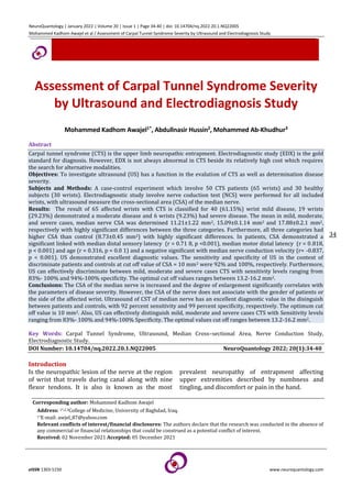
  • 4. NeuroQuantology | January 2022 | Volume 20 | Issue 1 | Page 34-40 | doi: 10.14704/nq.2022.20.1.NQ22005 Mohammed Kadhom Awajel et al / Assessment of Carpal Tunnel Syndrome Severity by Ultrasound and Electrodiagnosis Study eISSN 1303-5150 www.neuroquantology.com 37 Figure 1. CTS disease severity Association of MN CSA (median nerve cross-sectional area) with Disease Severity: The mean CSA of median nerve in mild, moderate, severe cases was 11.21±1.22 mm2, 15.09±0.1.14 mm2 and 17.88±0.2.1 mm2, respectively with highly significant differences between the three categories. Furthermore, all three categories had higher CSA than control (8.73±0.45 mm2) with highly significant differences (Figure 2). Figure 2. Association of CSA of median Nerve with Disease Severity **Highly significant difference. Analysis of test variance was to analyse data Diagnostic Performance of Ultrasound Value diagnostic investigated by a Relative Operating Characteristic (ROC) curve median CSA (calculated through US) in detection and differentiation of CTS. Generally, US demonstrated excellent diagnostic values. In the context distinguish between CTS patients groub and control group, area under the curve (AUC) was 0.950 and 95% CI = 0.903 - 0.996, p < 0.001. Sensitivity, Specificity of US at value cut off of CSA = 10 mm2 were 92% and 100 %, respectively (Figure 3). Figure 3. Relative operating characteristic curve for Ultrasound context of discriminate between CTS patients and controls In the context of discriminate between mild and moderate CTS, the AUC was 0 .9 74, 95 % CI = 0. 925-1.00, < 0.00 1. Sensitivity, Specificity of the Ultrasound at value cut-off of CSA = 13.1 mm2 were 95% and 97.5%, respectively (Figure 4). Figure 4. Relative operating characteristic curve for US in context of discrimination between mild and moderate CTS In the context of discrimination mild and severe CTS, AUC was 1 .0 0, 95 % CI = 1 .0 0 - 1 .0 0,  < 0 .001. Sensitivity and Specificity of US at cut off value of CSA = 14.0 mm2 100% for both (Figure 5).
  • 5. NeuroQuantology | January 2022 | Volume 20 | Issue 1 | Page 34-40 | doi: 10.14704/nq.2022.20.1.NQ22005 Mohammed Kadhom Awajel et al / Assessment of Carpal Tunnel Syndrome Severity by Ultrasound and Electrodiagnosis Study eISSN 1303-5150 www.neuroquantology.com 38 Figure 5. Relative operating characteristic curve for US in context of discriminate between mild and severe CTS In the context of discriminate between moderate and severe CTS, AUC 0. 8 68, 95 % CI = 0. 645-1.00, p = 0.008. Sensitivity, Specificity of US at cut off value of CSA = 16.2 mm2 83% and 94%, respectively (Figure 6). Figure 6. Relative operating characteristic curve for US in context of discriminate between moderate and severe CTS Discussion In the present study, patients were older than controls with a significant difference. This result corroborates with many previous studies. In A Turkish study including 547 patients with CTS, wrists were categorized into four groups normal, mild, moderate, sever. After adjusting for BMI, the CTS development risk raised by a factor of 1.86 for ages thirty–sixty years and by 4.17 for ages sixty when the age group eighteen–thirty-five years was used as the control group. Almost similar result regarding the risk of older age in development of CTS were reported by two other studies (Kouyoumdjian, 2002; Kouyoumdjian, 1999). The precise reasons beyond this association are not precisely known. However, it was assumed axon loss, development of nerve conduction, vessels abnormalities increase with age (Kommalage, 2011). In the present study, motor and sensory latency of the median nerve increased significantly in CTS, while the conduction velocity of this nerve significantly decreased compared with the controls. These electrophysiological changes are the basic for gold standard diagnosis of CTS. The median nerve is always affected with different degree of damage. Several mechanisms have been proposed that results in median nerve injury with an eventual increase sensory and motor latency and decrease conduction velocity (Arrori and Spence, 2008). An interesting finding in this study was median cross-sectional area was significantly different on CTS patients than controls, and the CSA of median was significantly linked with severity disease. Such results were frequently reported by a plethora of previous studies (Mohammadi 2012; Ajeena, 2013; El-Shintenawy 2019). (Tsai 2013) and (Chan 2011) identified a strong link between median nerve conduction anomalies detected by electro diagnostic testing and CSA measured by ultrasound. In the same context, there was correlating between CSA medain at the tunnel inlet, median motor distal latency (DML). As prolonged median DML is a marker of focal nerve demyelination across the carpal tunnel, this provides a biological basis for the US finding in CTS (Chan 2011). The most important finding in the present study were that the US had excellent diagnostic values either discrimination between control and patients with CTS in the context of CTS or in the context of discrimination between different severities of the CTS. In accordance with these results are many studies worldwide. In many of these studies the CSA of the median nerve yielded sensitivities range from sixty-seven to ninety four percent, and from fifty-seven to ninety seven percent to specificities, with value cut off 8 .5 to 9.5 percent 15mm2 (Wang 2008; Mondelli 2008; Padua 2008; Polykandriotis, 2007; Hobson-Webb, 2008). Also, in agreement with the present result is the result by (Karadag 2010) who claimed that
  • 6. NeuroQuantology | January 2022 | Volume 20 | Issue 1 | Page 34-40 | doi: 10.14704/nq.2022.20.1.NQ22005 Mohammed Kadhom Awajel et al / Assessment of Carpal Tunnel Syndrome Severity by Ultrasound and Electrodiagnosis Study eISSN 1303-5150 www.neuroquantology.com 39 ultrasound was helpful in determining the severity of CTS They came to the conclusion that ultrasound measurements of CSA could provide information regarding the severity of median involvement, it established value cutoff in the US to differentiate between different levels of CTS severity as follows: >thirteen mm2 for mild, thirteen to fifteen mm2 for moderate, and < fifteen mm2 for severe symptoms. In a Turkish study, (Kaymak 2008) conducted a cross-sectional study to clarification whether US or electrophysiologic testing is a preferable foreteller of grading severity and to estimated value diagnostic US in CTS patients. The median CSA at the tunnel entrance and proximal tunnel were 12. 5 ± 2.6 and 10.6 ±2.6, respectively in patients versus 15.6 ±4.2 and 11.5 ±3.2, respectively in control group, with highly significant differences. The optimum value cut off for median CSA was 11.2mm2 at the entrance tunnel and 11.9mm2 at the proximal carpal tunnel. Sensitivity and specificity at the proximal tunnel (eighty-eight%, sixty-six%, respectively) were higher than those at the carpal tunnel entrance (sixty-eight% and sixty-two%, respectively). The differences cut off value is mainly related to the severity of disease. However, these values are variable and not universally applicable as there are differences in study population, demographic differences and the “gold standard” used in the diagnosis of CTS. NCS is not always abnormal in CTS (Chan 2011), and in two studies, US revealed abnormal findings in CTS patients who had normal NCS (Altinok 2004; Koyuncuoglu 2005). On the other hand, US appear to be relatively quite specific for CTS (Pastare 2009). Furthermore, US may be useful in severe CTS, where NCS may be unrecordable (Chan 2011). Lower cost, non-invasive - test and shorter time for test are additional advantages of sonography over Electrodiagnostic for appraisal of CTS (Mohammadi 2013). Conclusions 1. The majority of CTS patients in the study had a mild to moderate disease. 2. The CSA median is largest in patients with CTS and degree of enlargement significantly correlates with the parameters of disease severity. However, the CSA of the nerve not associated with the gender of patients or the side of the affected wrist. 3. Ultrasound of CST of median nerve has an excellent diagnostic value in the discrimination between patients and controls, with Sensitivity, Specificity of 92%, 99%. The optimum value cut off is ten mm2. 4. Also, US can effectively discriminate between mild, moderate and severe cases of CTS with sensitivity ranges 83%- 100% and 94%-100% specificity. The optimal cut off values ranges between 13.2-16.2 mm2. Refrecnses Altinok T, Baysal O, Karakas HM. Ultrasonographic assessment of mild and moderate idiopathic carpal tunnel syndrome. Clinical radiology 2004; 59(10): 916-925. Aroori S, Spence RA. Carpal tunnel syndrome. Ulster Medical Journal 2008; 77(1): 6-17. Bland JD. A neurophysiological grading scale for carpal tunnel syndrome. Muscle & Nerve: Official Journal of the American Association of Electrodiagnostic Medicine 2000; 23(8): 1280-1283. McDonagh C, Alexander M, Kane D. The role of ultrasound in the diagnosis and management of carpal tunnel syndrome: a new paradigm. Rheumatology 2015; 54(1): 9-19. Chan KY, George J, Goh KJ, Ahmad TS. Ultrasonography in the evaluation of carpal tunnel syndrome: diagnostic criteria and comparison with nerve conduction studies. Neurology Asia 2011; 16(1): 57–64. Chen SF. Ultrasonographic median nerve cross-section areas measured by 8-point “inching test” for idiopathic carpal tunnel syndrome: A correlation of nerve conduction study severity and duration of clinical symptoms. BMC Medical Imaging 2011; 11: 1–9. http://doi.org/10.1186/1471-2342-11-22 Coraci D, Santilli V, De Franco P, Padua L. Comment to “ultrasonic assessment of females with carpal tunnel syndrome proved by nerve conduction study. Neural plasticity 2014. http://doi.org/10.1155/2014/893963 El Miedany YM, Aty SA, Ashour S. Ultrasonography versus nerve conduction study in patients with carpal tunnel syndrome: substantive or complementary tests. Rheumatology 2004; 43(7): 887–889. El-Shintenawy AA. Diagnostic potential of high resolution ultrasound and nerve conduction study in patients with idiopathic carpal tunnel syndrome. Egyptian Rheumatologist 2019; 41(1): 71–75. http://doi.org/10.1016/j.ejr.2018.04.001 Hobson-Webb LD, Massey JM, Juel VC, Sanders DB. The ultrasonographic wrist-to-forearm median nerve area ratio in carpal tunnel syndrome. Clinical neurophysiology 2008; 119(6): 1353-1357. Ibrahim I, Khan WS, Goddard N, Smitham P. Suppl 1: carpal tunnel syndrome: a review of the recent literature. The open orthopaedics journal 2012; 6: 69-70. Kang S. Ultrasonography of median nerve and electrophysiologic severity in carpal tunnel syndrome. Annals of Rehabilitation Medicine 2012; 36(1): 72–79. http://doi.org/10.5535/arm.2012.36.1.72 Karadag YS, Karadag O, Cicekli E. Severity of carpal tunnel syndrome assessed with high frequency ultrasonography. Rheumatology international 2010; 30: 761-765.
  • 7. NeuroQuantology | January 2022 | Volume 20 | Issue 1 | Page 34-40 | doi: 10.14704/nq.2022.20.1.NQ22005 Mohammed Kadhom Awajel et al / Assessment of Carpal Tunnel Syndrome Severity by Ultrasound and Electrodiagnosis Study eISSN 1303-5150 www.neuroquantology.com 40 Kaymak B, Özçakar L, Çetin A. A comparison of the benefits of sonography and electrophysiologic measurements as predictors of symptom severity and functional status in patients with carpal tunnel syndrome. Archives of physical medicine and rehabilitation 2008; 89(4): 743-748. Kommalage M, Pathirana KD. Influence of age and the severity of median nerve compression on forearm median motor conduction velocity in carpal tunnel syndrome. Journal of Clinical Neurophysiology 2011; 28(6): 642-646. Kouyoumdjian JA, Zanetta DM, Morita MP: Evaluation of age, body mass index, and wrist index as risk factors for carpal tunnel syndrome severity. Muscle & Nerve: Official Journal of the American Association of Electrodiagnostic Medicine 2002; 25(1): 93– 97. Kouyoumdjian JA. Carpal tunnel syndrome. Age, nerve conduction severity and duration of symptomatology. Arquivos de neuro-psiquiatria 1999; 57(2B): 382–386. Koyuncuoglu HR, Kutluhan S, Yesildag A, Oyar O, Guler K, Ozden A. The value of ultrasonographic measurement in carpal tunnel syndrome in patients with negative electrodiagnostic tests. European journal of radiology 2005; 56(3): 365-369. Mohammadi A, Ghasemi-Rad M, Mladkova-Suchy N, Ansari S. Correlation between the severity of carpal tunnel syndrome and color Doppler sonography findings. American Journal of Roentgenology 2012; 198(2): W181-W184. Mondelli M, Filippou G, Gallo A, Frediani B. Diagnostic utility of ultrasonography versus nerve conduction studies in mild carpal tunnel syndrome. Arthritis Care & Research 2008; 59(3): 357-366. Padua L, Pazzaglia C, Caliandro P, Granata G, Foschini M, Briani C, Martinoli, C. Carpal tunnel syndrome: ultrasound, neurophysiology, clinical and patient-oriented assessment. Clinical Neurophysiology 2008; 119(9):2064-2069. Pastare D, Therimadasamy AK, Lee E, Wilder‐Smith EP. Sonography versus nerve conduction studies in patients referred with a clinical diagnosis of carpal tunnel syndrome. Journal of Clinical Ultrasound 2009; 37(7): 380-393. Polykandriotis E, Premm W, Horch RE. Carpal tunnel syndrome in young adults-an ultrasonographic and neurophysiological study. Min-Minimally Invasive Neurosurgery 2007; 50(6): 328-334. Preston DC, Shapiro BE. Approach to pediatric electromyography. Electromyograghy and neuromuscular disorders, chapter 38, 3rd edition. China: Elsevier Inc, 2011: 597-600. Preston DC, Shapiro BE. Electromyograghy and neuromuscular disorders clinical – electrophysiological - ultrasound correlation, chapter 20, 4rd edition. China: Elsevier Inc, 2021: 342-328. Tsai NW, Lee LH, Huang CR, Chang WN, Wang HC, Lin YJ, Lu CH. The diagnostic value of ultrasonography in carpal tunnel syndrome: a comparison between diabetic and non-diabetic patients. BMC neurology 2013; 13(1):1-8. Wang LY, Leong CP, Huang YC, Hung JW, Cheung SM, Pong YP. Best diagnostic criterion in high-resolution ultrasonography for car-pal tunnel syndrome. Chang Gung Medical Journal 2008; 31(5): 469-476. Al-Mamoori MHK, Alshrefi SM, Jader MJ, Kodeary AK. Structural characteristics, synthesis of novel TiO2/VO (II) composites thin films decorated with chlorophyllvia solvothermal-laser dual technique. NeuroQuantology 2020; 18(3): 6-15.