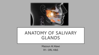
2. INTRODUCTION
• Humans have three pairs of major salivary glands – parotid,
submandibular and sublingual – and some 600–1000 minor
salivary glands, which are dispersed sub-mucosally throughout
the head and neck region.
• They are compound tubuloacinar exocrine glands found in oral
cavity that screate complex fluid known as Saliva
3. EMBRYOLOGY
• Each major gland develops from within the oral cavity (endoderm) and
matures through a series of stages.
• it is still debatable whether the epithelia of the salivary glands are
ectodermal or endodermal in origin. At least in the mouse model,
research revealed that the major glands are not derived from the
ectoderm. In contrast, some minor mucous glands of the tongue and
palate were fully or partially ectodermal-derived, respectively.
4.
5.
6. EMBRYOLOGY
• The parotid glands are the first to develop in the 4th week of
embryogenesis, followed by the submandibular glands at 6th weeks and
finally the sublingual glands at 8th weeks.
• Parenchymal tissue (secretory) of the glands arises from the proliferation
of oral epithelium.
• The stroma (capsule and septae) of the glands originates from
mesenchyme that may be mesodermal or neural crest in origin
8. BASED ON SIZE
• Major salivary glands:
Collection of secretory cells aggerated
into large bilaterally paired extra oral
gland with extend duct system through
which the gland secretion reaches the
mouth
• Parotid gland
• Submandibular gland
• Sublingual glands
• Minor salivary glands :
Collection of secretory cells scattered
throughout the mucosa & submucosa of
the oral cavity with short ducts opening
directly onto mucosal surface.
• Serous gland of Von Enber
• Anterior lingual glands
• Lingual , buccal , labial , palatal glands
, glossopalatine and retromolar
glands
9. BASED ON TYPE OF SECRETORY CELLS
• Serous: Parotid gland
• Mixed ( Seromucous): Submandibular gland
• Mucous : Sublingual & Minor salivary glands
10. PAROTID GLAND
• The largest of the major salivary glands ,
has an average weight of 25g.
• It produces between 25-30% of the total
daily salivary output which is released
through Stensen’s duct .
• They resemble a three-sided pyramid.
• Enveloped by the investing layer of deep
cervical fascia.
11.
12. STRUCTURE WITHIN THE GLAND
• Facial nerve, division of gland
• Retromandibular vein, anterior and
posterior divisions
• External carotid artery, terminal
branches
13.
14. BLOOD SUPPLY OF PAROTID GLAND
Arterial supply :
-The branches of the external
carotid artery
the superficial temporal,
maxillary, and (indirectly) the
transverse facial arteries.
15. Venous drainage :
-The maxillary and superficial
temporal veins.
-They unite to form
the retromandibular vein.
-The anterior division which
drains into the internal jugular
vein.
-The posterior division
draining into the external
jugular vein.
16. LYMPHATIC DRAINAGE OF PAROTID
GLAND
• Lymphatic drainage is to the
superficial and deep cervical
nodes
• Preauricular lymph nodes (LN)
in the superficial fascia drain
the temporal scalp, upper face,
anterior pinna
• LN within the gland drain the
parotid gland, nasopharynx,
palate, middle ear and external
auditory meatus
19. AUTONOMIC NERVE SUPPLY
Parasympathetic
Inferior salivatory nucleus
IX nerve
Lesser Petrosal nerve
Otic ganglion
Auriculotemporal nerve
PAROTID
Sympathetic
Superior cervical ganglion
Plexus around ECA
PAROTID
20. SUBMANDIBULAR GLAND
• he submandibular gland is the second
largest of the major salivary glands and
like all three of them it is a paired
gland.
• It produces by far the largest amount of
saliva up to 70%
• . Wharton’s duct (submandibular
duct) opens at the sublingual papilla
under the tongue.
21.
22. It separate from the parotid
gland by the
styelomandibular ligament
23. Divided by omohyoid muscle into
tow loops :
Superficial and deep ( deep
between hyoglossus and omohyoid
muscle
30. SUBLINGUAL GLAND
• the smallest of the major salivary glands
• it has several ductal openings that run along
the sublingual folds.
• It secretes the smallest portion of saliva per
day at just five percent.
31. RELATION OF SUBLINGUAL GLAND
• Sup – Oral floor mucosa
• Inf – Mylohyoid
• Post – Deep part Submandibular
gland
• Med – Lingual nerve, Submandibular
duct, Genioglossus
• Lat– Med surface of lower Mandible
32. • The ducts of the sublingual glands are
called Bartholin’s ducts.
• In most cases, Bartholin’s ducts consists of
8-20 smaller ducts of Rivinus. These ducts
are short and small in diameter.
• The ducts of Rivinis either open…
• individually into the FOM near the
punctum of Wharton’s duct
• on a crest of sublingual mucosa called
the plica sublingualis
• open directly into Wharton’s duct
33. BLOOD SUPPLY OD SUBLINGUAL GLAND
• The sublingual glands receive their
primary blood supply from the
sublingual and submental arteries, which
are branches of the lingual artery and
facial artery, respectively. These arteries
are both branches of the external carotid
artery.
• The sublingual vein drains into the
lingual vein, which then flows into the
internal jugular system.
34. INNERVATION OF SUBLINGUAL GLAND
• Via submandibular ganglion, which
receives autonomic fibres from the
chorda tympani, the lingual nerve and
sympathetic trunk.
35. MINOR SALIVARY GLANDS
• the minor salivary glands of mouth include
the buccal, labial,lingual,palatal and
palatoglossal glands.
• The buccal and labial glands contains both
mucous and serous elements.
• The palatal, lingual and palatoglossal
glands are mucous glands .
36. REFERANCES
• Orban’s oral histology and embryology , tenth edition.
• Logan Turner's Diseases of the Nose, Throat and Ear, 11th Edition
• Atlas of salivary gland disease
• Fastest Otolaryngology & Ophthalmology Insight Engine Salivary Gland
Embryology, Physiology, and Stem Cell Complexity | Ento Key
Editor's Notes
The content from superficial to deep are the facial nerve , the auriculotemporal nerve , the retro mandibular vein and the external carotid artery
Posteromedial surface is indented by the external carotid artery before it penetrates this surface of the gland.
nother important structure that passes through and divides within the substance of the parotid gland is the external carotid artery. As it passes through the posteromedial aspect of the gland, it gives off the maxillary artery, as well as the superficial temporal artery. Occasionally it also gives off the posterior auricular artery within the substance of the parotid gland as well.
Around 9 mm away from the posterior belly of digastric muscle and 11 mm from the bony part of EAC
Before entering the parotid it gave branch to Posterior auricular nerve
Then in the gland given temprofacial and cervicofacial that gives the 5 branches TZBMC
The external carotid artery travels parallel to the mandible after leaving the carotid bifurcation. It continues toward the parotid gland to enter the substance of the organ. Here, it gives off the superficial temporal and maxillary arteries (which are both terminal branches of the external carotid artery).
- The superficial temporal artery supplies
the superior aspect of the gland, while the maxillary artery supplies the medial aspect of the gland. The transverse facial artery arises off the superficial temporal artery. Not only does it supply the parotid gland, but also the duct and nearby masseter muscle as well.
Consist of a larger superficial part and a smaller deep part wrapped arround the posterior border of mylohyoid.
Each is irregular in shape.
About the size of a walnut.
Located in digasteric triangle , below and deep to the mandible
Deep to myelohyoid muscle