
Recommended
More Related Content
What's hot
What's hot (18)
Similar to The Palate and Salivary Glands
Similar to The Palate and Salivary Glands (20)
More from Hadi Munib
More from Hadi Munib (20)
Recently uploaded
Recently uploaded (20)
The Palate and Salivary Glands
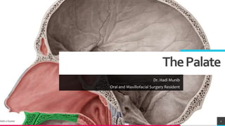
- 1. ThePalate Dr. Hadi Munib Oral and Maxillofacial Surgery Resident Add a footer 1
- 2. TREYresearch ThePalate • Forms the roof of the mouth and the floor of the nasal cavity. • It is divided into two parts: the hard palate in front and the soft palate behind. • Hard Palate; formed by the palatine processes of the maxillae and the horizontal plates of the palatine bones • It is continuous behind with the soft palate. • Soft Palate; a mobile fold attached to the posterior border of the hard palate. • Its free posterior border presents in the midline a conical projection called the uvula. • The soft palate is continuous at the sides with the lateral wall of the pharynx. • The soft palate is composed of mucous membrane, palatine aponeurosis, and muscles. • Mucous Membrane; covers the upper and lower surfaces of the soft palate. • Palatine Aponeurosis; a fibrous sheet attached to the posterior border of the hard palate. It is the expanded tendon of the tensor veli palatini muscle. Add a footer 2
- 3. TREYresearch MusclesoftheSoftPalate • The muscles of the soft palate are the tensor veli palatini, the levator veli palatini, the palatoglossus, the palatopharyngeus, and the musculus uvulae. • The muscle fibers of the tensor veli palatini converge as they descend from their origin to form a narrow tendon, which turns medially around the pterygoid hamulus. • The tendon, together with the tendon of the opposite side, expands to form the palatine aponeurosis. • When the muscles of the two sides contract, the soft palate is tightened so that the soft palate may be moved upward or downward as a tense sheet. • Nerve Supply of the Palate • The greater and lesser palatine nerves from the maxillary division of the trigeminal nerve enter the palate through the greater and lesser palatine foramina. • The nasopalatine nerve enters the front of the hard palate through the incisive foramen. • The glossopharyngeal nerve also supplies the soft palate. 3
- 4. TREYresearch ThePalate • Blood Supply of the Palate • The greater palatine branch of the maxillary artery, the ascending palatine branch of the facial artery, and the ascending pharyngeal artery • Lymph Drainage of the Palate; Deep Cervical Lymph Nodes • Palatoglossal Arch The palatoglossal arch is a fold of mucous membrane containing the palatoglossus muscle, which extends from the soft palate to the side of the tongue • The palatoglossal arch marks where the mouth becomes the pharynx. • Palatopharyngeal Arch The palatopharyngeal arch is a fold of mucous membrane behind the palatoglossal arch that runs downward and laterally to join the pharyngeal wall. • The muscle contained within the fold is the palatopharyngeus muscle. • The palatine tonsils, which are masses of lymphoid tissue, are located between the palatoglossal and palatopharyngeal arches 4
- 5. TREYresearch MovementsoftheSoftPalate • The pharyngeal isthmus (the communicating channel between the nasal and oral parts of the pharynx) is closed by raising the soft palate. • Closure occurs during the production of explosive consonants in speech. • The soft palate is raised by the contraction of the levator veli palatini on each side. • At the same time, the upper fibers of the superior constrictor muscle contract and pull the posterior pharyngeal wall forward. • The palatopharyngeus muscles on both sides also contract so that the palatopharyngeal arches are pulled medially, like side curtains. • By this means, the nasal part of the pharynx is closed off from the oral part. Add a footer 5
- 10. TREYresearch Add a footer 10
- 11. TREYresearch Add a footer 11
- 13. TREYresearch SalivaryGlands • Major and Minor • Major; Pairs of Parotid, Submandibular and Sublingual Glands • Minor; Palate and the Lips • Unstimulated Salivary Gland Secretions; Submandibular =65-70% • Stimulated Salivary Glands Secretions; Parotid = 45-50% Add a footer 13
- 14. TREYresearch ParotidGland • The largest salivary gland and is composed mostly of serous acini. • It lies in a deep hollow below the external auditory meatus, behind the ramus of the mandible and in front of the sternocleidomastoid muscle. • The facial nerve divides the gland into superficial and deep lobes. • The parotid duct emerges from the anterior border of the gland and passes forward over the lateral surface of the masseter. • It enters the vestibule of the mouth upon a small papilla opposite the upper second molar tooth. • Nerve Supply; Parasympathetic secretomotor supply arises from the glossopharyngeal nerve. • The nerves reach the gland via the tympanic branch, the lesser petrosal nerve, the otic ganglion, and the auriculotemporal nerve. Add a footer 14
- 15. TREYresearch Add a footer 15
- 16. TREYresearch Add a footer 16
- 17. TREYresearch Add a footer 17
- 18. TREYresearch SubmandibularGland • Consists of a mixture of serous and mucous acini. It lies beneath the lower border of the body of the mandible and is divided into superficial and deep parts by the mylohyoid muscle. • The deep part of the gland lies beneath the mucous membrane of the mouth on the side of the tongue. • The submandibular duct emerges from the anterior end of the deep part of the gland and runs forward beneath the mucous membrane of the mouth. • It opens into the mouth on a small papilla, which is situated at the side of the frenulum of the tongue • Nerve Supply; Parasympathetic secretomotor supply is from the facial nerve via the chorda tympani, and the submandibular ganglion. • The postganglionic fibers pass directly to the gland. Add a footer 18
- 19. TREYresearch Add a footer 19
- 20. TREYresearch SublingualGland • The sublingual gland lies beneath the mucous membrane (sublingual fold) of the floor of the mouth, close to the frenulum of the tongue. • It has both serous and mucous acini, with the latter predominating. • The sublingual ducts (8 to 20 in number) open into the mouth on the summit of the sublingual fold. • Nerve Supply; Parasympathetic secretomotor supply is from the facial nerve via the chorda tympani, and the submandibular ganglion. • Postganglionic fibers pass directly to the gland. Add a footer 20
- 21. TREYresearch References • Chapter 11: The Head and Neck Add a footer 21