
Recommended
More Related Content
Similar to Salivary gland and its importance in dentistry
Similar to Salivary gland and its importance in dentistry (20)
Recently uploaded
Recently uploaded (20)
Salivary gland and its importance in dentistry
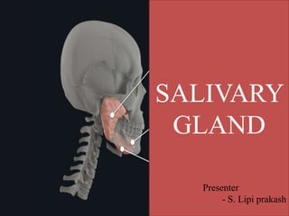
- 1. SALIVARY GLAND SALIVARY GLAND Presenter - S. Lipi prakash
- 2. CONTENTS 1. INTRODUCTION 2. DEVELOPMENT OF SALIVARY GLAND 3. STRUCTURES OF SALIVARY GLAND 4. MAJOR SALIVARY GLANDS 5. MINOR SALIVARY GLANDS 6. PAROTID GLAND 7. SUBMANDIBULAR GLAND 8. SUBLINGUAL GLAND 9. SALIVA 10. CLINICAL CONSIDERATION
- 3. INTRODUCTION Salivary glands are compound tubuloacinar exocrine glands that are found in and around the oral cavity that secrete a complex fluid known as saliva into the mouth Saliva is a complex fluid secreted from three major salivary glands (i.e., parotid, submandibular, and sublingual), as well as from the numerous minor salivary glands. The ducts of the salivary glands carry the saliva and discharge it into the oral cavity. The saliva forms a film of fluid which prevents the attachment of bacteria to the dentition and oral mucosa thereby creating and regulating a healthy environment in the oral cavity
- 4. DEVELOPMENT OF SALIVARY GLAND All salivary glands arises from ectoderm of oral cavity. The minor salivary glands arises from the oral ectoderm and nasopharyngeal ectoderm. The primordia of Parotid and submandibular – 6th week of intrauterine life Sublingual – 7th week of intrauterine life Minor salivary glands – 3rd month of intrauterine life Mesoderm forms the intervening connective tissue septa Signaling molecules include FGF, TGF -beta Ref Inderbir Singh’s human embryology eleventh edition
- 5. Salivary gland develops near the junctional area between the ectoderm of the stomatodeum and the endoderm of the foregut. During fetal life, each salivary gland is formed at a specific location in the oral cavity through the growth of a bud of oral epithelium into the underlying mesenchyme The epithelial bud grows into an extensively branched system of cords of cells that are first solid but gradually develop a lumen and become ducts. The secretory portions develop later than the duct system Ref Inderbir Singh’s human embryology eleventh edition
- 6. Ref Inderbir Singh’s human embryology eleventh edition
- 7. STRUCTURES OF SALIVARY GLAND The secretory units are known as acini and the secretions are carried by tubules, which open into the oral cavity- tubuloacinar in nature The terminal secretory units or acini are made up of secretory cells , which are either serous or mucous The secretions of the terminal end pieces pass through the intercalated duct Secretions from the intercalated duct passes through the striated duct Saliva passes from striated duct to terminal excretory duct Myoepithelial cells are the contractile cells with numerous processes associated with salivary gland. These are non secretory cells found in close association with terminal secretory end-piece and intercalated duct. Ref Orban’s oral histology and embryology
- 8. SEROUS CELL SEROUS ACINI •Pyramidal in shape with narrow apex near the lumen •Sperical nucleus near basal one third •Apical portion contains zymogen granules •Secretion of granules as string of pearls no - loss of cytoplasm •Spherical in shape •Smaller in size •Have smaller lumen •Intercellular canaliculi is present •Secretion – enzymatic activity- acid phosphatase, esterases, glucuronidase •Produce secretory proteins , carbohydrate content less. Produces thin watery fluid •Serous acini help digest starch Ref Orban’s oral histology and embryology
- 9. Ref Orban’s oral histology and embryology
- 10. •Pyramidal in shape •Flattened nucleus at base •Apical portion of cell appears empty •Apical cytoplasm not sealed – mucus spilled into lumen MUCOUS CELL MUCOUS ACINI •Elliptical in shape •Larger in size •Have larger lumen •Lacks intercellular canaliculi •Secretion – no enzymatic activity •Produces more carbohydrate components than proteins, thick viscous fluid that contains mucin, a glycoprotein •Mucous mainly serves as lubricant and protective layer Ref Orban’s oral histology and embryology
- 12. Ref Orban’s oral histology and embryology
- 13. Ref Orban’s oral histology and embryology
- 14. INTERCALATED DUCTS Location- Intralobular Cell morphology – single layer of low cuboidal cells Size- smallest in diameter Function – contribute to macromolecular components like lysozymes and lactoferrin
- 15. STRIATED DUCTS Location- intraglobular Cell morphology- single layer of tall columnar cells Size- bigger than intercalated duct Functions- site for electrolyte reabsorption- active transport These cells shows striations and they are due to deep infoldings of plasma membrane and contains such mitochondria Ref Orban’s oral histology and embryology
- 16. EXCRETORY DUCTS Location- Interlobular Cell morphology – pseudostratified columnar epithelium to stratified squamous Size – largest in diameter Function – no modification to saliva
- 17. Ref Orban’s oral histology and embryology
- 18. MAJOR SALIVARY GLANDS There are three bilaterally paired major salivary glands , which are located extraorally , and their secretions reach the mouth by variably long ducts. Parotid gland (purely serous) Submandibular gland ( mixed gland predominantly serous) Sublingual gland ( mainly mucous)
- 20. MINOR SALIVARY GLAND Labial and buccal glands Glossopalatine glands Palatine glands Anterior Lingual glands(Blandin-Nuhn glands) Posterior lingual glands (Von Ebner glands)
- 21. PAROTID GLAND Largest major salivary gland Superficial portion – lies in front of the external ear Deeper portion – lies behind the ramus of the mandible filling the retromandibular fossae It weighs between 14 and 28 g. The main excretory duct- Stenson’s duct which opens at buccal mucosa opposite to maxillary second molar .The duct measures 4–6 cm in length and 5 mm in diameter. Pure serous gland Purely serous secretion and contributes to 25% of saliva Flow rate – 0.4ml/min (unstimulated) - 1-2 ml/min ( stimulated ) Ref B D Chaurasia’s Human Anatomy Volume 3 ‘Head and Neck’ 7 th edition
- 23. The parotid gland receives its blood supply from the branches of the external carotid artery. Venous drainage is through external jugular vein and internal jugular vein. The parasympathetic nerve supply is derived mainly from the ninth cranial nerve reaching the gland via the otic ganglion and the auriculotemporal nerve. Sympathetic (vasomotor)- plexus around middle meningeal artery Sensory – auriculotemporal nerve and greater auricular nerve The lymphatic drainage is via paraparotid and intraparotid lymph nodes into the superficial and deep cervical lymph nodes. Ref B D Chaurasia’s Human Anatomy Volume 3 ‘Head and Neck’ 7 th edition
- 24. INFERIOR SALIVATORY NUCLEUS 9TH NERVE TYMPANIC BRANCH TYMPANIC PLEXUS LESSER PETROSAL RELAY IN OTIC GANGLION POSTGANGLIONIC FIBRES THROUGH AURICULOTEMPORAL NERVE PAROTID GLAND
- 25. Ref B D Chaurasia’s Human Anatomy Volume 3 ‘Head and Neck’ 7 th edition
- 26. STRUCTURES WITHIN THE PAROTID GLAND Most of the arteries and veins enter through the posteromedial surface and exit through the anteromedial surface External carotid artery Retromandibular vein Facial nerve
- 27. Ref B D Chaurasia’s Human Anatomy Volume 3 ‘Head and Neck’ 7 th edition
- 28. HISTOLOGY OF PAROTID GLAND Consists of lobular systems of ducts , seperated by connective tissue septa Surrounded by dense connective tissue capsule Pseudocapsule arises from investing layer of deep cervical fascia Rich in serous cells Tubules and ducts from acini contain myoepithelial cells
- 29. Clinical significance Gustatory sweating (Frey’s syndrome/ auriculotemporal syndrome) Occurs after parotid surgery/trauma- results in opening of parotid capsule Characterized by sweating, warmth, redness of the face as a result of salivary stimulation by smell or taste of food Treatment- symptomatic- includes SC infiltration of botox, anti- perspirant or denervation (worst case)
- 30. Parotitis Inflammation Mostly caused by mumps Cancers/tumors 80% are benign Most common- Pleomorphic Adenoma, Warthin’s tumor 20%are malignant Include MEC, ACC, adenocarcinoma
- 31. Parotid calculi (Sialolithiasis) Either within the gland/ duct Obstructs flow of saliva- swelling and pain Located by sialogram – These calculi can be located by injecting a radiopaque dye through its opening in the vestibule of the mouth Clinically , facial nerve palsy occurs after inferior alveolar nerve block. An injection with long needle in the very posterior position of the mandibular foramen could spread the anesthetic solution behind the mandibular ramus but inside the parotid gland, as the parotid gland envelops the facial nerve, thus leading to the direct anesthesia of the facial nerve Ref. case report on facial nerve palsy after inferior alveolar nerve block : A rare presentation of ocular complication and literature review by Glauco Chisci , Dafne Chisci, Enea Chisci
- 32. SUBMANDIBULAR GLAND The submandibular gland is the second largest salivary gland. It is placed posterior and superficial to the mylohyoid muscle. The main excretory duct- Wharton’s duct, runs forward above the mylohyoid muscle lying just below the mucosa of the floor of the mouth. The gland receives blood supply from the lingual and facial arteries The parasympathetic innervation is derived primarily from the VII cranial nerve reaching the gland through the lingual nerve after synapsing in the submandibular ganglion. The lymphatic drainage is to the deep cervical and jugular chain of nodes. Sensory fibers – lingual nerve Postganglionic sympathetic fibers (vasomotor) –plexus surrounding the facial artery
- 33. Contributes to 70% of saliva Flow rate – 0.1 ml/min( unstimulated) 0.8ml/min ( stimulated)
- 34. Superior salivatory nucleus Nervus intermedius Facial nerve Chorda tympani Joins lingual nerve , branch of mandibular nerve Relay in submandibular ganglion Postganglionic fibers Submandibular gland Ref B D Chaurasia’s Human Anatomy Volume 3 ‘Head and Neck’ 7 th edition
- 35. CLINICAL SIGNIFICANCE Submandibular duct calculi 80% of all salivary sialoliths Tortuous duct course, different nature of saliva predisposes to higher chances They are formed by deposition of calcium salts around a central nidus which consists of altered salivary mucin , desquamated epithelial cells, bacteria , foreign bodies Modrate to severe pain – meals – psychic stimulation of salivary flow associated with swelling of the gland Calculi in the Peripheral portion may be palpated or sialography
- 36. Sialolithiasis Ref Shafer’s textbook of oral pathology 8th edition
- 37. SUBLINGUAL GLAND Smallest of the main salivary glands which is almond shaped. The sublingual gland is also a mixed gland, but predominantly mucous secretory units. Pure serous acini are rare or absent The main duct, Bartholin’s duct opens with or near the submandibular duct and duct of rivinus opens independently in the floor of mouth Contributes to 5% of saliva Ref B D Chaurasia’s Human Anatomy Volume 3 ‘Head and Neck’ 7 th edition
- 38. The sublingual gland receives its blood supply from the sublingual and the submental arteries. The parasympathetic nerve supply is also derived from the VII cranial nerve.It reaches the gland via the lingual nerve after synapsing in the submandibular ganglion The lymphatic drainage is to the submandibular lymph nodes
- 39. CLINICAL SIGNIFICANCE Ranula Retention cyst When trauma occurs in the floor of the mouth and obstructs the drainage of the gland, ranula is formed Large, tense, bluish swelling, often displacing the tongue
- 40. Mucocele [Mucous extravasation cyst] It is mainly characterised by pooling of mucous in a cavity within the connective tissue, due to the rupture of salivary duct or acini Aetiology – Traumatic severance of a salivary duct , by lip or cheek biting or pinching of lips by extraction forceps Involves accessory salivary gland structures, Occurs most frequently in the lower lip, also occur on palate cheek tongue floor of mouth Treatment – Excision Ref Shafer’s textbook of oral pathology 8th edition
- 41. The saliva is mainly produced by major salivary gland differ from one another in composition. The parotid gland secretes a watery saliva rich in enzymes such as amylase, proline-rich protein, and glycoproteins. The submandibular gland contains higher proportion of glycosylated substances, such as mucin. The sublingual gland produces a viscous saliva rich in mucin The secretions of all the minor and major salivary glands contribute to whole saliva. SALIVA
- 43. FUNCTIONS OF SALIVA 1. Protection and lubrication- Mucin, proline-rich proteins, water. 2. Antimicrobial action Lysozyme, lactoferrin, lactoperoxidases, mucins, cystatin, histatins, IgM, PRP, IgA. 3. Mucosal integrity-Mucins, electrolytes, water 4. Dilution and cleansing -Water 5. Buffer capacity and remineralization-Bicarbonate, phosphate, calcium, statherin, proline-rich anionic proteins, fluoride 6. Deglutition -Water, mucins 7. Digestion-Amylase, lipase 8. Taste 9. Phonation 10. Excretion (Through) water
- 44. AGE CHANGES IN SALIVARY GLANDS Age changes in the salivary glands, particularly in the parotid, consist of a gradual replacement of parenchyma with fatty tissue. Since the parotid is the major source of serous saliva; with advancing age, patients often complain of dryness and an increase in the viscosity of saliva
- 45. CLINICAL CONSIDERATION Loss of salivary function or reduction in volume of saliva secreted is called xerostomia, which leads to dryness of the mouth. Oral tissues become susceptible to frequent oral infection; inflammation and ulceration of oral mucosa is commonly seen. In some instances, excessive salivary secretion is seen because of certain physiologic states and rare pathologies. This is called sialorrhea Increased salivary secretion (sialorrhea) can make isolation difficult during restorative procedures Decreased salivary production (xerostomia) can increase the risk of caries, tooth loss and infections in the mouth
- 46. Owing to the opening of the Stenson’s duct opposite the maxillary 2nd molar, there is a continuous discharge of saliva in this region, making isolation critical The maxillary incisors are most vulnerable to caries, while the mandibular incisors are the least vulnerable as they are protected by the saliva from the submandibular and the sublingual glands Ref Shafer’s textbook of oral pathology 8th edition
- 47. Xerostomia means dry mouth. It is due to hyposalivation or absence of salivary secretion (aptyalism). Causes i. Dehydration or renal failure. ii. Sjogren syndrome. iii. Radiotherapy. iv. Trauma to salivary gland or their ducts. v. Side effect of some drugs like antihistamines, antidepressants, monoamine oxidase inhibitors, antiparkinsonian drugs and antimuscarinic drugs. vi. Shock. vii. After smoking XEROSTOMIA
- 48. Xerostomia causes difficulties in mastication, swallowing and speech. It also causes halitosis (bad breath; exhalation of unpleasant odors). Signs of xerostomia, including the following: • Intraoral dryness • Burning sensation • Altered tongue surface • Dysphagia • Alterations in taste • Difficulty with speech • Root caries Xerostomia -Treated by saliva substitutes. Extensive dental caries is common so frequent fluoride applications are indicated.
- 49. SJOGREN’S SYNDROME Chronic, inflammatory, autoimmune disease affecting salivary, lacrimal and other exocrine glands 2 types- primary (dry eyes + dry mouth) and secondary (primary + RA/SLE) Could be due to auto-antibodies (B cell hyperactivity), increased IgG or IgM, CMV, lymph node malignancy Features- Keratoconjunctivitis sicca, xerostomia, stomatitis, enlargement of salivary glands, frothy saliva, mucosa is glazed, dry and wrinkled, severe dental caries, periodontal disease, bacterial sialadenitis,enlarged and tender lymph nodes Radiological features- Snowstorm/cherry blossom/branchless fruit laden tree appearance Treatment/management- Ocular lubricants, fluoride application, bromhexine, pilocarpine surgery (only when enlargement is causing discomfort)
- 50. BRANCHLESS FRUIT LADEN TREE SNOWSTORM Ref Shafer’s textbook of oral pathology 8th edition
- 51. 1. Standring Susan. 2016. Gray's Anatomy : The Anatomical Basis of Clinical Practice Forty-first ed. Philadelphia: Elsevier Limited 2. B D Chaurasia’s Human Anatomy Volume 3 ‘Head and Neck’ 7 th edition 3. Orban’s oral histology and embryology 4. Inderbir Singh’s human embryology eleventh edition 5. Shafer’s textbook of oral pathology 8th edition REFERENCE
Editor's Notes
- Exocrine glands- glands whose products are carried away by the ducts leading from the gland
- Photomicrographs of mucous salivary gland showing mucous acini with basally placed , flattened nucleus and basophilic cytoplasm
- Photomicrograph of mixed salivary gland showing mucous acini-1 serous acini-2 and serous demilunes -3
- These duct reabsorb sodium and chloride ions and sectrete potassium and bicarbonate ions this makes saliva hypotonic
- Ciliated cells goblet cells and dendritic cells may be present
- Parasympathetic supply of parotid gland
- Relations of the parotid gland
- Occlusion of the gland prevents free flow of saliva Rx by increasing salivation by sucking a lemon
- Superficial lesions – raised ,circumscribed vesicle – millimeters to centimeters Deeper lesions – swelling