Sideroblastic Anemia: Iron Accumulation and Ringed Erythroblasts
•Download as DOCX, PDF•
39 likes•9,416 views

Sideroblastic anemia is a disease where the bone marrow produces abnormal red blood cells called ringed sideroblasts that contain iron deposits around the nucleus, rather than healthy red blood cells. It can be caused by genetic disorders, myelodysplastic syndrome, or certain toxins and medications. Patients experience symptoms of pale skin, fatigue, and enlarged organs. The disease is diagnosed through identification of ringed sideroblasts in bone marrow samples and blood tests showing iron overload. Treatment depends on the severity of anemia and underlying cause, and may include blood transfusions, high dose vitamin supplements, chelation therapy to remove excess iron, and in severe cases, bone marrow transplant.

Recommended
More Related Content
What's hot
What's hot (20)
Viewers also liked
Similar to Sideroblastic Anemia: Iron Accumulation and Ringed Erythroblasts
Similar to Sideroblastic Anemia: Iron Accumulation and Ringed Erythroblasts (20)
More from Shaikho Elmuhagir
Recently uploaded
Recently uploaded (20)
Sideroblastic Anemia: Iron Accumulation and Ringed Erythroblasts
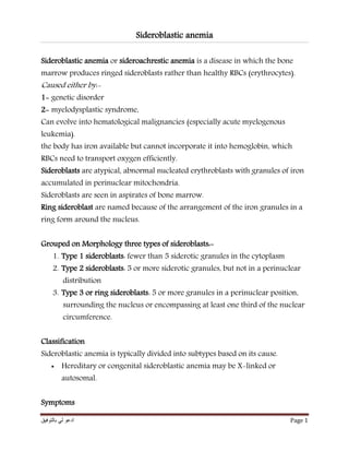
- 1. بالتوفيق لي ادعو Page 1 Sideroblastic anemia Sideroblastic anemia or sideroachrestic anemia is a disease in which the bone marrow produces ringed sideroblasts rather than healthy RBCs (erythrocytes). Caused either by:- 1- genetic disorder 2- myelodysplastic syndrome, Can evolve into hematological malignancies (especially acute myelogenous leukemia). the body has iron available but cannot incorporate it into hemoglobin, which RBCs need to transport oxygen efficiently. Sideroblasts are atypical, abnormal nucleated erythroblasts with granules of iron accumulated in perinuclear mitochondria. Sideroblasts are seen in aspirates of bone marrow. Ring sideroblast are named because of the arrangement of the iron granules in a ring form around the nucleus. Grouped on Morphology three types of sideroblasts:- 1. Type 1 sideroblasts: fewer than 5 siderotic granules in the cytoplasm 2. Type 2 sideroblasts: 5 or more siderotic granules, but not in a perinuclear distribution 3. Type 3 or ring sideroblasts: 5 or more granules in a perinuclear position, surrounding the nucleus or encompassing at least one third of the nuclear circumference. Classification Sideroblastic anemia is typically divided into subtypes based on its cause. Hereditary or congenital sideroblastic anemia may be X-linked or autosomal. Symptoms
- 2. بالتوفيق لي ادعو Page 2 Skin paleness, fatigue, dizziness, and enlarged spleen and liver. Heart disease, liver damage, and kidney failure can result from iron buildup in these organs. Causes failure to completely form heme molecules in the mitochondrion lead to deposits of iron in the mitochondria that form a ring around the nucleus of the developing RBC. Sometimes the disorder represents a stage in evolution of a generalized bone marrow disorder that may ultimately terminate in acute leukemia. Toxins: lead, copper, or zinc poisoning Drug- induced: ethanol, isoniazid, chloramphenicol, cycloserine, Linezolid, oral contraceptives Nutritional: pyridoxine (Vitamin B6) or copper deficiency Diseases: Rheumatoid arthritis or multiple myeloma Genetic: ALA synthase deficiency (X-linked, associated with ALAS2) Diagnosis 1. Ringed sideroblasts are seen in the bone marrow. 2. The anemia is moderate to severe 3. Dimorphic with marked anisocytosis and poikilocytosis.Basophilic stippling is marked and target cells are common. Pappenheimer bodies are present. 4. MCV is decreased (i.e., a microcytic anemia). 5. RDW is increased with the red blood cell histogram shifted to the left. 6. Leukocytes and platelets are normal. 7. Bone marrow shows erythroid hyperplasia with a maturation arrest. 8. In excess of 40% of the developing erythrocytes are ringed sideroblasts. 9. Serum iron, percentage saturation and ferritin are increased. The 10. TIBC is normal to decreased.
- 3. بالتوفيق لي ادعو Page 3 11. Stainable marrow hemosiderin is increased. Laboratory findings 1. Increased ferritin levels 2. Normal total iron-binding capacity 3. Hematocrit of about 20-30% 4. Serum Iron: High 5. High transferrin saturation 6. MCV is usually normal or low. 7. With lead poisoning, see coarse basophilic stippling of RBCs on peripheral blood smear 8. Specific test: Prussian Blue stain of RBC in marrow. Shows ringed sideroblasts. 9. Can also cause microcytic hypochromic anemia. Treatment 1. anemia is so severe that support with transfusion is required. 2. Patients usually do not respond to erythropoietin therapy. 3. improved heme level by moderate to high doses of Vitamin 4. Severe cases of SBA, bone marrow transplant with limited information about the success rate. 5. In the case of isoniazid-induced sideroblastic anemia, the addition of B6 is sufficient to correct the anemia. 6. Desferrioxamine is used to treat iron overload from transfusions. 7. Bone Marrow Transplant (BMT) is the last possible treatment. Course and prognosis Sideroblastic anemia’s are often described as responsive or non-responsive in terms of increased Hb level to pharmacological doses of vitamin B6.
- 4. بالتوفيق لي ادعو Page 4