
![• Many medications given to neonates have the
potential to injure when an extravasation
occurs.
• An extravasation is described by the Infusion
Nurses Society (INS) as the inadvertent
administration of a vesicant solution or
medication into the surrounding tissues.[1]](data:image/gif;base64,R0lGODlhAQABAIAAAAAAAP///yH5BAEAAAAALAAAAAABAAEAAAIBRAA7)
Recommended
More Related Content
What's hot
What's hot (20)
Viewers also liked
Viewers also liked (20)
Similar to Extravasation in neonate
Similar to Extravasation in neonate (20)
Recently uploaded
Recently uploaded (20)
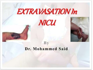
Extravasation in neonate
- 2. • Many medications given to neonates have the potential to injure when an extravasation occurs. • An extravasation is described by the Infusion Nurses Society (INS) as the inadvertent administration of a vesicant solution or medication into the surrounding tissues.[1]
- 3. A vesicant is defined as a solution or medication that causes the formation of blisters leading to tissue necrosis and sloughing.
- 4. Extravasation Inadvertent administration of vesicant medication or solution into the surrounding tissue. Infiltration the inadvertent administration of non- vesicant medication or solution into the surrounding tissue.
- 5. Interstitial • Any type of fluid in the interstitial space or the space between tissue layers and outside of the vein Occlusion Blockage that stops the passage of infusate or normal saline flush into the lumen.Often accompanied by an increase in pressure reading on the IV pump.
- 6. Infiltration Is Common Among Neonates • The peripheral intravenous (PIV) catheter is the most used vascular access device for the administration of medications in hospitalized neonates; however 95% of PIV catheters are removed due to complications such as leaking, occlusion and infiltration.[4] • Infiltration rates among neonates are as high as 57%–70% with extravasation occurring in 11– 23%.[5] Both infiltration and extravasation are destructive
- 10. • Extravasations have the potential to cause peripheral tissue injury depending on the type of vesicant, concentration of the vesicant, location, amount, and duration of exposure to the vesicant. • Damage from a vesicant may progress over time and become evident 48–72 hours after the extravasation occurs.
- 11. The preterm and sick neonate is more susceptible to skin injury and complications from extravasation injury than their mature, healthy counterparts. • Their immature skin structures, • flexible subcutaneous tissue, • small blood vessels and poor venous integrity increase the risk of complication from venipuncture and IV infusions.[5,8]
- 12. • The goal in neonatal care is to prevent skin breakdown whenever possible. Similarly, attention to thermoregulation, pain and stress that infants endure as a result of repeated IV attempts or restarts, and infiltrations and extravasations must be considered and managed
- 13. Inflammation in the Premature Infant • The neonatal immune system is poorly regulated compared to adults and dysregulation is magnified when neonates are born early.[11-13] While intravenous therapy is necessary in this population, it is not without its risks.
- 14. • Vesicants can harm the endothelial lining of the blood vessel, triggering production and release of oxygen free radicals that spur inflammation.[14,
- 15. • which is common in prematurity, or the inflammatory assault is severe, endothelial dysfunction leads to programmed cell death (apoptosis).[16] • The load of oxidative stress in premature infants is especially of concern as it has been linked to various neonatal morbidities including necrotizing enterocolitis,[16,17] retinopathy of prematurity,[18-20] and chronic lung disease.[16,21- 23]
- 16. Pathophysiology of Vascular Injury
- 17. The Neonatal Intensive Care Unit (NICU) Nurse's Role • NICU nurses monitor the PIV site with vigilance to aid in early identification of infiltration and extravasation and prevent this type of injury whenever possible. Identifying an infiltration may be difficult, even for the most experienced nurse.[10]
- 18. • The NICU nurse is aware of the subtle changes in heart rate, oxygen saturations, apnea, and the more obvious change in behavior such crying and agitation that may indicate problems with the PIV therapy.[4
- 19. Potential Origins of Infiltration • There is a supposition that an infiltration or extravasation is caused by IV catheter dislodgement or puncture of the vein during insertion or during handling of the infant. • Chemical composition of medications also impacts risk of vein rupture.[5] • The vein's tolerance to an infusion is affected by the osmolality and pH of the vesicant, the duration of the exposure, and irritation to the endothelial cells.[4] • An additional factor in causing a cannulated vessel to rupture and leak is the pressure in which the medication is being delivered by the infusion pump.[3,5]
- 20. Irritants and Vesicants Given to Neonates • ntravenous medications can be divided into three major subcategories: 1) non-vesicants, 2) irritants, and 3) vesicants. In order for an infiltration to be a true extravasation, the offending agent, by definition, must be a vesicant. There are a number of different qualities that affect the potential for a medication to result in tissue damage. These include, but are not limited to: osmolarity, pH, direct medication effects and solubility.[27]
- 21. • ntravenous medications can be divided into three major subcategories: 1) non-vesicants, 2) irritants, and 3) vesicants. In order for an infiltration to be a true extravasation, the offending agent, by definition, must be a vesicant. There are a number of different qualities that affect the potential for a medication to result in tissue damage. These include, but are not limited to: osmolarity, pH, direct medication effects and solubility.[27]
- 23. Nursing Actions to Prevent Vascular Injury • The best method to decrease complications of PIV therapy is to prevent them in the first place.[2] • Serious complications are not entirely preventable, but following recommended standards of IV therapy is the best approach for avoiding complications.[3] • The decision to place a peripherally inserted central catheters (PICC) or central venous lines (CVL) might be needed if vascular access is difficult or long-term parenteral therapy is planned. However
- 24. Recommendations for Practice to Prevent Vascular Injury. Peripheral IV Insertion and Maintenance Use small enough plastic/silicone catheter to avoid restriction of blood flow Avoid repeated use of a vein Avoid placing a PIV in an areas difficult to immobilize Use transparent tape to secure Cover the site with a sterile semi-permeable transparent dressing that will permit ongoing visualization of the insertion site Upper extremities less likely to infiltrate or leak compared with peripheral IV in lower extremities or scalp veins Place tape loosely over boney prominences to avoid restricting blood flow to the extremity Infusion Maintenance Limit PIV glucose to 12.5% Dilute medications as much as possible before administration are other solutions
- 27. Key Actions to Minimize Injury When Extravasation Occurs • Once an infiltration or extravasation is discovered, immediate treatment is the key to preventing progressive damage from the vesicant.[2] • Treatment decisions are based on the size and appearance of the injury, type of IV infusing, duration of exposure and location.[8] Protocols and algorithms can be used to assist nurses in the steps needed to minimize the potential damage and start the treatment process.
- 28. • Stopping the infusion and elevation of the extremity is the first actions followed by placement of a saline soaked gauze or prepackaged normal saline pad. • The saline draws out the vesicant, and impedes a scab from forming to allow fluid to leak out. • Gently squeezing the fluid from the open insertion site can also help to remove the offensive agent.[8] While the saline soaks are held in place, assistance with the various tasks that are required for treatment may require additional personnel.
- 29. • Documentation: Following an IV insertion, document the following information in the patient record: Date and time of procedure. Gauge and type of access device used. Site of insertion. Patient’s tolerance of the procedure. type of solution, additives and rate of infusion •
- 30. Wound care • Cleansing: · Normal saline. Any wound swabs should only be taken after cleansing. • Moisture and autolytic debridement: · Water-based wound gel (hydrogel). • Protection and Absorption: · An absorptive layer such as Aquacel® can be placed directly over the wound to prevent any dressing from sticking to the wound and to absorb necrotic material as it softens. · Cover with a hydrocolloid or clear acrylic dressing. A thin product conforms better with infant’s small limbs. Use only enough to give approximately a 1 cm border around the • Assess dressings a minimum of q4h and prn for integrity and drainage.
- 31. • Replace dressing q 7 days or PRN if the dressing being saturated with exudate or is lifting significantly. A hydrocolloid will have a “swiss cheese” appearance when saturated. If the edges begin to lift they can be reinforced with a transparent dressing. • If there are signs of systemic sepsis such as fever or temperature instability etc, or local inflammation around the area of the wound, remove the dressing and assess for wound infection evidenced by purulent drainage and peri-wound erethema. Send wound swab after cleansing the wound bed with saline and consult wound and skin assessment team for a dressing protocol for an infected wound.
- 32. • Saline Flush Technique: This procedure involves making small puncture marks around the edge of the area of extravasation and the inserting a cannula into each of the puncture sites in turn and flushing normal saline through each puncture site. The volume in the literature is 500 mls, although this should be modified down in the neonate. The goal is that the flush solution will exit out of the other puncture site.
- 42. Hyaluronidase • Hyaluronidase is an enzyme that breaks down hyaluronic acid, a compound best described as the "glue" which holds cells together. When this extracellular glue is dissolved, cells are separated. This is helpful in extravasations as it allows for the medication to distribute through a larger area by breaking down the walls that keep it localized. This not only helps to decrease the concentration effects of the extravasated product, but also exposes the medication to more capillary beds that allows for reduction of edema via more rapid reabsorption and removal of the product from the damaged area.
- 43. Phentolamine • Phentolamine is an antidote that will counteract the effect of vasoactive agents such as dopamine, epinephrine, norepinephrine and phenylephrine.[8] These medications result in vasoconstriction via stimulation of alpha-receptors. • Phentolamine acts to block the activity of alpha-receptors and subsequently will help relax vascular smooth muscle. This will improve circulation in the area of the extravasation and thus decrease ischemia and cell death. Phentolamine can also be utilized for vasopressin or dopamine extravasation.[28]Phentolamine should be administered within 12 hours of initial exposure but administration should occur as soon as possible. Prepare a 0.5 to 1 mg/mL solution and administer 0.1 mg/kg (to a max of 2.5 mg in neonates,
- 44. • Nitroglycerin Ointment (2%) • Nitroglycerin 2% is an option to treat extravasations.[8]Nitroglycerin acts to relax smooth muscle resulting in arteriolar, arterial and venous vasodilation that results in increased capillary blood flow, counteracting the effects of vasoactive medications. This will help to reverse tissue ischemia and cell death. In the neonatal population, there is a case report describing the use of 1 inch of 2% nitroglycerin ointment for treatment of a dopamine extravasation, located in the dorsum of the left hand, in a 1.8 kg 34 week preemie. This resulted in return of circulation within a few minutes. Of note, treatment was started almost 12 hours after the extravasation was initially noted and patient had no significant change in hemodynamics.[33]
- 46. References • References 1. Alexander M, Corrigan A, Gorski L, et al. Infusion Nurses Society (INS) Infusion nursing an evidence- based approach.St. Louis, Missouri: Saunders Elsevier; 2010: 366, 470–471, 481. 2. Sawatzky-Dickson D, Bodnaryk K. Neonatal Intravenous extravasation injuries: evaluation of a wound care protocol. Neonatal Network.2006;25:13–19. 3. Thigpen J. Peripheral intravenous extravasation: nursing procedure for initial treatment. Neonatal Network.2007;26:379–384. 4. Pettit J. Assessment of the infant with a peripheral intravenous device. Advances in Neonatal Care.2003;3:230–240. 5. McCullen K. A retrospective chart review of risk factors for extravasation among neonates receiving peripheral intravascular fluids. Journal of Wound, Ostomy and Continence Nurses.2006;33:133–139. 6. Amjad I, Murphy T, Nylander-Housholder L, Ranft A. A new approach to management of intravenous infiltration in pediatric patients. Journal of Infusion Nursing.2011;34:242–249. 7. Kuensting L. Treatment of intravenous infiltration in a neonate. Journal of Pediatric Health Care.2012;24:184–188. 8. Association of Women's Health, Obstetrics, and Neonatal Nurses. Neonatal Skin Care; Evidence-based Guidelines. 3rd Ed. Washington, DC: Association of Women's Health, Obstetric and Neonatal Nurses; 2013. p. 57–62. 9. Cisler-Cahill L. A protocol for the use of amorphous hydrogel to support wound healing in neonatal patients: an adjunct to nursing skin care. Neonatal Network.2006;25:267–273.
- 47. 10. Sundquist-Beauman S, Swanson A. Neonatal infusion therapy: complications and improving outcomes. Newborn and Infant Nursing Reviews.2006;6:193–201. 11. Jurges ES, Henderson DC. Inflammatory and immunological markers in preterm infants: correlation with disease. Clin Exp Immunol.1996;105:551–555. 12.. Wynn J, Cornell TT, Wong HR, Shanley TP, Wheeler DS. The host response to sepsis and developmental impact. Pediatrics.2010;125:1031–1041. 13. Matoba N, Yu Y, Mestan K, et al. Differential patterns of 27 cord blood immune biomarkers across gestational age. Pediatrics.2009;123:1320–1328. 14. Ng PC, Li K, Wong RP, Chui K, Wong E, Li G, Fok TF. Proinflammatory and antiinflammatory cytokine responses in preterm infants with systemic infections. Arch Dis Child Fetal Neonatal Ed.2003;88:F209-F213. 15. Kim M, Christley S, Alverdy JC, Liu D, An G. Immature oxidative stress management as a unifying principle in the pathogenesis of necrotizing enterocolitis: insights from an agent-based model. Surgical infections.2012;13:18–32. 16. Saugstad OD. Update on oxygen radical disease in neonatology. Curr Opin Obstet Gynecol.2001;13:147–153. 17. Aydemir C, Dilli D, Uras N, et al. Total oxidant status and oxidative stress are increased in infants with necrotizing enterocolitis. J Pediatr Surg.2011;46: 2096–2100. 18. Dani C, Cecchi A, Bertini G. Role of oxidative stress as physiopathologic factor in the preterm infant. Minerva Pediatr.2004;56:381–394. 19. Kumar P, SankarMJ, Deorari A, et al. Risk factors for severe retinopathy of prematurity in preterm low birth weight neonates. Indian J Pediatr.2011;78:812–816.
- 48. 20. Lin HJ, Lin CC, Tsai SW, Lin HC, Su BH. Risk factors for retinopathy of prematurity in very low birth- weight infants. Journal of the Chinese Medical Association: JCMA.2003;66:662–668. 21. Dennery PA. Role of redox in fetal development and neonatal diseases. Antioxidants & redox signaling.2004;6:147–153. 22.Bassiouny MR, Almarsafawy H, Abdel-Hady H, Nasef N, Hammad TA, Aly H. A randomized controlled trial on parenteral nutrition, oxidative stress, and chronic lung diseases in preterm infants. J Pediatr Gastroenterol Nutr.2009;48: 363–369. 23.Baird LL. Protecting TPN, and lipid infusions from light: reducing hydroperoxides in NICU patients. Neonatal Netw.2001;20:17–22. 24.Mordente A, Meucci E, Silverstrini A, Martorana GE, Giardina B. Anthracyclines and mitochondria. In: Scantena R, Bottoni P, Giardina B, eds. Advances in Mitochondrial Medicine. Netherlands: Springer; 2012. p. 385–419. 25.Sauerland C, Engelking C, Wickham R, Corbi D. Vesicant extravasation part 1: Mechanisms, pathogenesis, and nursing care to reduce risk. Oncology Nursing Forum.2006;33:1134–1141. 26.Schulmeister L. Extravasation management: clinical update. Seminars in Oncology Nursing.2011;27:82–90. 27.K, Cohen B. Reversal of dopamine extravasation injury with topical nitroglycerin ointment. Plastic and Reconstructive Surgery.1989;84:811–813.
- 49. 28. Ricthey K, Sorenson M. Extravasation Guidelines. Children's Oncology Group (COG) Pharmacy Committee; 2007. p. 1–11. 29.Beauleiu M. Hyaluronidase for extravasation management. Neonatal Network.2012;31:413–418. 30. Perez Fidalgo JA, Garcia Fabregat L, Cervantes A, Margulies A, Vidall C, Roila F, and on behalf of the ESMO Guidelines Working Group. Management of chemotherapy extravasation: ESMO-EONS Clinical Practice Guidelines. Annals of Oncology.2012;23(supplement 7):vii167-vii173. 31.Bey D, El-Chaar GM, Bierman F, Valderrama E. The use of phentolamine in the prevention of dopamine-induced tissue extravasation. Journal of Critical Care.1998;13:13–20. 32.Subhani M, Sridhar S, DeCristofaro JD. Clinical perinatal/neonatal case presentation: Phentolamine use in a neonate for the prevention of dermal necrosis caused by dopamine: A case report. Journal of Perinatology.2001;21:324–326. 33.Schummer W. Extravasation injury in the perioperative setting. Anesthesia and Analgesia.2005;100:722–727.