
Recommended
More Related Content
Similar to Chemical Nature of Carbohydrates: Classification, Structure and Functions
Similar to Chemical Nature of Carbohydrates: Classification, Structure and Functions (20)
More from Mohamed Afifi
More from Mohamed Afifi (16)
Recently uploaded
Recently uploaded (20)
Chemical Nature of Carbohydrates: Classification, Structure and Functions
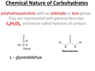
- 1. Chemical Nature of Carbohydrates polyhydroxyalcohols with an aldehyde or keto group. They are represented with general formulae Cn(H2O)n and hence called hydrates of carbons L - glyceraldehye
- 2. importance dietary calories storage form of energy in the body cell membrane components that mediate some forms of intercellular communication
- 3. Classification of Carbohydrates Monosaccharides: contain one monosaccharide unit Polysaccharides: contain more than 10 monosaccharide Disaccharides: contain 2 monosaccharide Oligosaccharides contain 3 -10 monosaccharide
- 4. MONOSACCHARIDES the general formula (CH2O)n classified according to:- The number of carbons in the molecule into trioses (3 carbons), tetroses (4 carbons), pentoses (5 carbons), hexoses (6 carbons) and heptoses (7 carbons) The presence of aldehyde or ketone group into aldoses and ketoses
- 5. Aldoses Aldotrioses( C3) e.g.D- glyceraldehyde Aldopentoses (C5) e.g. D-ribose and D-xylose. glceraldehyde is the mather of all aldoses Aldotetroses (C4) e.g. D-erythrose Most of the naturally occurring monosaccharid es are of the D type Aldohexoses (C6) e.g. D- glucose, D-mannose and D-galactose
- 6. Ketoses Ketotrioses (C3): dihydroxyacetone Ketopentoses (C5) e.g. D-ribulose dihydroxyacetoneis the mather of all ketoses Ketotetroses (C4) e.g. D-erythrulose D-Sedoheptulose is the only one in human that contains seven carbons; it is formed in the body from glucose Ketohexoses (C6) e.g. D- fructose
- 7. Asymmetric (chiral) carbon atoms Carbon atom that attached to 4 different atoms or groups Isomers are compounds which have the same molecular formula (the same number and types of atoms) but have different structural formula All monosaccharides contain asymmetric carbon atom(s) except dihydroxy acetone responsible for the isomerism and the optical activity The number of isomers = 2n ( n = number of asymmetric carbon atoms) Glucose has 4 asymmetric carbon atoms so has 16 isomers
- 8. Isomerism Optical isomerism dextrorotatory (d or+ve) sugar rotate PPL to the right levorotatory (l or –ve) sgere rotate PPL to the left Stereo isomerism Enantiomers: They are the D- and L- forms of the same compounds (mirror images) eg. D and L-glucose Aldose-Ketose Isomers (Functional Group Isomerism
- 9. Stereo isomerism Epimers compounds which have more than one asymmetric carbon and differ only in the configuration around one carbon Furanose and pyranose forms In solution, aldehyde group of glucose combines with hydroxyl group of 5th carbon atom forming 6 membered heterocyclic pyranose ring. And 5 membered furanose ring structure is formed from fructose when its keto group combines with hydroxyl group on 5th carbon atom Anomers α and β It refer to the orientation of the OH group of anomeric carbon atoms ring structure. If it to the right………. α- sugar If to the left ………… β-sugar
- 10. Important Monosaccharides Trioses and tetroses: • Glyceraldehyde 3-phosphate and dihydroxyacetonephosphate are intermediates during glucose oxidation in living cells. Hexoses : D-glucose (blood sugar)enters in the formation of many disaccharides and polysaccharides. D-fructose (fruit sgar) component of sucrose and inulin. D-galactose component of lactose (milk sugar). It is also found in glycosaminoglycans (GAGS), glycolipids and glycoproteins Pentoses : D-ribose in RNA. deoxyribose in DNA.
- 11. Monosacchari des derivatives Sugar acids: Uronic acids : The primary alcohol group of monosaccharides is oxidised to form the corresponding uronic acid. - Glucose is oxidised to form glucuronic acid (GlcUA). Aminosugars glucosamine (GluN), galactosamine (GlaN) and mannosamine (ManN) -constituents of glycosaminoglycans (GAGs) and some types of glycolipids and glycoproteins. -Several antibiotics contain aminosugars which are important for their activity Sugar alcohols Glucose …………. Sorbitol Manose…………....Mannitol Galactose………….Galactitol Fructose……..produces 2 alcohols (sorbitol and mannitol Ribose…….ribitol (componat of riboflavin B2) Deoxy Sugers as deoxy ribose: It is present in the structure of DNA. Reduction oxidation Oxygen removal Sugars with NH2 replaces the OH at C2
- 12. Monosaccharides derivatives Easter formation a- Phosphate esters: as glucose 1-P and glucose 6-P. b- Sulfate esters : They are present in certain types of polysaccharides and glycolipids (sulfolipids) e.g. β-D-galactose 3-sulfate Another sugar (Glycon): e.g. formation of disaccharides and polysaccharides. Non-Carbohydrate compound (Aglycon): such as alcohols, phenols or nitrogenous bases. -The glycosidic linkage is named according to the anomeric carbon to which it is attached (α & β) and according to the parent sugar e.g. glucosidic, galactosidic or fructosidic bond. - Examples of Glycosides: -Nucleosides are glycosides formed of ribose or deoxyribose and a nitrogenous base found in nucleic acids OH of sugar +Acid condensation of the anomeric carbon of the sugar with
- 13. DISACCHARIDES Reducing have a free anomeric carbon in the second sugar unit, so they exist in both α and β forms Maltose (Malt sugar) *main product of digestion of starch by amylase *composed of 2 D- glucopyranose connected by α - (1,4) glycosidic *hydrolyzed into 2 D glucos by Maltase enzyme or acids Non–Reducing the 2 anomeric carbon of 2 sugers are involved in the glycosidic link so not have free anomeric carbon Sucrose (Cane sugar) (Table sugar) *It is formed of β- D-fructofuranose and α -D- glucopyranose. It is united by α 1, 2-glucosidic linkage or β2,1-fructosidic linkage *Knowen as inverted suger * By hydrolysis give glucose + Fractose Isomaltose *hydrolysis products of starch and glycogen by amylase, as it represents the branching point of the molecule *composed of 2 D-glucopyranose connected by - (1,6) glycosidic bond Lactose ( Milk sugar): - It is formed of β- D-galactopyranose and D- glucopyranose united by β1,4- galactosidic linkage. -It is hydrolyzed by lactase enzyme or by acids into D- glucose and D- galactose
- 14. Polysaccharides All nonreducing Homopolysaccharides formed from one type of monosuccharids Glucans: formed of D- glucose units and include starch, dextrins, glycogen and cellulose heteropolysaccharides formed from more than one type of monosuccharids Proteoglycans *Formed of GAGs+ Protein core They are formed mainly of carbohydrates (95%) and only (5%) proteins Fructans: formed of D- fructose units e.g. inulin present in plants glycosaminoglycans (GAGs) formly called mucopolysaccharid es. are: Unbranched -Long chains (usually >50 sugar units) Composed of repeating disaccharide units, usually made up of an amino sugar and a uronic acid.
- 15. Homopolysaccharides Dextrins: -They are produced during the hydrolysis of starch by salivary or pancreatic amylase. Cellulose *In cell wall of plants. *formed of long non-branched chain of β-D- glucopyranose units connected together by β1,4- glucosidic linkage. * insoluble in water. It is non-hydrolysable by amylase because it contains a β1,4-glucosidic linkage. -The presence of cellulose in diet is important as it increases the bulk of food, which stimulates intestinal contractions and prevents constipation Starch : *storage form of carbohydrates in plants. *present in large amounts in cereals (rice and wheat), tubers (potatoes and sweet potatoes) and legumes. -*Starch granules contain two forms, amylose (15- 20%) in the inner part and amylopectin (80-85%) in the outer part. Glycogen: is the storage form of carbohydrates in animals (animal starch). -It is mainly present in skeletal muscles and liver.
- 16. Classification of Glycosaminoglycans 1- Sulfate free glycosaminoglycans: e.g. hyaluronic Acid. 2- Sulfate containing glycosaminoglycans: e.g. chondroitin sulphate, keratan sulphate, dermatan sulphate, heparin and heparan sulphate
- 17. Functions of GAGs and proteoglycans 1-They are important constituents of extracellular matrix, the proteoglycans interact with a variety of proteins in the matrix, such as collagen and elastin, and theses interactions are important in determining the structural organization of the matrix. 2-They are highly polar and attract water molecules, thereby creating a hydrated gel. This gel: A- Provides flexible mechanical support for the ECM. B-Is compressible: when a GAG solution is compressed, water is squeezed out and GAGs occupy a smaller volume. When the compression is released, their molecules regain their original hydrated size. This gives GAGs solutions the shock absorbing properties and explains their role as shock absorbents in joints and making the eyeball resilient. 3- Hyaluronic acid proteoglycans is essential for wound repair. It allows rapid migration of the cells to the site of connective tissue development.
- 18. 4-Heparin proteoglycan: is produced by mast cells present in the arteries, liver, lung and skin. Heparin function 1- anticoagulant i.e. prevents thrombus formation. It activates antithrombin & inactivates coagulation factors IX, XI. 2- Release lipoprotein lipase from the capillary wall to blood, this enzyme helps in removal and clearance of blood lipids (so lipoprotein lipase is known as clearing factor). 5-Keratan sulfate proteoglycan is important for transparency of the cornea. 6-Heparan sulfate proteoglycans are associated mainly with plasma membrane of cells and play an important role in cell membrane receptors and cell-cell interactions. 7- Aggrecan: the major proteoglycan present in cartilage contributes to its compressibility. It has a very complex structure containing many types of GAGs (hyaluronic, chondroitin sulfate and keratan sulfate). GAGs and aging: Structure of aggrecan changes with age: These changes may contribute to the development of osteoarthritis
- 19. Glycoproteins: • They are proteins to which oligosaccharide chains are covalently bound. • They are found in mucous fluids, tissues, blood and in cell membrane