
Recommended
More Related Content
What's hot
What's hot (20)
Similar to L3 PAROTID GLAND .pdf
Similar to L3 PAROTID GLAND .pdf (20)
Recently uploaded
Recently uploaded (20)
L3 PAROTID GLAND .pdf
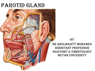
- 1. PAROTID GLAND By Dr Abulmaaty Mohamed Assistant professor Anatomy & Embryology Mutah University
- 2. PAROTID GLAND Def.: largest salivary gland (serous) parts: main part: will be discussed . accessory part: small detached that lies lie in front the main part position: lies in the gap bounded by above: ext. auditory meatus ant.: ramus of mandible & its muscles post. :mastoid process & its muscles medial: styloid process & its muscles
- 3. PAROTID GLAND shape: inverted 3 sided pyramid with 2 ends: upper(base):concave lower(apex):rounded 3 surfaces: lateral (superficial) anteromedial (ant.) posteromedial(deep) 3 borders: ant. :convex post. medial
- 4. PAROTID GLAND relations: structures inside: 1-ext. carotid art (deep). -enter through posteromedial surface -give 2 terminal brs maxillary art. that emerge from anteromedial surface & superficial temporal gives transverse facial a . then emerge from upper end 2- retronandibular v. (intermediate). -formed by union of superficial temporal with maxillary veins -give ant.& past. Divisions that emerge from lower end
- 5. PAROTID GLAND relations: structures inside: 3- facial n. (superficial). -enter through posteromedial surface -end in 5 terminal brs (temporal , zygomatic , buccal , marginal mandibular , cervical) That emerge from anterior border but (cervical br emerge from lower end) 4- auriculotemporal n. pass through the upper end 5- deep parotid L.Ns
- 6. PAROTID GLAND relations: post. border: related to sternomastoid ant. border: related to: masseter. give passage to. temporal , zygomatic branches of facial n. -transverse facial vessels -parotid duct -buccal , marginal mandibular of facial n. upper end: related to: -ext. auditory meatus. give passage to: -auriculotemporal n. -superficial temporal vessels (v.& art). lower end: related to post. belly of digastric give passage to cervical branch of facial n. -ant. & post. divisions of retro mandibular v.
- 7. PAROTID GLAND relations: lateral surface:- related to 1-skin. 2-superficial fascia with: -superficial parotid (preauricular) l.n. -great auricular n. -platysma. medial border: related to pharynx .
- 8. PAROTID GLAND relations: anteromedial surface:- related to 1-masseter. 2-ramus of mandible. 3-medial pterygoid give passage to -maxillary vessels posteromedial surface: related to 1-mastoid process & -sternomastoid.-post belly of digastric. 2-styloid process & the attached structures 3-styloid process separate the surface from Carotid sheath with:-int. carotid art. -int jugular v. Last 4 nerves: gives passage to: -facial n. & -ext. carotid art.
- 9. PAROTID GLAND parotid duct: -5 cm -emerge from middle of ant border. -pass forward: superficial to masseter ( ) the 2 buccal brs of facial n. below accessory part of the gland & tr. facial art. -after crossing ant. border of masseter it curves medially piercing. buccal pad of fat. buccopharyngeal fascia. buccinator. buccal m.m -open in vestibule of mouth opposite upper 2nd molar.
- 10. PAROTID GLAND capsule: Inner capsule:- condensed fibrous tissue Outer capsule derived from: investing fascia of Neck layers: superficial: on superficial surface. It attach to zygomatic arch deep: on deep surface. It attach to styloid process and post. border of mandible. The part of this layer that extends ( ) the the styloid process & angle of is thickened forming stylomandibular lig.
- 11. PAROTID GLAND Surface anatomy: main part: 4 points: 1-at head of mandible in front tragus. 2-center of mastoid. 3-point below & behind angle of mandible by 2 cm 4-center of masseter. upper end: Line Concave upward between 1& 2. post. border: Line between 2 & 3 lower end: 3 ant. border: line between 3,4 & 1 parotid duct: middle 1/3 of line between -tragus. -point midway between ala of nose & angle of mouth.
- 12. PAROTID GLAND arterial Supply: ext. carotid art. venous drainage: retromandibular v. lymphatic drainage: superficial& deep parotic L.Ns. deep cervical L.Ns
- 13. PAROTID GLAND nerve supply: branches from otic ganglion these branches contain 3 types of fibers: sensory: from auriculotemporal n. sympathetic: from middle meningeal plexus parasym. (secretomotor, secretory): from lesser petrosal n. from tympanic br. of glossopharyngeal n. from inferior salivary nucleus these fibers pass from otic ganglion to parotid gland through auriculotemporal n. N. B: the capsule of the gland is supplied by great auricular n.
- 14. PAROTID GLAND applied anatomy: The parotid capsule is very tight , and so in inflammation of the gland (e.g mumps) , the edema causes stretch of capsule leading to severe pain due to irritation of great auricular n. . The pain increases in eating due to increased salivation and movement of mandible. The pain is referred to auricle due to similarity in nerve supply
- 15. THANQ