
Recommended
More Related Content
What's hot
What's hot (20)
Similar to Cranial nerves
Similar to Cranial nerves (20)
More from Dr. Meenal Atharkar
More from Dr. Meenal Atharkar (20)
Recently uploaded
Recently uploaded (20)
Cranial nerves
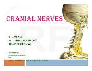
- 1. CRANIAL NERVES X - VAGUS 1 X - VAGUS XI -SPINAL ACCESSORY XII -HYPOGLOSSAL PRESENTED BY : DR. MEENAL ATHARKAR MDS DEPT OF ENDODONTICS AND CONSERVATIVE DENTISTRY
- 2. 2 INTRODUCTION OF NERVOUS SYSTEM - HISTORY -NEURON -CLASSIFICATION OF NERVE FIBERS - NEUROGLIA EMBRYONIC DEVELOPMENT OF CRANIAL NERVES CRANIAL NERVES ( X to XII ) CONCLUSION REFERENCES
- 3. HISTORY - By means of nerves, the pathways of the senses are distributed like the roots and fibers of a tree."--Alessandro Benedetti, 1497 3 -In the fourth century B.C.,the Greek philosopher Aristotle believed firmly that the nerves were controlled by and originated in the heart because it was, in his interpretation, the first organ of the body and the seat of all motion and sensation.
- 4. Galen saw the spinal cord as an extension of the brain which carried sensation to the limbs. He believed that the nerves controlled the actions of muscles in the limbs, and that the two principal functions of the nervous system, sensation and motion, were governed by two different types of nerves: respectively Soft and Hard. 4 Soft and Hard.
- 5. 5 Slightly more than a century later, Master Nicolaus offered a more precise vocabulary to express the new complexity of the nervous system, discarding the terms "soft" and "hard" for the more familiar idea of Sensory and Motor nerves.
- 6. Harvey wrote in his Lectures on the Whole of Anatomy: "For the nerves only carry down, they do not act, move, nor are they sentient by a faculty, but are organs." Descartes The essence of motor control is, then, the direction of animal 6 Descartes The essence of motor control is, then, the direction of animal spirits into the proper inter-filamentous channels for transmission to the proper nerve."
- 7. The enumerations of the modern 12 pairs of cranial nerves by a number of anatomists are reviewed in a continuous historical perspective using original texts and modern translations. The history of the numeration of the nerves is traced from Galen's classification into seven pairs (second century A.D.), through to Willis' nine pairs (1664) and Soemmerring's 12 pairs (1778). 7 Soemmerring's 12 pairs (1778).
- 8. One of the regulatory systems, made of millions of neurons in precise pathways to transmit electrochemical impulses, and of neuroglial cells that have several functions, including formation of myelin sheaths of neurons. NERVOUS SYSTEM myelin sheaths of neurons. 8
- 9. 9NERVOUS SYSTEM CENTRAL NERVOUS PERIPHERAL NERVOUS SYSTEM SYSTEM Brain Spinal Cord Neurons Their Process - Formed by neurons n neuroglia. - Cranial nerves and spinal nerves. Spinal Cord Their Process
- 10. . 10 PERIPHERAL NERVOUS SYSTEM SOMATIC NERVOUS SYSTEM (Includes nerves supplying the AUTONOMIC NERVOUS SYSTEM (regulation of visceral or(Includes nerves supplying the skeletal muscles ) Vegetative functions ) Sympathetic Parasympatheti c
- 11. NEURON : A nerve cell, the structural and functional unit of the nervous system. Neurons function in initiation and conduction 11 Neurons function in initiation and conduction of impulses.
- 12. Neuron is made up of 3 parts : 1. Nerve cell body 2.Dendrite 3. Axon 1.NERVE CELL BODY : - Irregular in shape - Neuroplasm contains large nucleus , nissle bodies , neurofibrils, mitochondria and golgi apparatus. 12 apparatus. 2. Dendrites : - Branched process . - Conductive nature . - Transmits the impulse towards the nerve cell body. .
- 13. 3. AXON : the length of longest axon is about one meter . - Axons are arranged in different bundles called fasciculi. -Tubular sheath covering around whole nerve formed by areolar membrane. 13 nerve formed by areolar membrane.
- 14. Axoplasm contain mitochondria, neurofibrils, axoplasmic vesicles. -Neurotransmitter substances are carried by axonal flow. -Axons might be insulated by myelin sheath and called 14INTERNAL STRUCTURE OF AXON AXIS CYLINDER : -Axons might be insulated by myelin sheath and called Myelinated Nerve Fibers. Axis cylinder of nerve fiber is covered by a membrane called neurilemma ,also called sheath of Schwan (non myelinated fibers).
- 15. - Does not form a continuous sheath and is absent in regular interval . - Node of Ranvier. - Segment of nerve fiber between two node is called internode. -Responsible for white colour of the nerve. CHEMISTRY : 15 CHEMISTRY : -Formed by concentric layers of proteins alternating with lipids.
- 16. A) DEPENDING UPON THE NUMBER OF POLES : 16 A) DEPENDING UPON THE NUMBER OF POLES :
- 17. 1. Depending upon structure : Myelinated Non Myelinated 2 Depending upon the distribution : 17 Somatic Nerve Fibers Visceral or Autonomic Nerve Fibers
- 18. 3. Depending upon the origin : Cranial Nerves Spinal Nerves 4. Depending upon functions : Sensory Nerve Fibers Motor Nerve Fibers 18 5. Depending upon secretion of neurotransmitter : Adrenergic Nerve Fibers Cholinergic Nerve Fibers
- 19. 6. DEPENDING UPON THE DIAMETER AND CONDUCTION ( EARLANGER –GASSER CLASSIFICATION ) 19 TYPE DIAMETER (µ) VELOCITY OF CONDUCTION (m/ sec ) 1. A alpha 12 TO 24 70 TO 120 2. A beta 6 TO 12 30 TO 70 - Except C fibers all the nerve fibers are myelinated. 2. A beta 6 TO 12 30 TO 70 3. A gamma 5 TO 6 15 TO 30 4. A delta 2 TO 5 12 TO 15 5. B 1 TO 2 3 TO 10 6. C < 1.5 0.5 TO 2
- 21. EMBRYONIC DEVELOPMENT Neural tube gives rise to brain and spinal cord . It is formed from the ectoderm overlying notochord (process is known as neurulation). Cranial is enlarged part of neural tube form brain. 21 Cranial is enlarged part of neural tube form brain. Caudal tubular part forms spinal cord.
- 22. Nervous Tissue : Neurons and many neuroglial cells are formed in neural tube. Neural tube is first lined by single layer. These cells proliferate to form several layers. Matrix cell layer/Germinal layer The cells of this layer give rise to nerve cells ,neuroglial cells . Mental layer 22 Mental layer Developing nerve cell -Neuroglial cells. Marginal zone It provides a frame work into which the processes of nerve cells developing in mantle layer can grow.
- 23. Stages : One of the germinal cell passes from germinal layer to mantle layer and become APOLAR NEUROBLAST . Two process develop and convert apolar neuroblast to a bipolar neuroblat . One of the process of neuroblast disappear and it can now be called a unipolar neuroblast . 23 and it can now be called a unipolar neuroblast . Multipolar neuron are formed. - And these neurons grow into the marginal layer and becomes the axon of nerve cell. Neurons may remain in CNS or May grow out as efferent nerve Fibers of peripheral nervous system.
- 24. Formation of Myelin Sheath : 24
- 25. Formation of Myelin Sheath : Formation of myelin sheath around axon is called Myelinogenesis Starts at 4th month of intrauterine life. Completed by 2nd year after the birth. The schwann cells wrap up and rotate around the axis cylinder in many concentric layers. 25 many concentric layers. Concentric layers fuse to produce the myelin sheath but cytoplasm. Outermost membrane of schwann cells remains as neurilemma . Nucleus of these cells remains in between myelin sheath and neurilemma.
- 26. CRANIAL NERVES Ⅰ Olfactory nerve Ⅱ Optic nerve Ⅲ Oculomotor nerve Ⅳ Trochlear nerve Ⅴ Trigeminal nerve Ⅵ Abducent nerve 26 Ⅵ Abducent nerve Ⅶ Facial nerve Ⅷ Vestibulocochlear nerve Ⅸ Glossopharyngeal nerve Ⅹ Vagus nerve Ⅺ Accessory nerve Ⅻ Hypoglossal nerve
- 27. 27
- 28. Extensive course. The fibers of cranial root of XI nerve is also distributed through it. 28 Motor division of the vagus nerve – basal plate of the embryonic medulla oblongata. Sensory division originates- cranial neural crests.
- 29. Two ganglia Superior Inferior -Round -Cylinderical 29 -Round -Cylinderical -Juglar foramen -Near the base of skull
- 30. Functional components 30 a) Special visceral efferent fibers : - Nucleus ambiguus - Muscles of palate, pharynx ,larynx. b) General visceral efferent fibers : - Dorsal motor nucleus - Thoracic and abdominal viscera . c) General visceral afferent fibers : - Inferior ganglion - Bring sensation from pharynx ,larynx ,trachea , oesophagus and from abdominal and thoracic viscera.
- 31. 31 - These are conveyed by central processes of ganglion cells to nucleus of tactus solitarius. - Terminate in dorsal nucleus of vagus. d) Special visceral afferent fibers : - Inferior ganglion - Carry sensation of taste from posterior most part of tongue and from epiglottis .from epiglottis . - Terminate in Tractus Solitarius e) General somatic afferent fibers : - superior ganglion. - Skin of external ear. - Terminate in the relation to spinal nucleus of trigeminal nerve.
- 32. 32 The vagus nerve includes axons which emerge from or converge onto three nuclei of the medulla
- 33. NUCLEI 33 1. NUCLEUS AMBIGUUS : A part of cranial root of accessary nerve : partly of vagus. 2. Dorsal nucleus of vagus (parasympathetic ) : - Mixed nucleus - Fibers from main bulk of nerve 3. Nucleus of tractus solitarius ( gustatory) : - Distributed through internal laryngeal nerve to the taste buds of epiglottis and vallecula. 4. Nucleus of spinal tract of trigeminal nerve
- 34. 34
- 35. 35
- 36. The vagus nerve descends vertically within the carotid sheath posterolateral to the internal and common carotid arteries and medial to the internal jugular vein (IJV) at the root of the neck. 36
- 37. Anterior and posterior gastric fibers are mainly formed by oesophageal complex. Anterior gastric fibers – left vagus Posterior gastric fibers – right vagus The gastric nerves supply all abdominal organs and the gastrointestinal tract ending just before the left colonic (splenic) 37 gastrointestinal tract ending just before the left colonic (splenic) plexure.
- 38. 38 Branches in Head And Neck Branches in juglar foramen Branches in neck - Meningeal - Pharyngeal - Auricular - Carotid- Auricular - Carotid - Communicating branches - Superior laryngeal - Right recurrent laryngeal - Cardiac
- 39. 39 Behind IJV Enters mastoid canaliculus It crosses facial canal 4mm above stylomastoid i) Meningeal branch : - Supply durameter of posterior cranial fossa . ii) Auricular branch : - Arise from superior ganglion. stylomastoid foramen It emerges through tympanomastoid fissure Ends by supplying the choncha,root of auricle , posterior half of EAM,tympanic membrane
- 40. 40 iii) Pharyngeal branch : - Inferior ganglion - Chiefly fibers : cranial root of accessary nerve. - Passes between ECA & ICA . -Reaches the upper border of middle constrictor pharyngeal plexus .-Reaches the upper border of middle constrictor pharyngeal plexus . - Supply muscle of pharynx and soft palate except tensor veli palatine.
- 41. 41 iv) Carotid branch : carotid sinus and carotid body. HUMAN ANATOMY , B.D. CHAURASIA 4TH EDITION
- 42. 42
- 43. 43 v) Superior laryngeal nerve : - Inferior ganglion - at middle constrictor it divides into two branches external laryngeal internal laryngeal - thin - thick - accomapanies Sup. Thyroid - above the Sup. Laryngeal- accomapanies Sup. Thyroid - above the Sup. Laryngeal artery vessel -branches to inf.constrictor - pierces thyrohyoid memb. and phyrngeal plexus - supply the mucous membrane - supply cricothyroid muscles of larynx upto level of vocal folds. .
- 44. vi) Recurrent laryngeal nerve : 44
- 45. 45 vi) Recurrent laryngeal nerve : -Winds backwards below the subclavian artery. -Runs upward and medially behind subclavian and common carotid arteries to reach the tracheo-oesophageal groove . -It is relatd to inferior thyroid artery. -It may be superficial or deep to artery. -The nerve passes deep to lower border of inferior constrictor and enters the larynx behind cricothyroid joint .
- 46. 46 It Supplies - a) Supplies all intrinsic muscles of larynx. b) Sensory nerves to larynx below vocal cords. c) Cardiac branches d) Branches to trachea, oesophagus. e) Inferior constrictor
- 47. 47 vii) Left recurrent laryngeal nerve : - Arise from vagus. - Crosses the left side of aorta . - Loops around the ligamentum arteriosum and reaches the tracheo- esophageal groove. - Does not have to pass behind the subclavian artery and carotid arteries. - Posterior to the inferior thyroid artery.
- 48. 48 vii) Cardiac branches : - 4 in number. SUPERIOR INFERIOR - 4 in number. - Left interior branch goes to superficial cardiac plexus. - The other 3 cardiac nerves go to the deep cardiac plexus.
- 49. 49 APPLIED ANATOMY : Tested clinically by comparing the palatal arches on two sides. On paralysed site –no arching and uvula is pulled to normal side. Paralysis produces : Paralysis produces : - Nasal regurgitation of swallowed liquids -Nasal twang in voice - Hoarseness of voice -Flattening of palatal arch -Dysphagia
- 50. 50 Irritation of auricular branch of vagus in external ear may cause persistent cough, vomiting or even death due to sudden cardiac inhibition. Stimulation of auricular branch –increased appetite. Sensory ganglion may have viral infection called Herpes zoster. Sensory ganglion may have viral infection called Herpes zoster. Vesicles over the skin of auricle.
- 51. Clinical anatomy 51 - Lesion of an entire vagus nerve is uncommon. - A)Pharyngeal branches : dysphagia - B)Superior laryngeal nerve : Anesthesia of Sup. part of larynx. Paralysis of cricothyroid muscle Paralysis of cricothyroid muscle Voice is weak and tires easily Lesion of LRN is more common (longer course) Proximal lesions of vagus nerve affect pharyngeal and superior laryngeal nerves , causing difficulty in swallowing.
- 52. 52 The two somatic sensory branches of the vagus nerve, the auricular branch and the superior laryngeal nerve, can also be the site of a rare pain syndrome that resembles that of TN. It is thought that compression of the upper fibers of the vagal nerve as they leave the brain stem and traverse the subarachnoid space to the jugular foramen is the cause of vagal neuralgia.
- 53. Vagus nerve neuralgia is characterized by paroxysms of shock-like pain in the side of the thyroid cartilage, pyriform sinus, angle of the jaw. The trigger zone is usually in the larynx; attacks are precipitated by talking, swallowing, yawning, or coughing. 53 by talking, swallowing, yawning, or coughing. When other portions of the vagus nerve are involved, the patient might have hiccups, excessive salivation, or coughing. The pain is similar to TN except for its location.
- 54. 54 The diagnosis is established by the history and by identifying the site of the trigger zone. Associated vagal nerve findings also pinpoint this nerve as the site of the pain. Laryngeal topical anesthesia or blockade of the superior laryngeal nerve stops the pain and is useful diagnostic and prognostic procedure.
- 55. Ramsay-Hunt Syndrome 55 Zoster infection of geniculate ganglion with involvement of external ear & oral mucosa. Facial paralysis, pain of external auditory meatus & pinna of ear. Vesicular eruptions in the oral cavity & oropharynx with Vesicular eruptions in the oral cavity & oropharynx with hoarseness, tinnitus & vertigo.
- 56. Vaso vagal syncope 56 Transient loss of consciousness due to cerebral ischemia, caused by reduction of blood supply to brain. Vasodilation Vagal stimulation dramatic fall in blood pressure Sign- nausea, weakness, sweating, hypotension Causes- pain/fear, postural changes, anoxia Dental consideration- Surgery in morning Minimize waiting Avoid anxiety Administer adequate pain control.
- 58. FUNCTIONAL COMPONENTS : 1) Cranial Root : - Special visceral (branchial) efferent. - Nucleus Amiguus - Distributed through the branches of vagus to muscles of palate, pharynx, larynx, possibly the heart. 58 pharynx, larynx, possibly the heart.
- 59. 59 2. Spinal Root : -Special visceral efferent -Long Spinal Nucleus -Supply to Sternocleidomastoid and Trapezius muscle .
- 60. 60 COURSE AND DISTRIBUTION OF CRANIAL ROOT Cranial root emerges in form of 4 to 5 rootlets which are attached to posterolateral sulcus of medulla . Rootlets soon join together to form a single trunk .
- 61. COURSE AND DISTRIBUTION OF SPINAL ROOT • C1- C5 • Foramen Magnum • Jugular Tubercle • Jugular Foramen 61 • Jugular Foramen • Unites &Separates • IJV & ICA • Deep To Parotid & Styloid Process • Angle Of Mandible & Mastoid Process • SCM EXTRACRANIAL
- 62. DEEP SCM OCCIPITAL ARTERY ANT. BORDER OF SCM 62 ANT. BORDER OF SCM POSTERIOR OF NECK LEVATOR SCAPULA ANT. TRAPEZIUS 5CM ABOVE CLAVICLE
- 63. 63 - Supplies : a) Sternocleidomastoid b) Trapezius - Cervical nerves provide a proprioceptive supply to these muscles .
- 64. APPLIED anatomy 64 - TEST : i) Asking the patient to shrug his shoulders (trapezius) against resistance and comparing the power on two sides. ii) Asking the patient to turn the chin to opposite side againstii) Asking the patient to turn the chin to opposite side against resistance and again comparing the power on two sides.
- 65. 65 - Lesions are accompanied by lesions of 9th ,10th nerve (close inter-relationship in cranium) . - Irritation of nerve by enlarge lymph nodes may produce torticollis or wry neck.
- 66. Superficial passage through the posterior triangle – spinal root is susceptible to injury.66
- 68. 68 FUNCTIONAL COMPONENTS/NUCLEAR COLUMNS : A) General somatic efferent column - Fibers arise from the hypoglossal nucleus which lies in medulla,in floor of fourth ventricle deep to hypoglossal triangle. B) General somatic afferent column - Nucleus is spinal nucleus of cranial nerve V where proprioceptive fibers from tongue end.
- 69. 69
- 70. 70 NUCLEUS : - 2 cm long , lies in floor of fourth ventricle beneath hypoglossal triangle. - Divides into parts for individual muscles innervated. - Connection of nucleus with opposite pyramidal tract forms supranuclear pathway of nerve.supranuclear pathway of nerve. - It is also connected to cerebellum,reticular formation of medulla , sensory nuclei of 5th nerve , and the nucleus of tractus solitarius.
- 71. 71 COURSE AND RELATIONS : INTRA NEURAL COURSE : - the fibers pass forwards lateral to medial longitudinal bundle , medial lemniscus and pyramidal tract , and medial to reticular formation and olivary nucleus . - Nerve is attached to anterolateral sulcus of medulla , between pyramid and olive by 10 to 15 rootlest .and olive by 10 to 15 rootlest . - Rootlets run laterally ( behind the vertebral artery and join to form two bundles which pierce the durameter seperately near hypoglossal canal. - The nerve leaves skull through the hypoglossal (anterior condylar ) canal.
- 72. Internal Juglar Vein Inclines Laterally Crosses The Vagus ( Laterally ) EXTRA CRANIAL COURSE 72 Crosses The Vagus ( Laterally ) B/W IJV &ICA Deep To Parotid Gland & Styloid Process, Post Belly Of Diagstric & Stylohyoid, Occipital artery. Hooks SCM branch of occipital artery
- 73. Crosses ICA & ECA Loops lingual artery Passes by digastric 73 Enters Submandibular region Continues forward Hyoglossus & Genioglossus, Mylohoid Enter tongue & supply its muscle
- 74. 74 BRANCHES AND DISTRIBUTION : -In addition to its own fibers , the nerve also carries some fibers that reach it from spinal nerve C1 and are distributed through it. Containing fibers of XII nerve Containing fibers of nerve C1
- 75. 75 A) FIBERS OF HYPOGLOSSAL NERVE PROPER Supply extrinsic and intrinsic muscles of tongue except palatoglossus which is supplied by fibers of cranial accessory nerve through vagus and pharyngeal plexus.
- 76. 76 b) FIBERS OF NERVE C1 Meningeal branch • Enters skull through hypoglossal canal • Supplies Descending branch • Continues as descendens hypoglossai or upper Other branches • Thyrohyoid • Geniohyoid muscles • Supplies bone an meninges or upper root of ansa cervicalis
- 77. APPLIED ANATOMY 77 - Hypoglossal nerve accompanies tonsillar artery on lateral wall at pharynx. - Because this wall is thin , CN XII is vulnerable to injury during during tonsillectomy . - Injury to CN XII paralyse the ipsilateral half of tongue . After some time tongue atrophies , making it appear shrunken- After some time tongue atrophies , making it appear shrunken and wrinkled . - When tongue protruded , its tip deviates toward the paralyzed side because of unopposed action of genioglossus in normal side of tongue.
- 78. TEST – Protrusion and other movements 78 The patient should be instructed to stick the tongue straight out. The tongue held in this position allows the clinician to visually inspect for lateral deviation, abnormal muscular mass, and aberrant movement.aberrant movement. An assessment should be made of lateral and vertical movements of the tongue.
- 79. 79 a) Infranuclear lesion : -Gradual atrophy of paralysed half of tongue. b) Supranuclear lesion :b) Supranuclear lesion : -Paralysis without wasting. -Tongue moves sluggishly resulting in defective speech. -On protrusion tongue deviates to opposite side.
- 80. Disorders of cranial nerve XII (hypoglossal nerve) cause weakness or wasting (atrophy) of the tongue on the affected side. As a result, people have difficulty speaking, chewing, and swallowing. Disorders of the hypoglossal nerve may result from a tumor at 80 Disorders of the hypoglossal nerve may result from a tumor at the base of the skull, a stroke, infections of the brain stem, or an injury to the neck, including that due to surgical removal of a blockage from an artery in the neck.
- 81. Damage due to amyotrophic lateral sclerosis produces a distinctive wormlike movement of the tongue. Magnetic resonance imaging (MRI) is usually performed to look for a tumor or evidence of a stroke. 81 look for a tumor or evidence of a stroke. Treatment depends on the elimination of the cause.
- 82. SUMMARY There are 12 pairs of cranial nerves and move to cover the needs of the cranium and face, rather than make their way down through the spinal cord .These nerves are important to consider ,as most are of critical importance to sensory data, yet do not pass through the central cord and 82 sensory data, yet do not pass through the central cord and so can not be intercepted at the same juncture.
- 83. REFERENCES 83 1. BURKET’S ORALMEDICINE 10TH EDITION 2.HUMAN ANATOMY, B.D. CHAURASIA , 4TH EDITION. 3.GRAY’S ANATOMY 39TH EDITION. 4.ESSENTIAL CLINICAL ANATOMY , SECOND EDITION ,KEITH N MOORE 5.HUMAN EMBRYOLOGY, 8TH EDITION, INDERBEER SINGH 6 . HUMAN PHYSIOLOGY, THIRD EDITION,A.K.JAIN 7.http://www.bmc.med.utoronto.ca/cranialnerves/images/stories/Accessory/xi -4_nolabels.jpg