03 bone non neoplastic part-1
•Download as PPT, PDF•
4 likes•941 views

Congenital Achondroplasia* Osteogenesis imperfecta* Osteopetrosis * Metabolic bone diseases Osteoporosis* Osteitis fibrosa cystica* Rickets and osteomalacia Renal osteodystrophy Disease caused by osteoclast dysfunction Paget’s disease* Osteonecrosis (avascular necrosis) ** Infections* Fractures Miscellaneous

Recommended
More Related Content
What's hot
What's hot (20)
Similar to 03 bone non neoplastic part-1
Similar to 03 bone non neoplastic part-1 (20)
More from med_students0
More from med_students0 (20)
Recently uploaded
Recently uploaded (20)
03 bone non neoplastic part-1
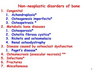
- 1. 11 Non-neoplastic disorders of bone 1. Congenital 1. Achondroplasia* 2. Osteogenesis imperfecta* 3. Osteopetrosis * 2. Metabolic bone diseases 1. Osteoporosis* 2. Osteitis fibrosa cystica* 3. Rickets and osteomalacia 4. Renal osteodystrophy 3. Disease caused by osteoclast dysfunction 1. Paget’s disease* 4. Osteonecrosis (avascular necrosis) ** 5. Infections* 6. Fractures 7. Miscellaneous
- 2. 22 Congenital disorders of bone 1. Achondroplasia • (long bones not formed properly) 2. Osteogenesis imperfecta • (Defective synthesis of type I collagen) 3. Osteopetrosis: • (Replacement of marrow cavity by bone due to defective osteoclast function)
- 3. 33 Achondroplasia • An autosomal dominant condition in which – there is impaired formation of the long bones due to – Impaired proliferation of cartilage at growth plate. • Is one of the MC causes of dwarfism • Clinical findings: – Normal sized head and vertebral column – Shortened arms and legs (dwarfism) – Normal intelligence, life span and reproductive ability. – Normal GH and insulin growth factor -1 levels.
- 4. 44 Achondroplasia • Molecular basis: – point mutation in gene coding for fibroblast growth factor receptor 3 (FGFR3) – Inhibits cartilage synthesis at growth plate – Decreased growth of long bones
- 5. 55 Osteogenesis imperfecta • AKA "brittle bone diseases“ or fragilitas ossium. • A group of diseases having in common defective synthesis of type I collagen. – Due to mutations in genes coding for α1 & α2 chains of collagen. • Pathology: – Generalized osteopenia (brittle bone) • Resulting in recurrent fractures and skeletal deformity. – Disease affects skeleton and other tissues rich in type I collagen.
- 6. 66 Osteogenesis imperfecta • Types: Several types are known (Type I-IV) – Most are autosomal dominant***. • Clinical findings: – pathological fractures** at birth – blue sclera** : due to reflection of underlying choroidal veins. – Deafness : involvement of bone of inner and middle ear – Thin dermis easy bruising. – Dental imperfections. – Type II is lethal in utero
- 8. 88
- 10. 1010 Osteopetrosis ("marble bone disease") • Group of genetic diseases characterized by: – decreased osteoclast function, leading to • Decreased resorption, and • Greatly increased density* of bone. • Pathology: – Increased bone density and thickening of bone cortex – Marrow cavity replaced by bone – The thickened bones are brittle and fracture easily.
- 11. 1111 Dense bones of the lower extremities Obliteration of the marrow space Absence of marrow
- 13. 1313 • Occurs in two forms – autosomal dominant (usually mild) or – autosomal recessive (usually severe). • Clinical findings: – Pathologic fractures – Pancytopenia and leukoerythroblastic blood picture: • Replacement of marrow cavity by bone. – Extramedullary hematopoiesis: hepatosplenomegaly – Cranial nerve compression: • Due to narrowing of cranial foramina • May result in blindness, deafness and facial nerve palsies. • Treatment: bone marrow transplant
- 14. 1414 Metabolic diseases of bone
- 15. 1515 Osteoporosis • Refers to increased porosity of skeleton. • Occurs due to: – Loss of organic bone matrix and minerals. • Resulting in : – decreased bone mass and density. – decreased thickness of cortical and trabecular bone. • Osteoporotic bones: – thin and fragile and are susceptible to fracture.
- 16. 1616 Osteoporosis • Pathogenesis: – Osteoporosis results from a slight excess of bone resorption over bone deposition, continuing over many years. • Epidemiology: – Is the most common metabolic abnormality of bone. – MC occurs in postmenopausal Caucasian women and the elderly.
- 17. 1717 TYPES of osteoporosis A: Localized : e.g. disuse OP of a limb B: Generalized: involves entire skeleton. Two types 1. Primary: – Old age (Senile) • Osteoblasts with diminished capacity to make new bone • Physical inactivity – Estrogen deficiency (postmenopausal) 2. Secondary: due to underlying disease – Cushing’s disease (hypercortisolism) – Drugs (heaprin and steroids) – Space travel
- 18. 1818 Postmenopausal osteoporosis Pathogenesis • Is secondary to estrogen deficiency. • Normally Estrogen: – Stimulates OPG production – Decreases production of M-CSF – Decreases response of osteoclasts to RANK ligand. – Net result: • Decreased formation of osteoclasts • Decreased bone resorption
- 19. 19 Postmenopausal osteoporosis Pathogenesis……… • Estrogen deficiency results in: • ↑production of IL1 and TNF by monocytes • Increased activity of RANK ligand and M- CSF • ↑ osteoclast activity bone loss.
- 20. 2020 Peak bone mass Osteoporosis Menopause ↓ estrogen ↑ IL1,TNF ↑ RANK ligand, M-CSF ↑ osteoclast activity Aging ↓ activity of Osteoprogenitor cells ↓ activity of osteoblasts ↓ physical activity Physical activity Nutrition Genetic factors
- 21. 2121
- 24. 2424 N L K
- 25. 25 Osteoporosis • Clinical findings: – Bone pain – Weight bearing bones predisposed to # • Compression # of vertebral bodies (most common) • Colles’ # of distal radius. • # femoral neck. – Loss of height and kyphosis 25
- 26. 2626
- 27. 2727 Pathology • thin cortical bone and thin trabecular bone
- 28. 2828 • Diagnosis: – Dual energy X ray absorptiometry (DEXA) • evaluate bone density. • Prevention of postmenopausal osteoporosis: – weight bearing exercises: walking, not swimming – calcium, vitamin D supplements – estrogen replacement therapy • Unopposed estrogen increases the risk of endometrial Ca. • Prevented by addition of progesterone. • Treatment: – Bisphosphonates: inhibit bone resorption. – Calcitonin: inhibits osteoclasts – SERM: selective estrogen receptor modulators
- 29. 2929 Osteitis Fibrosa Cystica ( von Recklinghausen disease of bone) • The cause is primary or secondary hyperparathyroidism 1. Primary hyperparathyroidism : – Parathyroid adenoma ↑ PTH – Parathyroid hyperplasia ↑ PTH 2. Secondary hyperparathyroidism : – prolonged hypocalcemia (renal failure) increased PTH. • Increased levels of PTH – Activates osteoblasts, which in-turn activates osteoclasts resulting in bone resorption and hypercalcemia.
- 30. 3030 Osteoclasts Fibrosis Osteoclasts in a fibrous stroma “Brown tumor” of hyperparathyroidism
- 31. 3131
- 32. 3232 • Characterized by: – Wide spread osteolytic lesions. – Predisposing to Deformity , microfractures and – Secondary hemorrhages with formation of cysts. – OFC can manifest as “Brown tumor” of bone characterized by: • Cystic spaces lined by multinucleated osteoclasts, filled with fibrous tissue, and with brown discoloration resulting from hemorrhage. Osteitis Fibrosa Cystica
- 33. 3333 Rickets and osteomalacia • Both diseases characterized by – Decreased mineralization of newly formed bone. – Usually caused by deficiency or abnormal metabolism of vitamin D. • Osteomalacia: – Cause is Vitamin D deficiency in adults • Rickets: – Cause is vitamin D deficiency in children • See nutrition lectures***.
- 34. 3434 Renal osteodystrophy • Osteomalacia secondary to chronic renal disease. • Pathogenesis: Chronic renal failure causes – Hypocalcemia • Due to lack of conversion of inactive vitamin D into active vit.D – Hypocalcemia results in secondary hyperparathyroidism – PTH secretion stimulates osteoclast activity
- 35. 3535 Paget’s disease of bone OSTEITIS DEFORMANS
- 36. 3636 Paget disease of bone (osteitis deformans) • Skeletal disease characterized by episodes of: – Excessive and disordered bone resorption by osteoclasts followed by – Exuberant but disorganized bone formation, producing • thickened but weak bone that is • susceptible to deformity and #. • Epidemiology: – Primarily occurs in elderly men (>40 yrs). • Etiology: – Cause unknown ( ? paramyxovirus)
- 37. 3737
- 38. 3838 Paget disease of bone • Forms of involvement: – Monostotic: involving one bone – Polystotic : involving multiple bones. – Common bones include: • pelvis >skull> femur.
- 39. 3939 Pathogenesis • Three stages of Paget disease: 1.Osteolytic stage 2.Mixed osteolytic-osteoblastic stage 3.Osteosclerotic (burnt out stage)
- 40. 4040
- 41. 4141 Paget disease of bone : Stages • Osteolytic Stage: – osteoclastic resorption of bone predominates • Mixed osteolytic and osteoblastic stage: characterized by – Osteolytic and increased osteoblastic bone formation (usually woven bone). – New bone: poorly mineralized, soft and weak. • vulnerable to # and deformation. • the bone is deposited in a tile like or mosaic pattern which is pathognomonic of Paget disease. • increased alkaline phosphatase levels. • Osteosclerotic stage: – Osteoblastic activity predominates
- 42. 4242 • Clinical findings: – Asymptomatic in most cases – Elevated alkaline phosphatase* – Bone pain from Pathologic fractures* – Skeletal deformities* (tibial bowing, skull enlargement; increased hat size) – Hearing loss:* narrowing of foramina – Warmth of overlying skin due to bone hypervascularity. – High output cardiac failure (due to AV connections in vascular bone)* – Increased risk of osteogenic sarcoma*.
- 43. 4343 Pathology • Microscopic features: – Haphazard arrangement of cement lines creating a “mosaic pattern” .
- 44. 4444Normal bone
- 45. 4545 Mosaic pattern of lamellar bone
- 46. 4646Paget's disease of bone: Mosaic pattern
- 47. 4747 Osteonecrosis (Avascular necrosis) • Ischemic infarction of bone & bone marrow. • Causes of ischemia: – Vascular interruption (fracture) • (femoral neck # bleeds into capsule compromise medial femoral circumflex artery avascular necrosis) – Corticosteroids* (most common) (SLE patients) – Sickle cell disease, Caisson disease. • Common sites include – femoral head – Scaphoid bone.
- 48. 4848 Osteonecrosis: Morphology • Osteonecrosis: Can involve – The medullary cavity or – The subchondral region • Medullary infarct: – involves marrow and the cancellous bone. – usually clinically silent. – Remains stable • Subchondral infarct: – Involves the subchondral bone • Wedge shaped area of necrosis with viable overlying articular cartilage – Causes chronic pain – Collapse predispose to osteoarthritis
- 50. 5050
- 51. 5151 Osteonecrosis in children • May involve characteristic sites: • Legg-Calve-Perthes disease: – Aseptic necrosis involving the ossification center in the femoral head. – Occurs most often in boys 3-10 years of age. – Pain in knee or limp – secondary osteoarthritis is common. • Kohler bone disease: – Aseptic necrosis of navicular bone
- 52. 5252 Osgood-Schlatter disease • Inflammation of proximal tibial apophysis at insertion of patellar tendon. • Affects physically active boys 11-15 yrs of age • Produces permanent knobby appearing knees.