Laryngeal myxoma resembling a laryngeal polyp
•
1 like•389 views

Laryngeal myxoma resembling a laryngeal polyp

Recommended
Recommended
More Related Content
What's hot
What's hot (20)
Similar to Laryngeal myxoma resembling a laryngeal polyp
Similar to Laryngeal myxoma resembling a laryngeal polyp (20)
More from Mario Fernando Dueñas Patólogo.
More from Mario Fernando Dueñas Patólogo. (17)
Recently uploaded
Recently uploaded (20)
Laryngeal myxoma resembling a laryngeal polyp
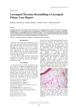
- 1. Van Tıp Dergisi: 20(2): 100-102, 2013 Laryngeal myxoma Olgu Sunumu Van Tıp Dergisi, Cilt:20, Sayı:2, Nisan/2013 100 Laryngeal Myxoma Resembling a Laryngeal Polyp: Case Report Mehmet Fatih Garca* , Hakan Cankaya* , Mahfuz Turan** , Mustafa Kosem*** Abstract Laryngeal myxoma is an extremely rare benign tumor of mesenchymal cell origin. The tumor is frequently misdiagnosed as a vocal polyp because its appearance resembles vocal polyp on laryngeal examination. Definite diagnosis of laryngeal myxoma is obtained by biopsy. A 61-year-old man presented with a laryngeal myxoma arising from the left vocal cord. Laryngeal myxoma can be confused with laryngeal polyp clinically and must be considered in the differential diagnosis of laryngeal masses. Key words: Benign laryngeal mass, diagnosis, laryngeal myxoma, pathology Introduction Myxoma is a benign tumor appearing on the bone, in the soft tissue, and in the internal organs, particularly the heart. The term myxoma was first described by Virchow in 1871 as a tumor resembling the mucinous part of the umbilical cord histologically (1). Many myxomas that rarely appear on the head and the neck regions are seen on the maxilla and the mandible and they are of odontogenic origin. The case that has been discussed in this study presented to our clinic with the complaint of dysphonia, for which the diagnosis of laryngeal myxoma was made. Case Report A 61-year-old male patient presented to our clinic with the complaint of dysphonia that had been present for 3 years. The patient did not have a history of any intubations or laryngeal trauma. The patient’s profession could not explain any features about any diseases of the voice. The patient had been smoking one pack of cigarettes per day for 40 years, but did not consume alcohol. On examination of the ear, nose, and * Yuzuncu Yil University, Department of Otorhinolaryngology, Van, TURKEY ** Lokman Hekim Hospital, Department of Otorhinolaryngology, Van, TURKEY *** Yuzuncu Yil University School of Medicine, Department Pathology, Van, TURKEY Corresponding Address: M. Fatih Garca, MD. Yuzuncu Yil Universitesi, Tip Fakultesi, Arastirma Hastanesi, KBB A.D. Maras Cad. 65100, Van, TURKEY Phone: 00 90 530 327 74 96 E-mail: fatihgarca@hotmail.com Makalenin Geliş Tarihi: 26.07.2011 Makalenin Kabul Tarihi: 31.10.2012 throat, there was a pediculated polypoid lesion on the mid 1/3 region of the right vocal cord of approximately 0.5x0.5 cm in diameter. Under general anesthesia, the polypoid lesion was excised completely from the pediculated region by suspension direct laryngoscopy, leaving behind no residual tissue. The result of the histopathological examination of the specimen was reported as laryngeal myxoma. Microscopically, the tumor was covered by squamous epithelium and comprised cytologically bland spindles and occasional satellite cells arranged randomly within a pale-staining basophilic myxoid matrix (Figure 1). The basophilic matrix stained positive for Alcian- blue. The immunoperoxidase staining performed on the paraffin sections of the tumor using the standard technique were negative for CD34, S- 100 protein, desmin, and muscle-specific actin. Fig. 1. FIG 1 Photomicrograph of laryngeal myxoma showing bland neoplastic spindle cells arranged haphazardly in a pale-staining myxoid matrix. Neoplastic spindle cells are covered with non- neoplastic squamous epithelium (upper left corner) (H- E X 100).
- 2. Garca ve ark. Van Tıp Dergisi, Cilt:20, Sayı:2, Nisan/2013 101 The patient’s complaint of dysphonia completely improved postoperatively. The patient remains under control by laryngoscopic examination periodically every three months. Discussion Benign vocal cord mass lesions are encountered as cases of dysphonia that should usually be surgically intervened. Benign vocal cord masses include lesions like vocal polyps, vocal cysts, vocal nodules and Reinke edema (2). Myxomas occur mostly in the subcutaneous soft tissue, intramuscular tissue or in the cardiac chambers. When they occur in the head and neck regions, they mainly involve the maxilla and the mandible, subcutaneous tissue and the parotid gland (3). Myxomas most frequently occur in the third or fourth decades of life, although individuals of all ages can be affected. In the literature, all the patients were male except for one case (3). The larynx is an unusual location for soft tissue myxomas. Within the larynx, the vocal cords (3- 6), the aryepiglottic folds (7, 8) and the epiglottis (9) have been described as locations. Common clinical presentations of laryngeal myxomas include hoarseness, dysphagia and airway obstruction related to tumor size and localization. A large myxoma may require tracheotomy due to airway obstruction and difficulty in breathing (3, 4). The laryngoscope and radiological instruments can be useful in the diagnosis, but the definite diagnosis is made by biopsy. The tumor shows a densely myxoid homogenous transparent and gelatinous appearance. The histological appearance comprises loosely dispersed satellite to spindle-shaped cells, with long, interlaced cytoplasmic processes lying in an abundant, poorly vascularized mucopolysaccaride-rich stroma and a variable meshwork of reticulin and collagen (4). The main diagnosis within the differential diagnoses is myxoid degeneration of laryngeal polyps. In laryngeal polyps, there are always two features which are different from myxomas: 1) significant number of blood vessels, and 2) a connective tissue that expresses a cell reaction. New bleeding and hemosiderin-laden macrophages are also frequently observed (10). In our case, the endoscopic laryngeal image of the lesion was consistent with a laryngeal polyp. Low-grade myxoid liposarcoma and chondrosarcomas must be included in the differential diagnosis. Immunohistochemical staining with S -100 protein is consistently negative for myxoma, but positive for lipoblast and chondroblasts (3). The treatment of myxoma is simple excision of the visible part of the tumor, since it is not capsulated. Finally, laryngeal myxomas are benign mesenchymal tumors that can clinically be confused with laryngeal polyp and must be considered in the differential diagnosis of laryngeal masses. Laringeal polipe benzer laringeal mixoma olgusu Özet Laringeal mixoma mezenkimal hüceden kaynaklanan odukça nadir bening tümördür. Bu tümör laringeal muayenede vokal kord poliplerine benzer görünümünden dolayı sıklıkla vokal kord polibi olarak yanlış tanı alırlar. Laringeal mixomada kesin tanı biyopsi ile elde edilir. Sol vokal kord kaynaklı laringeal mixomalı 61 yaşında bir hasta sunuldu. Laringeal mixoma klinik olarak laringeal polip ile karışabilir ve laringeal kitlelerin ayırıcı tanısında göz önünde bulundurulmalıdır. Anahtar kelimeler: Benign laringeal kitle, tanı, laringeal miksoma, patoloji References 1. Nakamura A, Iguchi H, Kusuki M, Yamane H, Matsuda M, Osako S. Laryngeal myxoma. Acta Otolaryngol 2008; 128(1):110-112. 2. Thomas G, Mathews SS, Chrysolyte SB, Rupa V. Outcome analysis of benign vocal cord lesions by videostroboscopy, acoustic analysis and voice handicap index. Indian J Otolaryngol Head Neck Surg 2007; 59:336-340. 3. Idrees MT, Hessler R, Terris D, Mixson C, Wang BY. Unusual polypoid laryngeal myxoma. Mt Sinai J Med 2005; 72(4):282-284. 4. Kim KM, Kim SC, Jeong HJ, Kie JH. Myxoma. Life-threatening Benign Nonepihelial Tumor of the Larynx. Yonsei Medical Journal 1997; 38(3):187-189. 5. Hadley J, Gardiner Q, Dilkes M, Boyle M. Myxoma of the larynx: a case report and a review of the literature. J Laryngol Otol 1994; 108(9):811-812. 6. Tsunoda K, Nosaka K, Housui M, Murano E, Ishikawa M, Imamura Y. A rare case of laryngeal myxoma. J Laryngol Otol 1997; 111(3):271-273. 7. Baruah P, Jha DN, Karak AK, Kumar R. Laryngeal myxoma. J Laryngol Otol 2001; 115(3):231-232.
- 3. Van Tıp Dergisi: 20(2): 100-102, 2013 Laryngeal myxoma Olgu Sunumu Van Tıp Dergisi, Cilt:20, Sayı:2, Nisan/2013 102 8. Sena T, Brady MS, Huvos AG, Spiro RH. Laryngeal myxoma. Arch Otolaryngol Head Neck Surg 1991; 117(4):430-432. 9. Chen KT, Ballecer RA. Laryngeal myxoma. Am J Otolaryngol 1986; 7(1):58-59. 10. Song YS, Jang SH, Min KW, Na W, Jang SM, Jun YJ, et al. Myxoma of the larynx presenting as a nodule. The Korean Journal of Pathology 2008; 42(5):306-307.