Vip Call Girls Anna Salai Chennai 👉 8250192130 ❣️💯 Top Class Girls Available
aiims ppt.pptx
1. 1. Kamal SA Othman EO.Granular cell tumor of the larynx.J Laryngol Otol 1998;112:83-85
2. Ordonez NG .Granular cell tumor: a review and update.Adv Ana Pathol 1999;6:186-203
3. Hwang JS,Aw CY .Case reort of a granular cell tumor in the nasal septum of a child.Singapore Med J 2001;42:378-9.
4. Sasaki T ,Yamamoto K,Akashi T.Granular cell tumor arising from the Kiesselbachs area of the nasal septum.J Laryngol Otol 2007;121:170-3
About 50% occur in the head and neck
Tongue is the most common site (30%).
Skin & subcutaneous tissues (30%), Breast (15%), Respiratory (10%), GIT and the biliary system.
Common in 3rd to 5th decades with slight female preponderance
GCTs have a neural origin, documented ultrastructurally and immunohistochemically.
We report the case because of its rarity.
Dr. Mamta Kumari, Dr Iffat Jamal, Dr. Punam Prasad Bhadani, Dr. Jitendra Singh Nigam
Department of Pathology, Indira gandhi instMedical Sciences Patna
RETROPERITONEAL FIBROSARCOMA- CLOSE MIMICKER OF MALIGNANT GASTROINTESTINAL
STROMAL TUMOR A RARE CASE REPORT
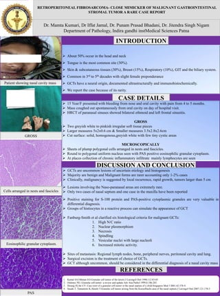
Patient showing nasal cavity mass.
GROSS
Cells arranged in nests and fascicles
Eosinophilic granular cytoplasm.
PAS
INTRODUCTION
CASE DETAILS
DISCUSSION AND CONCLUSION
REFERENCES
15 Year/F presented with bleeding from nose and oral cavity with pain from 4 to 5 months.
Mass coughed out spontaneously from oral cavity on day of hospital visit.
HRCT of paranasal sinuses showed bilateral ethmoid and left frontal sinusitis.
GROSS
Two greyish white to pinkish irregular soft tissue pieces
Larger measures 5x2x0.6 cm & Smaller measures 3.5x2.8x2.6cm
Cut surface: solid, homogenous,greyish white with few tiny cystic areas
MICROSCOPICALLY
Sheets of plump polygonal cells arranged in nests and fascicles.
Round to polygonal uniform nucleus seen with PAS positive eosinophilic granular cytoplasm.
At places collection of chronic inflammatory infiltrate mainly lymphocytes are seen
GCTs are uncommon lesions of uncertain etiology and histogenesis
Majority are benign and Malignant forms are rarer accounting only 1-2% cases
Clinically, malignancy is suggested by local recurrence, rapid growth, tumors larger than 5 cm
Lesions involving the Naso-paranasal areas are extremely rare.
Only two cases of nasal septum and one case in the maxilla have been reported
Positive staining for S-100 protein and PAS-positive cytoplasmic granules are very valuable in
differential diagnosis.
Groups of histiocytes in a reactive process can simulate the appearance of GCT
Fanburg-Smith et al clarified six histological criteria for malignant GCTs:
1. High N/C ratio
2. Nuclear pleomorphism
3. Necrosis
4. Spindling
5. Vesicular nuclei with large nucleoli
6. Increased mitotic activity.
Sites of metastasis: Regional lymph nodes, bone, peripheral nerves, peritoneal cavity and lung.
Surgical excision is the treatment of choice of GCTs.
GCT although uncommon, should be considered in the differential diagnosis of a nasal cavity mass