SlideShare a Scribd company logo
1 of 43
LECTURE 14 ATOMIC STRUCTURE: ELECTRONS,
PROTONS and NEUTRONS
The above figure displays a cathode-ray tube (CRT). Today, a
CRT is described as a vacuum tube that contains one or
more electron guns and a phosphorescent screen, and is used to
display images. It modulates, accelerates, and deflects electron
beams onto a screen tocreate the images. The images may
represent electrical waveforms (in an oscilloscope), pictures (a
television screen, computer monitor), radar targets, or other
phenomena.
We now know that cathode rays are streams of electrons
observed in discharge tubes. If an evacuated glass tube (upper
image) is equipped with two electrodes and a voltage is applied,
glass behind the positive electrode is observed to glow (lower
image), due to electrons emitted from the negative cathode.
The above “official” account presupposes that one knows what
an electron is and what are its physical properties (mass and
charge). The discovery of the electron opened up a whole new
chapter in the understanding of matter. This led to the
realization that light and matter could not be fully understood
using the classicallaws of physics, and that a totally different
way of understanding nature was needed. Thus emerged,
beginning in the last years of the 19th century, a completely
new description of light and matter. This new description
became known as quantum mechanics, and resulted in the
quantum theory of atoms, molecules and the chemical bond.
This is the historical journey on which we shall embark in this
Lecture.
Cathode rays were discovered by Julius Plücker (1801-1868)
and Johann Wilhelm Hittorf(1824-1914). Their experimental
apparatus depended on two earlier inventions: 1) Volta’s
battery; and, 2) a sealed glass tube in which a partial vacuum
was maintained. The latter was invented by a German physicist
and glassblower, Heinrich Geissler, in 1857.
Hittorf observed that some unknown rays were emitted from
the cathode (negative electrode) which could cast shadows on
the glowing wall of the tube, indicating the rays were traveling
in straight lines. In 1890, Arthur Schuster demonstrated cathode
rays could be deflected by electric fields, and William
Crookes showed they could be deflected by magnetic fields.
It was these experiments on cathode rays inside the cathode ray
tube that drew the attention of Röntgen. After repeating the
above experiments, he began to study the radiation emitted
outside the cathode ray tube, using fluorescent chemical
sensors, e.g., barium platinocyanide, to detect radiation. His
discovery of x-rays on November 8, 1895 was communicated to
the Physico-Medical Society of Würzburg later in November,
1895. A translation of his paper appeared two months later on
January 23, 1896 in the English journal, Nature. (You can dial
up this article on Gallica and read it for yourself).
Paraphrasing Louis XV(1710 – 1774) of France, were he not
such a humble, unassuming man,Röntgenmight have said "Après
nous, le déluge"
A controversy sprang up surrounding the interpretation of
cathode rays. Most German physicists believed that cathode
rays were some form of light (electromagnetic radiation).
Thomson, a British physicist and head of the Cavendish
Laboratory at Cambridge University (Newton’s alma mater),
believed otherwise on the grounds that cathode rays could be
deflected by both electric and magnetic fields, whereas
electromagnetic radiation could not.
Thomson believed that cathode rays were a stream of charged
particles and designed a variation on the earlier design of the
cathode ray tube in which the cathode ray was subjected to
combined electric and magnetic fields. See his apparatus
below. In 1897, he showed that the cathode ray consisted of
negatively charged particles smaller than atoms, the first
"subatomic particles", which were later named electrons.
In the 19th century, the charge-to-mass ratios of ions were
measured by electrochemical methods, using an apparatus first
introduced by Faraday. In measuring the charge-to-mass ratio,
Thompson showed that charged particles in the cathode ray
were characterized by a charge-to-mass ratio much smaller than
the ion of the smallest element, the hydrogen ion H+, by a
factor of nearly 2000. Importantly, he realized that the
previously-held view that the atom was the smallest, elemental
“building block” of Chemistry could not be correct. He
concluded that there must exist a subatomic structure of matter.
Thomson realized that the accepted model of a neutral atom did
not account for negatively or positively charged particles.
Therefore, he proposed a new model of the atom which he
likened to English plum pudding. The negative electrons
represented the raisins in the pudding and the dough contained
the positive charge. Thomson's model of the atom did explain
some of the electrical properties of the atom due to the
electrons, but it failed to account correctly for the positive
charges in the atom .
Over the years, I realized that a better metaphor than “plum
pudding” for students is the watermelon. The black seeds are
electrons, and the delicious red “stuff” the uniform background
(cloud) of positive charge.
.
The Nobel Prize in Physics 1906 was awarded to Joseph John
Thomson "in recognition of the great merits of his theoretical
and experimental investigations on the conduction of electricity
by gases."
At this point, the storyline takes an unexpected twist. While
Thompson was undertaking his experiments, Röntgen, on New
Years Day, 1896, mailed his manuscript (together with the
photograph of his wife’s hand), to five scientists, one of whom
was the celebrated French theoretical physicist, engineer, and
philosopher of science, Jules Henri Poincaré, at that time
President of the Académie française(French Academy).
Poincaré took Röntgen’s manuscript to the next meeting of the
Académie, sharing its contents with those that attended.
Poincaré was fascinated with the results of Röntgen’s study, and
speculated that the unknown radiation might be similar to the
kind emitted by fluorescent rocks and minerals. One of the
members of the Académie who showed up that day was Henri
Becquerel. Reports of that meeting comment that Becquerel
was dozing off during the meeting, but woke up when Poincaré
mentioned fluorescent minerals. Becquerel, following in the
footsteps of his Dad and Grandfather, had been studying these
interesting minerals for years. There is a great display of
fluorescent minerals at Chicago’s Field Museum.
Becquerel went back to his lab and began using naturally
fluorescent minerals intending to determine whether the
radiation emitted by these materials was, in fact, the x-rays
discovered by Röntgen. He exposed potassium uranyl sulfate
to sunlight and then placed it on photographic plates wrapped in
black paper, believing that the uranium absorbed the Sun’s
energy and then emitted it as x-rays. His hypothesis was
disproved on the 26th-27th of February, when his experiment
"failed" because it was overcast and rained in Paris on those
days. But, for some reason, Becquerel decided to develop his
photographic plates anyway. To his surprise, the images were
strong and clear, proving that the uranium emitted radiation
without an external source of energy (such as the Sun).
Becquerel had discovered radioactivity.
Becquerel used an apparatus similar to that displayed below to
show that the radiation he discovered could not be x-rays. X-
rays are neutral and can not be bent in a magnetic field. The
new radiation was bent by the magnetic field so that the
radiation must be charged and different from x-rays. When
different radioactive minerals were put in the magnetic field,
they deflected in different directions or not at all, showing that
there were three classes of radioactivity: negative, positive, and
electrically neutral.
The term radioactivity was actually coined by Marie
Skłodowska Curie (1867-1934), born in Warsaw, Poland and
later a naturalized French citizen. Together with her husband
Pierre, she began investigating the phenomenon discovered by
Becquerel. The Curies extracted uranium from ore and to their
surprise, found that the leftover ore showed greater
radioactivity than the pure uranium. They concluded that the ore
contained radioactive elements other than uranium. This led to
their discoveries of the elements polonium and radium. It took
four more years of processing tons of ore to isolate enough of
each element to determine their chemical and physical
properties.
BTW, if you discover a new comet or a new element, you can
name it whatever you please.
The first element discovered by the Curies was polonium,
named after the country of Marie’s birth. If you go to the
oldest quarter in Warsaw where she grew up, on the front of the
apartment building where she lived, there are two large discs,
one inscribed with the chemical symbol for polonium (Po) and
the other for radium (Ra).
Together with Becquerel, they received the 1903 Nobel Prize in
Physics "in recognition of the extraordinary services they have
rendered by their joint researches on the radiation phenomena
discovered by Professor Henri Becquerel."
Later she was awarded the Nobel Prize in Chemistry in 1911
"in recognition of her services to the advancement of chemistry
by the discovery of the elements radium and polonium, by the
isolation of radium and the study of the nature and compounds
of this remarkable element."
Following is the press release of her visit to Chicago:
June 3, 1921–Marie Curie, on her first trip to the United States,
visits Chicago for two hours and is “besieged by newspaper men
and women anxious to get her ideas on the fashions, the war,
radium, woman suffrage, the political situation.” [Chicago
Daily Tribune, June 4, 1921]. All Curie, the winner of two
Nobel Prizes in Physics, wants to discuss, though, is Lake
Michigan. The Tribune reports, “To her the engineering feat of
reversing the flow of the Chicago river to dispose of the city’s
sewage was a problem far more interesting than a comparison of
American styles with French creations.” By nightfall she is off
to Colorado with a topographical map of the western states in
hand, a gift of Mrs. W. Lee Lewis of Northwestern University.
So, what are the mysterious rays emitted from radioactive
minerals? Enter the story, Ernest Rutherford (1871-1937), a
New Zealand-born British physicist who came to be known as
the father of nuclear physics. Einstein considered Rutherford to
be the greatest experimentalist since Michael Faraday (1791–
1867).
In the years 1899 and 1900, physicists Rutherford (working in
McGill University in Montreal, Canada) and Paul
Villard (working in Paris) separated radiation into three types:
eventually named alpha (α), beta(β) , and gamma (γ) rays by
Rutherford, based on their penetration of objects and deflection
by an electric field. A sketch of his experimental apparatus is
shown below. Alpha rays were defined by Rutherford as those
having the lowest penetration of ordinary objects.
Rutherford's work also included measurements of the ratio of an
alpha particle's charge to mass ratio, which led him to
hypothesize that alpha particles were doubly charged helium
ions (later shown to be “bare” helium nuclei).
In 1907, Ernest Rutherford and Thomas Royds finally proved
that alpha particles were indeed ions of Helium, the second
element in the Periodic Table of elements.
To do this they allowed alpha particles to penetrate a very thin
glass wall of an evacuated tube, thus capturing a large number
of the hypothesized helium ions inside the tube. They then
caused an electric spark inside the tube, which provided a
shower of electrons that were taken up by the ions to form
neutral atoms of a gas. Subsequent study of the spectra of the
resulting gas showed that it was Helium and that the alpha
particles were indeed the hypothesized helium ions.
During this period, Rutherford also identified a signature of
radioactive elements, the half-life , and discovered the
radioactive element Radon. This work was performed at McGill
University in Montreal, Quebec, Canada. It was the basis for
the Nobel Prize in Chemistry which he was awarded in 1908 (at
age 37) "for his investigations into the disintegration of the
elements, and the chemistry of radioactive substances.” Today,
you can visit a museum on the McGill campus which displays
his experimental equipment. In contrast to the Röntgen museum
in Würzburg, you can benefit from a guided tour by a most
enthusiastic and engaging curator.
The next piece in the puzzle was provided by Robert Millikan
(1868-1953), an American experimental physicist. Millikan
graduated from Oberlin College in 1891 and obtained his
doctorate at Columbia University in 1895. In 1896 he became
an assistant professor at the University of Chicago. In 1909
Millikan began a series of experiments to determine the electric
charge carried by a single electron. His apparatus is shown
below.
He began by measuring the downward trajectory of
charged water droplets in an electric field. The results
suggested that the charge on the droplets is a multiple of an
elementary electric charge, but the experiment was not accurate
enough to be convincing. He obtained more precise results in
1910 with his famous “oil-drop” experiment in which he
replaced water (which tended to evaporate too quickly) with oil
(which was “heavier”).
Since Thomson had already determined the charge-to-mass ratio
of the electron,
q/m = −1.75882001076(53)×1011 C⋅ kg−1
where q is the charge measured in Coulombs (C) and m is the
mass measured in kilograms (kg). Once Millikan had
determined the charge on a single electron, 1.602 x 10⁻¹⁹ C, he
could calculate the mass m of the subatomic particle, the
electron, 9.109×10⁻³¹ kg, and compare this with the mass of
the smallest atom, Hydrogen, 1.67377 10-27 kg, the difference
a factor of 1838.
The 1923 Nobel Prize in Physics was awarded to Robert
Andrews Millikan "for his work on the elementary charge of
electricity and on the photoelectric effect." Later, Millikan left
the University of Chicago to become President of the newly
founded Throop University in Pasadena, CA, known today as
California Institute of Technology (Caltech), arguably the best
university in the World (according to the Chinese).
Now, back to Rutherford. Rutherford designed an experiment to
use alpha particles emitted by a radioactive element as probes to
the unseen world of atomic structure. See a sketch of his
experimental apparatus below.
The target was a gold foil, hence the name, “gold foil”
experiment. From Lecture 2, you recall that Gold is the most
malleable of elements. It can be hammered into sheets of such
thinness that you can see light passing through. If Thomson
was correct, the beam of alpha radiation should go straight
through the gold foil. Indeed, most of the beams went through
the foil, but a few were deflected at wide angles, even
backwards.
This was crazy! If, in Thomson’s model, you had electrons
embedded in a uniform background of positive “stuff,” and the
electron has a mass ~1/2000 the mass of the smallest element
(H), given that the mass of Helium is four times greater than
the mass of H, how on Earth could an electron scatter an alpha
particle that was ~ 8000 times heavier?
Rutherford presented, eventually, a new physical model for
subatomic structure to interpret this unexpected experimental
result. In this model, the atom is made up of a central charge
(this is the modern atomic nucleus) surrounded by a cloud of
orbiting electrons.
For definiteness, consider the passage of a high speed α particle
through an atom having a positive central charge Ne, and
surrounded by a compensating charge of N electrons.
From purely energetic considerations of how far charged
particles of known speed would be able to penetrate toward a
central charge, Rutherford was able to calculate that the radius
of the gold central charge (later called the nucleus of the atom)
would have to be less than 3.4 × 10⁻¹⁴ meters. The radius of a
single gold atom is ~ 10−10 meters or so. Comparison of these
two radii revealed the astonishing result that the nucleus was a
factor less than 1/3000 the diameter of the atom.
Today, Rutherford’s model of the atom is (sometimes) called
the “Solar System” model of the atom, representations of which
you probably saw in grade school.
While the Rutherford model concentrated the atom's positive
charge and a great deal of the mass in a very small core (the
nucleus), thus counterbalancing negatively-charged electrons,
so that the atom was electrostatically neutral, it did not account
for the atomic mass.
Rutherford (and others) believed that there must be an
elementary particle with a charge exactly opposite the charge on
the electron, but having a (much) heavier mass.
In a series of experiments performed by Rutherford and his
student James Chadwick, they were able to change one element
into another by hitting atoms with high energy alpha particles.
They noticed that nitrogen (N), oxygen(O), and aluminum(Al),
when hit with an alpha particle, disintegrated and emitted a fast
particle of positive charge. To be more specific, hydrogen
nuclei were always emitted in the process. In a dark room, they
were able to observe flashes of light when alpha particles hit a
florescent screen. They realized that the positive charge of any
nucleus could be accounted for by a whole (integer) number of
positively charged hydrogen nuclei, which were named protons
by Rutherford in 1920.
Knowing the mass of the Hydrogen atom (essentially the mass
of the proton, since the electron mass is ~ 2000 times smaller)
poses a problem. Helium, the second element in the Periodic
Table, has a mass four times the mass of a single proton. So,
the Helium atom, must have more than “just” protons to account
for the factor of four in mass.
This conundrum was solved by a student of Rutherford, James
Chadwick, with the discovery in 1932 of a new elementary
particle, distinct from the proton, the neutron. The neutron has
no charge, and a mass slightly greater than the mass of the
proton. The conclusion reached was that the Helium nucleus
has two protons and two neutrons, and is orbited by two
electrons.
Further, it was this discovery that opened up the understanding
of isotopes: two or more forms of the same element that
contain equal numbers of protons but different numbers of
neutrons in their nuclei, and hence differ in relative atomic
mass but not in chemical properties. This point will be crucial
in understanding nuclear chemistry, nuclear medicine,
radiography, nuclear power plants and the atomic bomb. See
Lecture 16.
The Nobel Prize in Physics 1935 was awarded to James
Chadwick "for the discovery of the neutron."
Two additional experiments, both involving the electron, need
to be discussed before we can take up the quantum theory of the
chemical bond, molecular structure, and chemical reactivity.
The first deals with the “spin” of the electron. This involves
consideration of a quantity called angular momentum. Angular
momentum is the rotational equivalent of linear momentum, the
latter defined as mass x velocity. In physics, mass is given the
symbol m, velocity v, linear momentum p, and angular
momentum the symbol L.
Spin is one of two types of angular momentum in quantum
mechanics (the other being orbital angular momentum). The
existence of spin angular momentum was inferred from
experiments, such as the Stern–Gerlach experiment (see below),
in which silver atoms were observed to possess two, discrete
values of the angular momenta and no orbital angular
momentum. The effect was attributed to “electron spin.”
Electrons have an intrinsic angular momentum characterized by
a (soon to be called) quantumnumbers.
In Rutherford’s “Solar System” model of the atom, planets
circling the Sun correspond to electrons orbiting the nucleus.
Just as the Earth revolves on its axis, some describe electron
spin as electrons spinning on their axis. Note that you can only
spin in two directions, clockwise and counterclockwise. The
spin quantum number s can take on only two values, s = +1/2
and s = − 1/2. In textbooks, these two values of the spin are
shown as arrows, ↑↓ .
Stern–Gerlach experiment: Silver atoms travelling through an
inhomogeneous magnetic field, and being deflected up or down
depending on their spin; (1) furnace, (2) beam of silver atoms,
(3) inhomogeneous magnetic field, (4) classically expected
result, (5) observed result.
Otto Stern was a German-American physicist, awarded the
Nobel Prize in Physics 1943 "for his contribution to the
development of the molecular ray method and his discovery of
the magnetic moment of the proton."
The second experiment is the Davisson-Germer experiment.
Between 1923 and 1927, Clinton Davisson and Lester
Germer at Western Electric (later Bell Labs), discovered that
electrons scattered by the surface of a crystal of nickel (Ni)
metal displayed a diffraction pattern. This confirmed
the hypothesis, advanced by Louis de Broglie in 1924, that an
electron can “behave” like a wave or a particle, a foundational
concept of quantum mechanics called wave-particle duality.
At the same time George Paget Thomson independently
demonstrated the same effect firing electrons through metal
films to produce a diffraction pattern. Davisson and Thomson
shared the Nobel Prize in Physics in 1937.
Given the avalanche of unexpected experimental results,
starting with Röntgen’s discovery of x-rays in 1895 and the
demonstration of wave-particle duality in the 1920’s, the natural
question is: What does this tell us about atoms, molecules and
the chemical bond?
Everything.
The theoretical understanding of these phenomena sprang from
analyzing two, apparently unrelated, experiments, both
involving radiation.
The first experiment centered on Black-body radiation,
thermalelectromagnetic radiation emitted by a black body (an
idealized opaque, non-reflective body). It has a specific
spectrum of wavelengths, inversely related to intensity, that
depend only on the body's temperature.
As the temperature (in degrees Kelvin, K) decreases, the peak
of the intensity of black-body radiation (vertical axis) moves to
lower intensities and longer wavelengths. Conversely, when the
temperature increases the radiation curve moves to higher
frequencies. Theories to explain the behavior of the radiation
curve using classical physics worked perfectly well at long
wavelengths (lower frequencies) but failed completely at higher
frequencies. The failure of classical theories in the regime of
high frequencies was called the ultraviolet catastrophe.
In 1900, the German physicist Max Planck (1858–1947)
explained the ultraviolet catastrophe by proposing that the
energy of electromagnetic waves is quantized rather than
continuous. This means that for each temperature, there is a
maximum intensity of radiation that is emitted in a blackbody
object, corresponding to the peaks in the above figure, so the
intensity does not follow a smooth curve as the temperature
increases, as predicted by classical physics. Rather, Planck
hypothesized that energy could only be gained or lost in integral
multiples of some smallest unit of energy, a quantum (the
smallest possible unit of energy). Energy can be gained or lost
only in integral multiples of a quantum.
Recall that in Lecture 13, we noted that in the classical theory
of waves, the energy is given by
E = ½ k A2 ( k = a
constant, A = amplitude )
In Planck’s quantum theory of radiation, the energy is given
by
E = h ν ( h = a
constant, ν = frequency)
The constant h in the above equation is now called Planck’s
constant.
The Nobel Prize in Physics 1918 was awarded to Max Karl
Ernst Ludwig Planck "in recognition of the services he
rendered to the advancement of Physics by his discovery of
energy quanta."
The second experiment is called the photoelectric effect.
The photoelectric effect is the emission of electrons or
other free carriers when electromagnetic radiation, like light,
hits a material.According to classical electromagnetic theory,
the photoelectric effect can be attributed to the transfer
of energy from the light to an electron. From this perspective,
an alteration in the intensity of light should induce changes in
the kinetic energy (Kinetic Energy = ½ mv2, where m is mass
and v is velocity) of the electrons emitted from the metal.
According to classical theory, a sufficiently dim light is
expected to show a time lag between the initial shining of its
light and the subsequent emission of an electron.
Sadly, the experimental results did not correlate with either of
the two predictions made by classical theory. Instead,
experiments showed that electrons are dislodged only by the
impingement of light when it reached or exceeded a
threshold frequency ν. Below that threshold, no electrons are
emitted from the material, regardless of the light intensity or
the length of time of exposure to the light.
In 1905, Albert Einstein (1879-1955) solved this apparent
paradox by describing light as composed of discrete quanta,
now called photons, rather than continuous waves. By
assuming that light actually consisted of discrete energy
packets, with the energy given by Planck’s E = h ν ,
Einstein wrote an equation for the photoelectric effect that
agreed with experimental results.
Albert Einstein in 1904 (age 25)
The Nobel Prize in Physics 1921 was awarded to
Albert Einstein "for his services to Theoretical Physics, and
especially for his discovery of the law of the photoelectric
effect."
In 1911 the BelgianindustrialistErnest Solvay organized an
invitation-only meeting at the Hotel Metropole in Brussels.
Considered a turning point in physics, in the following year
Solvay founded Conseil Solvay, the International Solvay
Institutes for Physics and Chemistry and, after WW I,
sponsored a series of historic meetings in Physics, Chemistry
and Biology. In the iconic photograph below, you will
recognize a few of the physicists and chemists noted in Lectures
13 and 14. Madame Curie is seated next to Henri Poincaré,
Rutherford is standing just behind them, Einstein is second from
the right in the second row, Planck and de Broglie are at the
other end of the row.
Photograph of the first conference in 1911 at the Hotel
Metropole. Seated (L–R): W. Nernst, M. Brillouin, E.
Solvay, H. Lorentz, E. Warburg, J. Perrin, W. Wien, M. Curie,
and H. Poincaré. Standing (L–R): R. Goldschmidt, M.
Planck, H. Rubens, A. Sommerfeld, F. Lindemann, M. de
Broglie, M. Knudsen, F. Hasenöhrl, G. Hostelet, E. Herzen, J.
H. Jeans, E. Rutherford, H. Kamerlingh Onnes, A.
Einstein and P. Langevin.
LECTURE 13. LIGHT
Hubble space telescope observations have taken advantage of
gravitational lensing to reveal the largest sample of the faintest
and earliest known galaxies in the universe. Some of these
galaxies formed just 600 million years after the Big Bang.
Space, mass, light and time are fundamental descriptors of our
Universe. Captured by the poetry in Genesis, “In the beginning,
when God created the heavens and earth, the earth was a
formless wasteland, and darkness covered the abyss, while a
mighty wind swept over the waters. Then God said “Let there
be light,” and there was light.
The cosmological model of the “Big Bang” describes how
the universe expanded from an initial state of very
high density and high temperature. If observed conditions
today areextrapolated backwards in time using the known laws
of physics, the prediction is that our Universe emerged from a
singularity, a point of infinite density, and that before this
event, space and time did not exist.
Current knowledge is insufficient to determine if anything
existed prior to the singularity. Sixteen centuries ago, in his
Confessions, Saint Augustine (354-430) posed the obvious
question in biblical terms: What was God doing before he
created the Universe?
ISAAC NEWTON: COLOR SPECTRUM and the
CORPUSCULAR THEORY of LIGHT
Our modern understanding of light and color begins with Isaac
Newton (1642-1726) and a series of experiments that he
published in 1672. He was the first to understand the rainbow.
He refracted white light with a glass prism, resolving it into its
component colors: red, orange, yellow, green, blue and violet.
In the graphic below, light enters the prism from the top right,
and is refracted by the glass. The violet is bent more than the
yellow and red, so the colors separate.
In the 1660s, Newton began experimenting with his “celebrated
phenomenon of colors.” At the time, people thought that color
was a mixture of light and darkness, and that
prisms colored light. Hooke was a proponent of this theory of
color, and had a scale that went from brilliant red, which was
pure white light with the least amount of darkness added, to dull
blue, the last step before black, which was the complete
extinction of light by darkness. Newton believed this theory was
false.
Newton set up a prism near a window at his boyhood home in
Woolsthorpe, England ( site of the famous apple tree), and
projected a beautiful spectrum 22 feet onto the far wall. Further,
to prove that the prism was not coloring the light, he refracted
the spectral light back together, producing white light.
Incidentally, he was at home because all the students at
Cambridge University where he was a student were sent home
because of an epidemic of the bubonic plague. In 1665, it was a
version of “social distancing.” BTW, Newton did his best work
working from home.
On a personal note, my wife and I once visited the hometown of
William Shakespeare (1564-1616), Stratford-upon-Avon.
Access to Anne Hathaway’s cottage was severely hampered by
rows of tour buses, cars, and floods of pedestrians. Later, we
drove to
Woolsthorpe by Colsterworth (took about an hour on country
roads) to visit Newton’s home, now a museum. When we arrived
the museum was closed, and had been for some time. I asked a
local gentleman “why,” and he responded “because no one ever
came.”
The diagram below isfrom Sir Isaac Newton’s crucial
experiment, 1666-72. A ray of light is divided into its
constituent colors by the first prism (left), and the resulting
bundle of colored rays is reconstituted into white light by the
second prism (right).
Artists were fascinated by Newton’s clear demonstration that
light alone was responsible for color. His most useful idea for
artists was his conceptual arrangement of colors around the
circumference of a circle, which allowed the painters’ primaries
(red, yellow, blue) to be arranged opposite their complementary
colors (e.g. red opposite green), as a way of denoting that each
complementary color would enhance the other’s effect through
optical contrast.
In optics, the corpuscular theory of light, arguably set forward
by Descartes in 1637, states that light is made up of small
discrete particles called "corpuscles" (little particles) which
travel in a straight line with a finite velocity and possesses
momentum (mass x velocity).
Isaac Newton was a pioneer of this theory, an elaboration of his
view of reality as interactions of material points through forces.
The following passage gives Albert Einstein's description of
Newton's conception of physical reality:
“[Newton's] physical reality is characterized by concepts
of space, time, the material point and force (interaction
between material points). Physical events are to be thought of
as movements according to law of material points in space.
The material point is the only representative of reality in so far
as it is subject to change.”
This early conception of the particle theory of light was an early
forerunner to the modern understanding of the photon. See next
Lecture 14.
CHRISTIAAN HUYGENS: WAVE THEORY of LIGHT
The corpuscular theorycannot
explain refraction, diffraction and interference, which require
an understanding of the wave theory of light of Christiaan
Huygens.
refraction: the change in direction of a wave passing from one
medium to another or from a gradual change in the medium.
Refraction of light is the most commonly observed
phenomenon, but other waves such as sound waves and water
waves also show refraction.
diffraction: Diffraction refers to various phenomena that occur
when a wave encounters an obstacle or a slit. It is defined as the
bending of waves around the corners of an obstacle or through
an aperture into the region of geometrical shadow of the
obstacle/aperture.
interference: interference is a phenomenon in which two waves
superimpose to form a resultant wave of greater (constructive
interference), lower (destructive interference), or the same
amplitude.
To explain the origin of color, Robert Hooke (1635–1703)
developed a "pulse theory" and compared the spreading of light
to that of waves in water in his 1665 Micrographia. In 1672
Hooke suggested that light's vibrations could
be perpendicular to the direction of propagation.
Christiaan Huygens (1629–1695) worked out a mathematical
wave theory of light in 1678, and published it in his Treatise on
light in 1690. He proposed that light was emitted in all
directions as a series of waves in a hypothetical medium called
the Luminiferous ether(invisible to the senses, no “weight,”
color, odor or taste) and that, as waves are not affected by
gravity, he assumed that they slowed down upon entering a
denser medium.
That light waves could interfere with each other like sound
waves was first noted around 1800 by Thomas Young, a
physician. Young demonstrated by means of a diffraction
experiment that light behaved as waves. He also proposed that
different colors were caused by different wavelengths of light,
and explained color vision in terms of three color receptors in
the eye.
.
Thomas Young's sketch of a double-slit
experimentshowing diffraction. Young's experiments supported
the theory that light consists of waves.
Coincidentally, Young’s report of the double-slit experiment
appeared in the Proceedings of the Royal Society of London in
the same year (1800) as Volta’s paper on his invention of the
battery.
In earlier lectures in the course, examples of the atomic
structure of inorganic, organic and biochemical entities were
shown [ e.g., NaCl (Lecture 1) , diamond (Lecture 5), COVID-
19 (Lecture 12) ]. Did you ever ask yourself (or me) how
those crystal structures were determined? Thank you, Thomas
Young.
Let’s get to the bottom of this.
ELECTROMAGNETIC WAVES and POLARIZATION
One speaks of the wave nature of light, but we have not
specified what it is that oscillates or undulates. It is easy to
visualize waves in a fluid medium such as water, where we can
see the motion of the water as the wave passes by.
wavelength (λ) = the distance, measured in the direction of
propagation of a wave, from any given point to the next point
in the same phase.
frequency (ν) = the number of oscillations (or cycles) of a wave
per unit of time
We now know that electromagnetic waves are very different
from water waves or sound waves. NO MEDIUM is
REQUIRED. Light waves can travel through a total vacuum,
unlike other waves with which we are familiar. Recognizing the
significance of this difference, established experimentally by
Michaelson and Morley in 1887 at what is now Case Western
University in Cleveland, is one of the two cornerstones on
which Einstein developed his Theory of Special Relativity,
published in 1905. We shall pursue this in Lecture 16 on
nuclear chemistry, nuclear medicine and radiography.
The Nobel Prize in Physics 1907 was awarded to Albert
Abraham Michelson "for his optical precision instruments and
the spectroscopic and metrological investigations carried out
with their aid."
Importantly, when watching water waves lap against the shore
on Lake Michigan, it appears that water is “moving” toward
shore. Not so. The water in the lake is not moving. What is
being transported is energy. In the classical theory of waves,
the energy is given by
E = ½ k A2 ( k = a
constant, A = amplitude )
In the quantum theory of light (to be presented in the next
Lecture 14), the energy is given by
E = h ν ( h = a
constant, ν = frequency)
Rather than having physical motions, electromagnetic (light)
waves consist of electric and magnetic fields that oscillate back
and forth at right angles to the direction of motion. The electric
and magnetic fields lie in planes that are also perpendicular to
each other, so that the electric field flips back and forth in one
plane, with the magnetic field doing so in the plane that lies
perpendicular to it, and both are traverse to the direction of
travel.
Electromagnetic (light) waves can be imagined as a self-
propagating, transverse, oscillating wave of electric and
magnetic fields. The above diagram shows a plane, linearly
polarized wave propagating from left to right. The electric field
is in a vertical plane and the magnetic field in a horizontal
plane.
A photon (a quantum or Newtonian “corpuscle” of light, to be
introduced in Lecture 14) can be viewed as a packet of
electromagnetic energy consisting of alternating fields, which,
by virtue of their wavelength and frequency, provide the photon
with its wave properties.
Summarizing the above discussion, light is energy that
radiates, or travels, in waves. Light is a wave of vibrating
electric and magnetic fields. Visible light is one small part of a
larger range of vibrating electromagnetic fields. This range is
called the electromagnetic spectrum. By contrast, sound is a
wave of vibrating air.
The energy of the radiation depends on its wavelength and
frequency. As noted above, wavelength is the distance between
the tops (crests) [ or bottoms (troughs) ] of the waves. And,
frequency is the number of waves that pass by each second. The
longer the wavelength of the light, the lower the frequency, and
the less energy it contains.
Recalling that
Velocity (v) = distance /time
= λ ν
electromagnetic waves travel through space at 299,792 km/sec
(186,282 miles/sec). This is
called the velocity (speed) of light. In Astronomy, the distance
a beam of light travels in one year is called a light year.
The light emitted at the first moment of the Big Bang (see the
photo taken with the Hubble telescope at the top of this Lecture)
is 46.1 billion (46.1x10⁹) light-years distant.
Light from most sources (such as stars) consists of vast numbers
of photons, each with a specific, constant orientation of its
electric and magnetic planes. Ordinarily the orientation of the
various photons is random, but under some circumstances it is
not. If light passes through or reflects from a medium with a
certain orientation, what is left is light in which all the photons
are aligned. That is, the planes of the electric and magnetic
fields of the photons are parallel. The light is said to be
polarized.
Many of the sunglasses sold today consist of polarizing filters,
and they have the effect of screening out light in a given
orientation. In Astronomy, polarization occurs naturally,
sometimes because a natural filtering occurs, as in the
interstellar medium, and in other cases because the source of the
light emits radiation with a preferred orientation.
Next, we need to understand the following phenomena. It
happens that one of the things about waves is the following: If
they encounter an obstacle which is smaller than their
wavelength, the wave just kind of wraps around the object and
keeps on going. This is why you can hide behind a big tree
trunk and the voices of people on the other side of the tree will
still reach you (because the long waves of the sound wrap
around the tree). BUT, light from their flash light won't
(because the short waves of the light can't wrap around such a
big object).
Sound and light act similarly when they reach a corner of a
house - the sound waves are long and kind of wrap around the
corner and then spread out from there, whereas the very short
light waves can't bend around the corner. As you might have
guessed already, this means that light does wrap around very
small things (things smaller than a millionth of a foot), and does
bend a tiny bit around corners, but this bending is so small it is
impossible to see with your eye. See, however, the following
discussion.
X-rays are light waves of very short wavelength and high
energy discovered by Wilhelm Röntgen (1845-1923) who, on
November 8, 1895, produced and detected electromagnetic
radiation in a wavelength range known today as x-rays.
Below is a photo of his modest laboratory in Würzburg,
Germany.
No guided tours. You can see his experimental equipment by
yourself, “up close and personal.”
Roentgen’s experiment involved studying the radiation emitted
outside a cathode ray tube (see description in the following
Lecture 14). He also used as sensors a series of compounds that
were sensitive to light of high frequencies, the principal one of
which was the barium salt of the platinocyanide ion. The ion
exists in two isometric forms:
Pt is located at the center of the plane in the above diagram.
Barium platinocyanide, Ba[Pt(CN)4] isa phosphor and a
scintillator. It fluoresces in the presence of light of high
frequencies (ν)
Röntgen baptized the mysterious radiation that caused a film of
Ba[Pt(CN)4] on a support to glow, “x-rays,” after the name of
the unknown in algebra. In Germany, they were called
Röntgen rays (which he discouraged).
One of the things Röntgen discovered about x-rays was their
incredible penetrating power. They could pass through paper,
glass, wood and human flesh (but not lead). He asked his wife
Bertha to put her hand between the source of x-rays and a
photographic plate. The result was one of the most famous of
all photographs.
Notice the dark circle on her finger, her wedding ring.
Bertha’s reaction to the almost supernatural character of this
picture, was “I have seen my own death.”
Within months x-rays were used in medicine worldwide to
diagnose fractures. X-rays remain today one of the most
important diagnostic tools in medicine. The first Nobel Prize in
Physics was awarded in 1901 to Wilhelm Conrad Röntgen "in
recognition of the extraordinary services he has rendered by the
discovery of the remarkable rays subsequently named after
him."
The discovery of x-rays launched a new era in experimental
physics and chemistry which led to the quantum theory of atoms
and molecules. See the next Lecture 14.
Max von Laue (1879 – 1960) discovered the diffraction of x-
rays by crystals. The idea came to him that estimates of the size
of atoms, and their spacing in a crystal, were about thesame as
the wavelength of Röntgen’s x-rays. He suggested that the
much shorter electromagnetic rays, which x-rays were supposed
to be, would cause in a crystal some kind of diffraction or
interference phenomena. Knipping, an experimentalist, tested
out the idea and, after some failures, succeeded in proving it to
be correct. Von Laue worked out the mathematical formulation
and the discovery was published in 1912. For this, he was
awarded the Nobel Prize in 1914. It established the fact that x-
rays are electromagnetic in nature and it opened the way to the
later theoretical and experimental work of Sir William and Sir
Lawrence Bragg.
Sir William Lawrence Bragg 1890 – 1971) was an Austalian-
born British physicist, discoverer in 1912 of Bragg's law of x-
ray diffraction, which is basic for the determination of crystal
structure. He was joint recipient (with his father, William Henry
Bragg) of the Nobel Prize in Physics in 1915.
Bragg was the director of the Cavendish Laboratory,
Cambridge, when the discovery of the structure of DNA (the
double helix, see Lecture 6 ), based on the x-ray diffraction
imagesof DNA by Rosalind Franklin, was reported by James D.
Watson and Francis Crick in February 1953. Watson, Crick
and Maurice Wilkins received the Nobel Prize in Physiology or
Medicine in 1962. Also at the Cavendish Laboratory was Max
Perutz (1914-2002), an Austrian-born British molecular
biologist, who shared the 1962 Nobel Prize for
Chemistry with John Kendrew, for their studies of the crystal
structures of hemoglobin and myoglobin (See Lecture 6 )
Another at Cambridge was Aaron Klug, a Lithuanian-born,
South African-educated, British biophysicist. He solved the
crystal structures of macromolecules such as viruses (see
Lecture 12) working with Rosalind Franklin. He found the rules
of the geometrical form of poliovirus and other spherical
viruses. His invention of electron tomography, in which a 3D
image of a virus is obtained from many electron micrographs,
led to the Nobel Prize in Chemistry in 1982.Rosalind Franklin
died at the age of 37 from ovarian cancer. Watson, among many
others, suggested that Franklin should have been awarded
a Nobel Prize in Chemistry, along with Wilkins, but, although
there was not yet a rule against posthumous awards, the Nobel
Committee generally did not make posthumous nominations.The
experimental technique of x-ray crystallography is the common
thread which links together much of what has been highlighted
in earlier lectures, and provides a good review of some of the
most important discoveries in inorganic chemistry, biochemistry
and medicinal chemistry.
Officially, X-ray crystallography is defined as the experimental
science determining the atomic and molecular structure of
a crystal, in which the crystalline structure causes a beam of
incident x-rays to diffract into many specific directions. By
measuring the angles and intensities of these diffracted beams,
a crystallographer can produce a three-dimensional picture of
the density of electrons within the crystal. From this electron
density, the mean positions of the atoms in the crystal can be
determined, as well as their chemical bonds and their
crystallographic disorder.
COLORS of LIGHT
Visible light is the part of the electromagnetic spectrum that our
eyes can see. Light from the sun or a light bulb may look white,
but it is actually a combination of many colors. We can see the
different colors of the spectrum by splitting the light with a
prism. The spectrum is also visible when you see a rainbow in
the sky.
The colors blend continuously into one another. At one end of
the spectrum are the reds and oranges. These gradually shade
into yellow, green, blue, indigo and violet. The colors have
different wavelengths, frequencies, and energies. Violet has the
shortest wavelength in the visible spectrum. That means it has
the highest frequency and energy. Red has the longest
wavelength, and lowest frequency and energy.
PERCEPTION of COLOR
When a sample absorbs visible light, an object will selectively
absorb specific wavelengths of light from the complete
spectrum of possibilities. The color we perceive is the sum of
the remaining colors that are reflected or transmitted by an
object and strike our eyes.
An object which reflects light is said to be opaque.
An object which transmits all light is said to be transparent.
An object which absorbs all wavelengths of visible light
appears black (because no light reaches our eyes). An object
which absorbs no visible light appears white or colorless.
In case you’re wondering, ….
What is a rainbow?
A rainbow occurs when light hits the water drops in the
atmosphere at a certain angle. It is an atmospheric phenomenon
that is formed through optical processes such as refraction,
dispersion, internal reflection and secondary refraction. If the
light source is the sun, then the rainbow will be colorful and
bright.
Why is water blue?
Water does absorb a tiny amount of light, but in such small
amounts, we don’t notice it. Water actually absorbs light that
has a reddish color. When there’s a lot of water, like Lake
Michigan, we notice that red light from the Sun has actually
been absorbed. So, what color do we see when reddish light is
gone?
Blue!
Blue is the color absorbed the least by water, so it’s the one we
see the most.
Blue light also scatters more than other light. That’s the reason
why the sky is blue. That scattering affects the color of water
too.
It just takes a big enough sample of water to notice these things.
That’s why you won’t see color in the glass you’re drinking.
Why is the grass green?
The blades of grass and the leaves of trees contain the molecule
chlorophyll (see Lecture 5 ) a molecule able to absorb in the
red (and blue) regions of the spectrum.
When white sunlight is reflected from leaves, what we see is the
light not absorbed by the plant. The color vegetation appears to
our senses, the color which is not absorbed, is green. Red and
green are complementary colors.
Why is the color of an orange, orange ?
Orange and blue are complementary colors.
The removal of blue light from white light makes the light look
orange. [Conversely, the removal of orange light from white
light makes the light look blue.]
If light of all colors except blue strikes our eyes, we perceive
an orange color. If an orange absorbs all light except orange,
in the reflected light, the fruit appears orange.
In his book “Origins,” Richard Leaky notes that the perception
of color in primates was a key factor in human evolution. He
displays a color photograph of an orange tree, leaves in green
and oranges in orange, and compares this with a black-and-
white photo. If you’re looking for fruit to eat, the advantage of
color vision is obvious.
Why are minerals colored?
ANALCIME
REALGAR
CROCOITE
TYUYAMUNITE
MALACHITE
TURQUOISE
CAVANSITE
AZURITE
FLUORITE
AMETHYST
KAEMMERERITE
BIXBY
Color in minerals is caused by the absorption, or lack of
absorption, of various wavelengths of light. The color of light is
determined by its wavelength. When pure white light
(containing all wavelengths of visible light) enters a crystal,
some of the wavelengths might be absorbed while other
wavelengths may be emitted. If this happens, then the light that
leaves the crystal will no longer be white but will have some
color. Small amounts of impurities can also change the color of
crystals. Recall the Hope Diamond (Lecture 5).
My colleague, Harry Gray at Caltech, pointed out the
remarkable fact that only small differences in Cr(III)O6 ligand
fields make emeralds green and rubies red !
COLOR and the HUMAN BRAIN
Color vision is the capacity of an organism or machine to
distinguish objects based on the wavelengths (or frequencies) of
the light they reflect, emit, or transmit. Colors can be measured
and quantified in various ways. A human's perception of colors
is a subjective process whereby the brain responds to the stimuli
that are produced when incoming light reacts with the several
types of photoreceptors in the eye.
Rods and Cones are the photoreceptors found in the retina.
Rods have rod-like structure and provide twilight vision.
Cones are of the cone shape, fewer in number and provide
vision in the day or bright light. Rods are found around the
boundary of the retina, whereas cones are there in the center of
the retina.
Color vision relies on a brain perception mechanism that treats
light with different wavelengths as different visual stimuli (e.g.,
colors). Usual color insensitive photoreceptors (the rods in
human eyes) only react to the presence or absence of light and
do not distinguish between specific wavelengths.
NOTE: Colors are not “real.” They are “synthesized” by our
brain to distinguish light with different wavelengths. While rods
give us the ability to detect the presence and intensity of light
(and thus allow our brain to construct the picture of the world
around us), specific detection of different wavelengths through
independent channels gives our view of the world additional
high resolution. For instance, red and green colors look like
near identical shades of grey in black and white photos.
An animal with black and white vision alone won’t be able to
make a distinction between, let’s say, a green and red apple, and
won’t know which one tastes better before trying them both
based on color. Evolutionary biologists believe that human
ancestors developed color vision to facilitate the identification
of ripe fruits, which would obviously provide a selective
advantage in the competitive natural world. The same
conclusion had been reached by the anthropologist, Richard
Leaky.
Why certain wavelengths are paired with certain colors remains
a mystery. As mentioned above, technically, color is an illusion
created by our brain. Therefore, it is not clear if other animals
see colors the same way we see them. It is likely that, due to
shared evolutionary history, other vertebrates see the world
colored similarly to how we see it. Color vision is quite
common across the vast animal kingdom: insects, arachnids, and
cephalopods are able to distinguish colors.
What kind of colors do these animals see?
As was first proposed by Thomas Young, human color vision
relies on three photoreceptors that detect primary colors: red,
green, and blue. However, some people lack red photoreceptors
(they are “bichromates”) or have an additional photoreceptor
that detects somewhere between red and green colors
(“tetrachromates”). Having only three photoreceptors doesn’t
limit our ability to distinguish other colors.
Each photoreceptor can absorb a rather broad range of
wavelengths of light. To distinguish a specific color, the brain
compares and quantitatively analyses the data from all three
photoreceptors. Our brain does this remarkably successfully.
Some research indicates that we can distinguish colors that
correspond to wavelength differences of just one nanometer (10-
9 meters)
This scheme works in largely the same way in most higher
vertebrate animals that have color vision. Although the ability
to distinguish between specific shades varies significantly
between the species, with humans having one of the best color
distinguishing abilities.
However, invertebrates that have developed color vision (and
vision in general) completely independently from us
demonstrate remarkably different approaches to color detection
and processing. These animals can have an exceptionally large
number of color receptors. The mantis shrimp, for instance, has
12 different types of photoreceptors. The common bluebottle
butterfly has even more, 15 receptors.
Does it mean that these animals can see additional colors
unimaginable to us? Perhaps yes. Some of their photoreceptors
operate in a rather narrow region of light spectrum. For
instance, they can have 4-5 photoreceptors sensitive in the
green region of the visual spectrum. This means that for these
animals the different shades of green may appear as different as
blue and red colors appear to our eyes! Again, the evolutionary
advantages of such adaptations are obvious for an animal living
among the trees and grasses where most objects, as we see
them, are colored in various shades of green.
Researchers tried to test if a more complicated set of visual
receptors provide any advantages for animals when it comes to
the distinguishing between main colors. The findings show that
this is not necessarily the case, at least not for the mantis
shrimp.
The mantis shrimp has an impressive array of receptors
detecting light in a much broader part of the electromagnetic
spectrum compared to humans. They determine the colors fast.
This is important for practical purposes, as mantis shrimps are
predators. A large number of photoreceptors allows for their
quick activation at specific wavelengths of light and thus
communicate directly to the brain what specific wavelength was
detected. In comparison, humans have to assess and quantify the
signals from all three photoreceptors to decide on a specific
color. This requires more time and energy.
Apart from employing a different number of photoreceptors to
sense light of specific wavelengths, some animals can detect
light that we humans are completely unable to see. For example,
many birds and insects can see in the UV part of the spectrum.
Bumblebees, for instance, have three photoreceptors absorbing
in the UV, blue, and green regions of the spectrum. This makes
them trichromates, like humans, but with the spectral sensitivity
shifted to the blue end of the spectrum. The ability to detect UV
light explains why some flowers have patterns visible only in
this part of the spectrum. These patterns attract pollinating
insects, which have an ability to see in this spectral region.
A number of animals can detect infrared light (the long
wavelength radiation) emitted by heated objects and bodies.
This ability significantly facilitates hunting for snakes that are
usually looking for small warm-blooded prey. Seeing them
through IR detecting receptors is a great tool to detect slow-
moving reptiles. The photoreceptors sensitive to IR radiation in
snakes are located not in their eye but in “pit organs” located
between the eyes and nostrils. The result is still the same:
Snakes can color objects according to their surface temperature.
Wavelength and hue detection
As noted earlier, Isaac Newton discovered that white light splits
into its component colors when passed through a prism, but that
if those bands of colored light pass through another and rejoin,
they make a white beam. The characteristic colors are, from low
to high frequency: red, orange, yellow, green, cyan, blue, violet.
Sufficient differences in frequency give rise to a difference in
perceived hue. The just noticeable difference in wavelength
varies from about 1 nm in the blue-green and yellow
wavelengths, to 10 nm and more in the red and blue. Though the
eye can distinguish up to a few hundred hues, when those pure
spectral colors are mixed together or diluted with white light,
the number of distinguishable chromaticities can be quite high.
In very low light levels, vision is scotopic. Light is detected by
rod cells of the retina. Rods are the principal players.
In brighter light, such as daylight, vision is photopic. Light is
detected by cone cells which are responsible for color vision.
Cones are sensitive to a range of wavelengths, but are most
sensitive to wavelengths near 555 nm (or, 5550 Å).
Between these regions, mesopic vision comes into play and both
rods and cones provide signals to the retinal ganglion cells. The
shift in color perception from dim light to daylight gives rise to
differences known as the Purkinje effect.
The perception of “white” is formed by the entire spectrum of
visible light, or by mixing colors of just a few wavelengths,
such as red, green, and blue, or by mixing just a pair of
complementary colors such as blue and yellow.
PHUSIOLOGY of COLOR PERCEPTION
Perception of color begins with specialized retinal cells
containing pigments with different spectral sensitivities, known
as cone cells. In humans, there are three types of cones
sensitive to three difference spectra, resulting in trichromatic
color vision.
The cones are conventionally labeled according to the ordering
of the wavelengths of the peaks of their spectral sensitivities:
short (S), medium (M), and long (L) cone types. These three
types do not correspond well to particular colors as we know
them. Rather, the perception of color is achieved by a complex
process that starts with the differential output of these cells in
the retina and it will be finalized in the visual cortex and
associative areas of the brain.
For example, while the L cones have been referred to simply as
red receptors, their peak sensitivity is in the greenish-yellow
region of the spectrum. Similarly, the S- and M-cones do not
directly correspond to blue and green, although they are often
depicted as such.
The peak response of human cone cells varies, even among
individuals with normal color vision. In non-human species this
polymorphic variation is even greater, and it may well be
adaptive.
Normalized response spectra of human cones, S, M, and L
types, to monochromatic spectral stimuli, with wavelength
given in nanometers.
The same figures as above represented here as a single curve in
three (normalized cone response) dimensions.
Relative brightness sensitivity of the human visual system as a
function of wavelength.
Theories of color vision
Two complementary theories of color vision are the trichromatic
theory and the opponent process theory. The trichromatic
theory, or Young–Helmholtz theory, proposed in the 19th
century by Thomas Young and Hermann von Helmholtz, states
that the retina's three types of cones are preferentially sensitive
to blue, green, and red. Ewald Hering proposed the opponent
process theory in 1872. It states that the visual system
interprets color in an antagonistic way: red vs. green, blue vs.
yellow, black vs. white. We now know both theories to be
correct, describing different stages in visual physiology.
Cone cells in the human eye
Cone type
Name
Range
Peak wavelength
S
β
400–500 nm
420–440 nm
M
γ
450–630 nm
534–555 nm
L
ρ
500–700 nm
564–580 nm
A range of wavelengths of light stimulates each of these
receptor types to varying degrees. Yellowish-green light, for
example, stimulates both L and M cones equally strongly, but
only stimulates S-cones weakly. Red light, on the other hand,
stimulates L cones much more than M cones, and S cones hardly
at all. Blue-green light stimulates M cones more than L cones,
and S cones a bit more strongly, and is also the peak stimulant
for rod cells; and blue light stimulates S cones more strongly
than red or green light, but L and M cones more weakly. The
brain combines the information from each type of receptor to
give rise to different perceptions of different wavelengths of
light.
The opsins (photopigments) present in the L and M cones are
encoded on the X chromosome; defective encoding of these
leads to the two most common forms of color blindness. The
OPN1LW gene, which codes for the opsin present in the L
cones, is highly polymorphic (a recent study found 85 variants
in a sample of 236 men).
A very small percentage of women may have an extra type of
color receptor because they have different alleles for the gene
for the L opsin on each X chromosome. X chromosome
inactivation means that only one opsin is expressed in each cone
cell, and some women may therefore show a degree of
tetrachromatic color vision. Variations in OPN1MW, which
codes the opsin expressed in M cones, appear to be rare, and the
observed variants have no effect on spectral sensitivity.
Now, for the brain per se.
Visual pathways in the human brain. The ventral stream (purple)
is important in color recognition. The dorsal stream (green) is
also shown. They originate from a common source in the visual
cortex.
Color processing begins at a very early level in the visual
system (even within the retina) through initial color opponent
mechanisms. Both the Young-Helmholtz trichromatic theory,
and Hering's opponent process theory are therefore correct, but
trichromacy arises at the level of the receptors, and opponent
processes arise at the level of retinal ganglion cells and beyond.
In Hering's theory opponent mechanisms refer to the opposing
color effect of red–green, blue–yellow, and light–dark.
However, in the visual system, it is the activity of the different
receptor types that are opposed. Some midget retinal ganglion
cells oppose L and M cone activity, which corresponds loosely
to red–green, but actually runs along an axis from blue-green to
magenta. Small bistratified retinal ganglion cells oppose input
from the S cones to input from the L and M cones. This is often
thought to correspond to blue–yellow ,opponency, but actually
runs along a color axis from lime green to violet.
Visual information is then sent to the brain from retinal
ganglion cells via the optic nerve to the optic chiasma, a point
where the two optic nerves meet and information from the
temporal (contralateral) visual field crosses to the other side of
the brain. After the optic chiasma (the X-shaped structure
formed at the point below the brain where the two optic nerves
cross over each other), the visual tracts are referred to as the
optic tracts, which enter the thalamus to synapse at the lateral
geniculate nucleus (LGN).
The LGN is divided into laminae (zones), of which there three
types: the M-laminae, consisting primarily of M-cells, the P-
laminae, consisting primarily of P-cells, and the koniocellular
laminae. M- and P-cells received relatively balanced input from
both L- and M-cones throughout most of the retina, although
this seems to not be the case at the fovea, with midget cells
synapsing in the P-laminae. The koniocellular laminae receive
axons from the small bistratified ganglion cells.
After synapsing at the LGN, the visual tract continues on back
to the primary visual cortex (V1) located at the back of the
brain within the occipital lobe. Within V1 there is a distinct
band (striation). This is also referred to as "striate cortex", with
other cortical visual regions referred to collectively as
"extrastriate cortex". It is at this stage that color processing
becomes much more complicated.
In V1 the simple three-color segregation begins to break down.
Many cells in V1 respond to some parts of the spectrum better
than others, but this "color tuning" is often different depending
on the adaptation state of the visual system. A given cell that
might respond best to long wavelength light if the light is
relatively bright might then become responsive to all
wavelengths if the stimulus is relatively dim.
Because the color tuning of these cells is not stable, some
believe that a different, relatively small, population of neurons
in V1 is responsible for color vision. These specialized "color
cells" often have receptive fields that can compute local cone
ratios. Such "double-opponent" cells were initially described in
the goldfish retina by Nigel Daw. Their existence in primates
was suggested by David H. Hubel and Torsten Wiesel and
subsequently proven by Bevil Conway. As Margaret Livingstone
and David Hubel showed, double opponent cells are clustered
within localized regions of V1 called blobs, and are thought to
come in two flavors, red–green and blue–yellow. Red–green
cells compare the relative amounts of red–green in one part of a
scene with the amount of red–green in an adjacent part of the
scene, responding best to local color contrast (red next to
green). Modeling studies have shown that double-opponent cells
are ideal candidates for the neural machinery of color constancy
explained by Edwin H. Land in his retinex theory.
The Nobel Prize in Physiology or Medicine 1981 was divided,
one half awarded to Roger W. Sperry "for his discoveries
concerning the functional specialization of the cerebral
hemispheres", the other half jointly to David H. Hubel and
Torsten N. Wiesel "for their discoveries concerning information
processing in the visual system."
From the V1 blobs, color information is sent to cells in the
second visual area, V2. The cells in V2 that are most strongly
color tuned are clustered in the "thin stripes" that, like the blobs
in V1, stain for the enzyme cytochrome oxidase (separating the
thin stripes are interstripes and thick stripes, which seem to be
associated with other visual information like motion and high-
resolution form). Neurons in V2 then synapse onto cells in the
extended V4. This area includes not only V4, but two other
areas in the posterior inferior temporal cortex, anterior to area
V3, the dorsal posterior inferior temporal cortex, and posterior
TEO.
Color processing in the extended V4 occurs in millimeter-sized
color modules (globs). This is the first part of the brain in
which color is processed in terms of the full range of hues
found in color space.
Anatomical studies have shown that neurons in extended V4
provide input to the inferior temporal lobe . The "IT" cortex is
thought to integrate color information with shape and form,
although it has been difficult to define appropriate criteria for
this claim. Despite this murkiness, it has been useful to
characterize this pathway (V1 > V2 > V4 > IT) as theventral
stream or the "what pathway", anddistinguished from the dorsal
stream("where pathway") that is thought to analyze motion.

More Related Content

Similar to LECTURE 14 ATOMIC STRUCTURE ELECTRONS, PROTONS and NEUTRONS.docx

Physics Holi
Physics HoliPhysics Holi
Physics Holirockstarr
 
Light - The Electromagnetic Spectrum
Light - The Electromagnetic SpectrumLight - The Electromagnetic Spectrum
Light - The Electromagnetic SpectrumVictor Andrei Bodiut
 
Atomic_spectra.ppt.ppt
Atomic_spectra.ppt.pptAtomic_spectra.ppt.ppt
Atomic_spectra.ppt.pptcarybenson
 
Atomic theory chelsie
Atomic theory chelsieAtomic theory chelsie
Atomic theory chelsieChemrcwss
 
2018 HM WORLD OF PHYSICS
2018 HM WORLD OF PHYSICS2018 HM WORLD OF PHYSICS
2018 HM WORLD OF PHYSICSHarsh Mohan
 
JJ Thomson (student preso)
JJ Thomson (student preso)JJ Thomson (student preso)
JJ Thomson (student preso)Roppon Picha
 
Matter and the Atomic Theory
Matter and the Atomic TheoryMatter and the Atomic Theory
Matter and the Atomic TheoryFatima_Carino23
 
Discovery of the atom
Discovery of the atomDiscovery of the atom
Discovery of the atomChemrcwss
 
ZS9dGETTN83Lp039346.docx
ZS9dGETTN83Lp039346.docxZS9dGETTN83Lp039346.docx
ZS9dGETTN83Lp039346.docxHajsgdbHdbdjd
 
Quantum Theory
Quantum TheoryQuantum Theory
Quantum Theoryjpsellanes
 
Dear Democritus...
Dear Democritus...Dear Democritus...
Dear Democritus...camilaisern
 
Oral radiology lecture 1
Oral radiology lecture 1Oral radiology lecture 1
Oral radiology lecture 1Lama K Banna
 
HISTORY OF RADIOLOGY ppt.pptx
HISTORY OF RADIOLOGY  ppt.pptxHISTORY OF RADIOLOGY  ppt.pptx
HISTORY OF RADIOLOGY ppt.pptxnisha dua
 

Similar to LECTURE 14 ATOMIC STRUCTURE ELECTRONS, PROTONS and NEUTRONS.docx (20)

Physics Holi
Physics HoliPhysics Holi
Physics Holi
 
Light - The Electromagnetic Spectrum
Light - The Electromagnetic SpectrumLight - The Electromagnetic Spectrum
Light - The Electromagnetic Spectrum
 
Atomic_spectra.ppt.ppt
Atomic_spectra.ppt.pptAtomic_spectra.ppt.ppt
Atomic_spectra.ppt.ppt
 
Atomic theory chelsie
Atomic theory chelsieAtomic theory chelsie
Atomic theory chelsie
 
ADVANCED NUCLEAR PHYSICS
ADVANCED NUCLEAR PHYSICSADVANCED NUCLEAR PHYSICS
ADVANCED NUCLEAR PHYSICS
 
2018 HM WORLD OF PHYSICS
2018 HM WORLD OF PHYSICS2018 HM WORLD OF PHYSICS
2018 HM WORLD OF PHYSICS
 
JJ Thomson (student preso)
JJ Thomson (student preso)JJ Thomson (student preso)
JJ Thomson (student preso)
 
Scienceproj
ScienceprojScienceproj
Scienceproj
 
Werner heisenber1
Werner heisenber1Werner heisenber1
Werner heisenber1
 
Matter and the Atomic Theory
Matter and the Atomic TheoryMatter and the Atomic Theory
Matter and the Atomic Theory
 
Discovery of the atom
Discovery of the atomDiscovery of the atom
Discovery of the atom
 
Quantum theory
Quantum theoryQuantum theory
Quantum theory
 
ZS9dGETTN83Lp039346.docx
ZS9dGETTN83Lp039346.docxZS9dGETTN83Lp039346.docx
ZS9dGETTN83Lp039346.docx
 
Quantum Theory
Quantum TheoryQuantum Theory
Quantum Theory
 
The atom (handouts)
The atom (handouts)The atom (handouts)
The atom (handouts)
 
Dear Democritus...
Dear Democritus...Dear Democritus...
Dear Democritus...
 
Oral radiology lecture 1
Oral radiology lecture 1Oral radiology lecture 1
Oral radiology lecture 1
 
HISTORY OF RADIOLOGY ppt.pptx
HISTORY OF RADIOLOGY  ppt.pptxHISTORY OF RADIOLOGY  ppt.pptx
HISTORY OF RADIOLOGY ppt.pptx
 
Phy 310 chapter 2
Phy 310   chapter 2Phy 310   chapter 2
Phy 310 chapter 2
 
Phy 310 chapter 2
Phy 310   chapter 2Phy 310   chapter 2
Phy 310 chapter 2
 

More from manningchassidy

Living in a Sustainable WorldImagine a future in which human bei.docx
Living in a Sustainable WorldImagine a future in which human bei.docxLiving in a Sustainable WorldImagine a future in which human bei.docx
Living in a Sustainable WorldImagine a future in which human bei.docxmanningchassidy
 
LO Analyze Culture and SocialDiscuss the concepts in this c.docx
LO Analyze Culture and SocialDiscuss the concepts in this c.docxLO Analyze Culture and SocialDiscuss the concepts in this c.docx
LO Analyze Culture and SocialDiscuss the concepts in this c.docxmanningchassidy
 
Literature Review Project.Assignment must comply with APA 7th ed.docx
Literature Review Project.Assignment must comply with APA 7th ed.docxLiterature Review Project.Assignment must comply with APA 7th ed.docx
Literature Review Project.Assignment must comply with APA 7th ed.docxmanningchassidy
 
lobal Commodity Chains & Negative ExternalitiesThe worldwide n.docx
lobal Commodity Chains & Negative ExternalitiesThe worldwide n.docxlobal Commodity Chains & Negative ExternalitiesThe worldwide n.docx
lobal Commodity Chains & Negative ExternalitiesThe worldwide n.docxmanningchassidy
 
LMP1 IO and Filesystems=========================Welcome .docx
LMP1 IO and Filesystems=========================Welcome .docxLMP1 IO and Filesystems=========================Welcome .docx
LMP1 IO and Filesystems=========================Welcome .docxmanningchassidy
 
Livy, ​History of Rome​ 3.44-55 44. ​[What is Appius plot t.docx
Livy, ​History of Rome​ 3.44-55 44. ​[What is Appius plot t.docxLivy, ​History of Rome​ 3.44-55 44. ​[What is Appius plot t.docx
Livy, ​History of Rome​ 3.44-55 44. ​[What is Appius plot t.docxmanningchassidy
 
Literature, Culture & SocietyLecture 4 Solitary readingDr C.docx
Literature, Culture & SocietyLecture 4 Solitary readingDr C.docxLiterature, Culture & SocietyLecture 4 Solitary readingDr C.docx
Literature, Culture & SocietyLecture 4 Solitary readingDr C.docxmanningchassidy
 
Live Your MissionDescribe how your organizations mission st.docx
Live Your MissionDescribe how your organizations mission st.docxLive Your MissionDescribe how your organizations mission st.docx
Live Your MissionDescribe how your organizations mission st.docxmanningchassidy
 
Literature ReviewYou are to write a 1200 word literature revie.docx
Literature ReviewYou are to write a 1200 word literature revie.docxLiterature ReviewYou are to write a 1200 word literature revie.docx
Literature ReviewYou are to write a 1200 word literature revie.docxmanningchassidy
 
Literature Evaluation TableStudent Name Vanessa NoaChange.docx
Literature Evaluation TableStudent Name Vanessa NoaChange.docxLiterature Evaluation TableStudent Name Vanessa NoaChange.docx
Literature Evaluation TableStudent Name Vanessa NoaChange.docxmanningchassidy
 
LITERATURE ANALYSIS TOPIC IDENTIFICATION & BIBLIOGRAPHY TEMPLATE.docx
LITERATURE ANALYSIS TOPIC IDENTIFICATION & BIBLIOGRAPHY TEMPLATE.docxLITERATURE ANALYSIS TOPIC IDENTIFICATION & BIBLIOGRAPHY TEMPLATE.docx
LITERATURE ANALYSIS TOPIC IDENTIFICATION & BIBLIOGRAPHY TEMPLATE.docxmanningchassidy
 
Literature ReviewThis paper requires the student to conduct a sc.docx
Literature ReviewThis paper requires the student to conduct a sc.docxLiterature ReviewThis paper requires the student to conduct a sc.docx
Literature ReviewThis paper requires the student to conduct a sc.docxmanningchassidy
 
literary Research paper12 paragraph paper central argument.docx
literary Research paper12 paragraph paper central argument.docxliterary Research paper12 paragraph paper central argument.docx
literary Research paper12 paragraph paper central argument.docxmanningchassidy
 
Literature Review about Infection prevention in ICU with CVC lines a.docx
Literature Review about Infection prevention in ICU with CVC lines a.docxLiterature Review about Infection prevention in ICU with CVC lines a.docx
Literature Review about Infection prevention in ICU with CVC lines a.docxmanningchassidy
 
Literature Evaluation You did a great job on your PICOT and .docx
Literature Evaluation You did a great job on your PICOT and .docxLiterature Evaluation You did a great job on your PICOT and .docx
Literature Evaluation You did a great job on your PICOT and .docxmanningchassidy
 
Literature Evaluation Table In nursing practice, accurate identi.docx
Literature Evaluation Table In nursing practice, accurate identi.docxLiterature Evaluation Table In nursing practice, accurate identi.docx
Literature Evaluation Table In nursing practice, accurate identi.docxmanningchassidy
 
Listen to the following; (1st movement of the Ravel)Ravel Pi.docx
Listen to the following; (1st movement of the Ravel)Ravel Pi.docxListen to the following; (1st movement of the Ravel)Ravel Pi.docx
Listen to the following; (1st movement of the Ravel)Ravel Pi.docxmanningchassidy
 
Listen perceptively to the Kyrie from Missa O Magnum Mysteri.docx
Listen perceptively to the Kyrie from Missa O Magnum Mysteri.docxListen perceptively to the Kyrie from Missa O Magnum Mysteri.docx
Listen perceptively to the Kyrie from Missa O Magnum Mysteri.docxmanningchassidy
 
Literary Analysis on Mending Wall” by Robert Frost The .docx
Literary Analysis on Mending Wall” by Robert Frost The .docxLiterary Analysis on Mending Wall” by Robert Frost The .docx
Literary Analysis on Mending Wall” by Robert Frost The .docxmanningchassidy
 

More from manningchassidy (20)

Living in a Sustainable WorldImagine a future in which human bei.docx
Living in a Sustainable WorldImagine a future in which human bei.docxLiving in a Sustainable WorldImagine a future in which human bei.docx
Living in a Sustainable WorldImagine a future in which human bei.docx
 
LO Analyze Culture and SocialDiscuss the concepts in this c.docx
LO Analyze Culture and SocialDiscuss the concepts in this c.docxLO Analyze Culture and SocialDiscuss the concepts in this c.docx
LO Analyze Culture and SocialDiscuss the concepts in this c.docx
 
Literature Review Project.Assignment must comply with APA 7th ed.docx
Literature Review Project.Assignment must comply with APA 7th ed.docxLiterature Review Project.Assignment must comply with APA 7th ed.docx
Literature Review Project.Assignment must comply with APA 7th ed.docx
 
lobal Commodity Chains & Negative ExternalitiesThe worldwide n.docx
lobal Commodity Chains & Negative ExternalitiesThe worldwide n.docxlobal Commodity Chains & Negative ExternalitiesThe worldwide n.docx
lobal Commodity Chains & Negative ExternalitiesThe worldwide n.docx
 
LMP1 IO and Filesystems=========================Welcome .docx
LMP1 IO and Filesystems=========================Welcome .docxLMP1 IO and Filesystems=========================Welcome .docx
LMP1 IO and Filesystems=========================Welcome .docx
 
Livy, ​History of Rome​ 3.44-55 44. ​[What is Appius plot t.docx
Livy, ​History of Rome​ 3.44-55 44. ​[What is Appius plot t.docxLivy, ​History of Rome​ 3.44-55 44. ​[What is Appius plot t.docx
Livy, ​History of Rome​ 3.44-55 44. ​[What is Appius plot t.docx
 
Liu Zhao 1 Liu Zh.docx
Liu Zhao 1                                              Liu Zh.docxLiu Zhao 1                                              Liu Zh.docx
Liu Zhao 1 Liu Zh.docx
 
Literature, Culture & SocietyLecture 4 Solitary readingDr C.docx
Literature, Culture & SocietyLecture 4 Solitary readingDr C.docxLiterature, Culture & SocietyLecture 4 Solitary readingDr C.docx
Literature, Culture & SocietyLecture 4 Solitary readingDr C.docx
 
Live Your MissionDescribe how your organizations mission st.docx
Live Your MissionDescribe how your organizations mission st.docxLive Your MissionDescribe how your organizations mission st.docx
Live Your MissionDescribe how your organizations mission st.docx
 
Literature ReviewYou are to write a 1200 word literature revie.docx
Literature ReviewYou are to write a 1200 word literature revie.docxLiterature ReviewYou are to write a 1200 word literature revie.docx
Literature ReviewYou are to write a 1200 word literature revie.docx
 
Literature Evaluation TableStudent Name Vanessa NoaChange.docx
Literature Evaluation TableStudent Name Vanessa NoaChange.docxLiterature Evaluation TableStudent Name Vanessa NoaChange.docx
Literature Evaluation TableStudent Name Vanessa NoaChange.docx
 
LITERATURE ANALYSIS TOPIC IDENTIFICATION & BIBLIOGRAPHY TEMPLATE.docx
LITERATURE ANALYSIS TOPIC IDENTIFICATION & BIBLIOGRAPHY TEMPLATE.docxLITERATURE ANALYSIS TOPIC IDENTIFICATION & BIBLIOGRAPHY TEMPLATE.docx
LITERATURE ANALYSIS TOPIC IDENTIFICATION & BIBLIOGRAPHY TEMPLATE.docx
 
Literature ReviewThis paper requires the student to conduct a sc.docx
Literature ReviewThis paper requires the student to conduct a sc.docxLiterature ReviewThis paper requires the student to conduct a sc.docx
Literature ReviewThis paper requires the student to conduct a sc.docx
 
literary Research paper12 paragraph paper central argument.docx
literary Research paper12 paragraph paper central argument.docxliterary Research paper12 paragraph paper central argument.docx
literary Research paper12 paragraph paper central argument.docx
 
Literature Review about Infection prevention in ICU with CVC lines a.docx
Literature Review about Infection prevention in ICU with CVC lines a.docxLiterature Review about Infection prevention in ICU with CVC lines a.docx
Literature Review about Infection prevention in ICU with CVC lines a.docx
 
Literature Evaluation You did a great job on your PICOT and .docx
Literature Evaluation You did a great job on your PICOT and .docxLiterature Evaluation You did a great job on your PICOT and .docx
Literature Evaluation You did a great job on your PICOT and .docx
 
Literature Evaluation Table In nursing practice, accurate identi.docx
Literature Evaluation Table In nursing practice, accurate identi.docxLiterature Evaluation Table In nursing practice, accurate identi.docx
Literature Evaluation Table In nursing practice, accurate identi.docx
 
Listen to the following; (1st movement of the Ravel)Ravel Pi.docx
Listen to the following; (1st movement of the Ravel)Ravel Pi.docxListen to the following; (1st movement of the Ravel)Ravel Pi.docx
Listen to the following; (1st movement of the Ravel)Ravel Pi.docx
 
Listen perceptively to the Kyrie from Missa O Magnum Mysteri.docx
Listen perceptively to the Kyrie from Missa O Magnum Mysteri.docxListen perceptively to the Kyrie from Missa O Magnum Mysteri.docx
Listen perceptively to the Kyrie from Missa O Magnum Mysteri.docx
 
Literary Analysis on Mending Wall” by Robert Frost The .docx
Literary Analysis on Mending Wall” by Robert Frost The .docxLiterary Analysis on Mending Wall” by Robert Frost The .docx
Literary Analysis on Mending Wall” by Robert Frost The .docx
 

Recently uploaded

CARE OF CHILD IN INCUBATOR..........pptx
CARE OF CHILD IN INCUBATOR..........pptxCARE OF CHILD IN INCUBATOR..........pptx
CARE OF CHILD IN INCUBATOR..........pptxGaneshChakor2
 
How to Configure Email Server in Odoo 17
How to Configure Email Server in Odoo 17How to Configure Email Server in Odoo 17
How to Configure Email Server in Odoo 17Celine George
 
internship ppt on smartinternz platform as salesforce developer
internship ppt on smartinternz platform as salesforce developerinternship ppt on smartinternz platform as salesforce developer
internship ppt on smartinternz platform as salesforce developerunnathinaik
 
18-04-UA_REPORT_MEDIALITERAСY_INDEX-DM_23-1-final-eng.pdf
18-04-UA_REPORT_MEDIALITERAСY_INDEX-DM_23-1-final-eng.pdf18-04-UA_REPORT_MEDIALITERAСY_INDEX-DM_23-1-final-eng.pdf
18-04-UA_REPORT_MEDIALITERAСY_INDEX-DM_23-1-final-eng.pdfssuser54595a
 
How to Make a Pirate ship Primary Education.pptx
How to Make a Pirate ship Primary Education.pptxHow to Make a Pirate ship Primary Education.pptx
How to Make a Pirate ship Primary Education.pptxmanuelaromero2013
 
Solving Puzzles Benefits Everyone (English).pptx
Solving Puzzles Benefits Everyone (English).pptxSolving Puzzles Benefits Everyone (English).pptx
Solving Puzzles Benefits Everyone (English).pptxOH TEIK BIN
 
Pharmacognosy Flower 3. Compositae 2023.pdf
Pharmacognosy Flower 3. Compositae 2023.pdfPharmacognosy Flower 3. Compositae 2023.pdf
Pharmacognosy Flower 3. Compositae 2023.pdfMahmoud M. Sallam
 
भारत-रोम व्यापार.pptx, Indo-Roman Trade,
भारत-रोम व्यापार.pptx, Indo-Roman Trade,भारत-रोम व्यापार.pptx, Indo-Roman Trade,
भारत-रोम व्यापार.pptx, Indo-Roman Trade,Virag Sontakke
 
Final demo Grade 9 for demo Plan dessert.pptx
Final demo Grade 9 for demo Plan dessert.pptxFinal demo Grade 9 for demo Plan dessert.pptx
Final demo Grade 9 for demo Plan dessert.pptxAvyJaneVismanos
 
Proudly South Africa powerpoint Thorisha.pptx
Proudly South Africa powerpoint Thorisha.pptxProudly South Africa powerpoint Thorisha.pptx
Proudly South Africa powerpoint Thorisha.pptxthorishapillay1
 
CELL CYCLE Division Science 8 quarter IV.pptx
CELL CYCLE Division Science 8 quarter IV.pptxCELL CYCLE Division Science 8 quarter IV.pptx
CELL CYCLE Division Science 8 quarter IV.pptxJiesonDelaCerna
 
Software Engineering Methodologies (overview)
Software Engineering Methodologies (overview)Software Engineering Methodologies (overview)
Software Engineering Methodologies (overview)eniolaolutunde
 
POINT- BIOCHEMISTRY SEM 2 ENZYMES UNIT 5.pptx
POINT- BIOCHEMISTRY SEM 2 ENZYMES UNIT 5.pptxPOINT- BIOCHEMISTRY SEM 2 ENZYMES UNIT 5.pptx
POINT- BIOCHEMISTRY SEM 2 ENZYMES UNIT 5.pptxSayali Powar
 
Alper Gobel In Media Res Media Component
Alper Gobel In Media Res Media ComponentAlper Gobel In Media Res Media Component
Alper Gobel In Media Res Media ComponentInMediaRes1
 
Enzyme, Pharmaceutical Aids, Miscellaneous Last Part of Chapter no 5th.pdf
Enzyme, Pharmaceutical Aids, Miscellaneous Last Part of Chapter no 5th.pdfEnzyme, Pharmaceutical Aids, Miscellaneous Last Part of Chapter no 5th.pdf
Enzyme, Pharmaceutical Aids, Miscellaneous Last Part of Chapter no 5th.pdfSumit Tiwari
 

Recently uploaded (20)

CARE OF CHILD IN INCUBATOR..........pptx
CARE OF CHILD IN INCUBATOR..........pptxCARE OF CHILD IN INCUBATOR..........pptx
CARE OF CHILD IN INCUBATOR..........pptx
 
ESSENTIAL of (CS/IT/IS) class 06 (database)
ESSENTIAL of (CS/IT/IS) class 06 (database)ESSENTIAL of (CS/IT/IS) class 06 (database)
ESSENTIAL of (CS/IT/IS) class 06 (database)
 
How to Configure Email Server in Odoo 17
How to Configure Email Server in Odoo 17How to Configure Email Server in Odoo 17
How to Configure Email Server in Odoo 17
 
internship ppt on smartinternz platform as salesforce developer
internship ppt on smartinternz platform as salesforce developerinternship ppt on smartinternz platform as salesforce developer
internship ppt on smartinternz platform as salesforce developer
 
18-04-UA_REPORT_MEDIALITERAСY_INDEX-DM_23-1-final-eng.pdf
18-04-UA_REPORT_MEDIALITERAСY_INDEX-DM_23-1-final-eng.pdf18-04-UA_REPORT_MEDIALITERAСY_INDEX-DM_23-1-final-eng.pdf
18-04-UA_REPORT_MEDIALITERAСY_INDEX-DM_23-1-final-eng.pdf
 
How to Make a Pirate ship Primary Education.pptx
How to Make a Pirate ship Primary Education.pptxHow to Make a Pirate ship Primary Education.pptx
How to Make a Pirate ship Primary Education.pptx
 
9953330565 Low Rate Call Girls In Rohini Delhi NCR
9953330565 Low Rate Call Girls In Rohini  Delhi NCR9953330565 Low Rate Call Girls In Rohini  Delhi NCR
9953330565 Low Rate Call Girls In Rohini Delhi NCR
 
TataKelola dan KamSiber Kecerdasan Buatan v022.pdf
TataKelola dan KamSiber Kecerdasan Buatan v022.pdfTataKelola dan KamSiber Kecerdasan Buatan v022.pdf
TataKelola dan KamSiber Kecerdasan Buatan v022.pdf
 
Model Call Girl in Bikash Puri Delhi reach out to us at 🔝9953056974🔝
Model Call Girl in Bikash Puri  Delhi reach out to us at 🔝9953056974🔝Model Call Girl in Bikash Puri  Delhi reach out to us at 🔝9953056974🔝
Model Call Girl in Bikash Puri Delhi reach out to us at 🔝9953056974🔝
 
Solving Puzzles Benefits Everyone (English).pptx
Solving Puzzles Benefits Everyone (English).pptxSolving Puzzles Benefits Everyone (English).pptx
Solving Puzzles Benefits Everyone (English).pptx
 
Pharmacognosy Flower 3. Compositae 2023.pdf
Pharmacognosy Flower 3. Compositae 2023.pdfPharmacognosy Flower 3. Compositae 2023.pdf
Pharmacognosy Flower 3. Compositae 2023.pdf
 
भारत-रोम व्यापार.pptx, Indo-Roman Trade,
भारत-रोम व्यापार.pptx, Indo-Roman Trade,भारत-रोम व्यापार.pptx, Indo-Roman Trade,
भारत-रोम व्यापार.pptx, Indo-Roman Trade,
 
Final demo Grade 9 for demo Plan dessert.pptx
Final demo Grade 9 for demo Plan dessert.pptxFinal demo Grade 9 for demo Plan dessert.pptx
Final demo Grade 9 for demo Plan dessert.pptx
 
Proudly South Africa powerpoint Thorisha.pptx
Proudly South Africa powerpoint Thorisha.pptxProudly South Africa powerpoint Thorisha.pptx
Proudly South Africa powerpoint Thorisha.pptx
 
CELL CYCLE Division Science 8 quarter IV.pptx
CELL CYCLE Division Science 8 quarter IV.pptxCELL CYCLE Division Science 8 quarter IV.pptx
CELL CYCLE Division Science 8 quarter IV.pptx
 
Software Engineering Methodologies (overview)
Software Engineering Methodologies (overview)Software Engineering Methodologies (overview)
Software Engineering Methodologies (overview)
 
POINT- BIOCHEMISTRY SEM 2 ENZYMES UNIT 5.pptx
POINT- BIOCHEMISTRY SEM 2 ENZYMES UNIT 5.pptxPOINT- BIOCHEMISTRY SEM 2 ENZYMES UNIT 5.pptx
POINT- BIOCHEMISTRY SEM 2 ENZYMES UNIT 5.pptx
 
OS-operating systems- ch04 (Threads) ...
OS-operating systems- ch04 (Threads) ...OS-operating systems- ch04 (Threads) ...
OS-operating systems- ch04 (Threads) ...
 
Alper Gobel In Media Res Media Component
Alper Gobel In Media Res Media ComponentAlper Gobel In Media Res Media Component
Alper Gobel In Media Res Media Component
 
Enzyme, Pharmaceutical Aids, Miscellaneous Last Part of Chapter no 5th.pdf
Enzyme, Pharmaceutical Aids, Miscellaneous Last Part of Chapter no 5th.pdfEnzyme, Pharmaceutical Aids, Miscellaneous Last Part of Chapter no 5th.pdf
Enzyme, Pharmaceutical Aids, Miscellaneous Last Part of Chapter no 5th.pdf
 

LECTURE 14 ATOMIC STRUCTURE ELECTRONS, PROTONS and NEUTRONS.docx

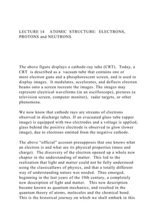
  • 1. LECTURE 14 ATOMIC STRUCTURE: ELECTRONS, PROTONS and NEUTRONS The above figure displays a cathode-ray tube (CRT). Today, a CRT is described as a vacuum tube that contains one or more electron guns and a phosphorescent screen, and is used to display images. It modulates, accelerates, and deflects electron beams onto a screen tocreate the images. The images may represent electrical waveforms (in an oscilloscope), pictures (a television screen, computer monitor), radar targets, or other phenomena. We now know that cathode rays are streams of electrons observed in discharge tubes. If an evacuated glass tube (upper image) is equipped with two electrodes and a voltage is applied, glass behind the positive electrode is observed to glow (lower image), due to electrons emitted from the negative cathode. The above “official” account presupposes that one knows what an electron is and what are its physical properties (mass and charge). The discovery of the electron opened up a whole new chapter in the understanding of matter. This led to the realization that light and matter could not be fully understood using the classicallaws of physics, and that a totally different way of understanding nature was needed. Thus emerged, beginning in the last years of the 19th century, a completely new description of light and matter. This new description became known as quantum mechanics, and resulted in the quantum theory of atoms, molecules and the chemical bond. This is the historical journey on which we shall embark in this
  • 2. Lecture. Cathode rays were discovered by Julius Plücker (1801-1868) and Johann Wilhelm Hittorf(1824-1914). Their experimental apparatus depended on two earlier inventions: 1) Volta’s battery; and, 2) a sealed glass tube in which a partial vacuum was maintained. The latter was invented by a German physicist and glassblower, Heinrich Geissler, in 1857. Hittorf observed that some unknown rays were emitted from the cathode (negative electrode) which could cast shadows on the glowing wall of the tube, indicating the rays were traveling in straight lines. In 1890, Arthur Schuster demonstrated cathode rays could be deflected by electric fields, and William Crookes showed they could be deflected by magnetic fields. It was these experiments on cathode rays inside the cathode ray tube that drew the attention of Röntgen. After repeating the above experiments, he began to study the radiation emitted outside the cathode ray tube, using fluorescent chemical sensors, e.g., barium platinocyanide, to detect radiation. His discovery of x-rays on November 8, 1895 was communicated to the Physico-Medical Society of Würzburg later in November, 1895. A translation of his paper appeared two months later on January 23, 1896 in the English journal, Nature. (You can dial up this article on Gallica and read it for yourself). Paraphrasing Louis XV(1710 – 1774) of France, were he not such a humble, unassuming man,Röntgenmight have said "Après nous, le déluge" A controversy sprang up surrounding the interpretation of cathode rays. Most German physicists believed that cathode rays were some form of light (electromagnetic radiation). Thomson, a British physicist and head of the Cavendish Laboratory at Cambridge University (Newton’s alma mater), believed otherwise on the grounds that cathode rays could be
  • 3. deflected by both electric and magnetic fields, whereas electromagnetic radiation could not. Thomson believed that cathode rays were a stream of charged particles and designed a variation on the earlier design of the cathode ray tube in which the cathode ray was subjected to combined electric and magnetic fields. See his apparatus below. In 1897, he showed that the cathode ray consisted of negatively charged particles smaller than atoms, the first "subatomic particles", which were later named electrons. In the 19th century, the charge-to-mass ratios of ions were measured by electrochemical methods, using an apparatus first introduced by Faraday. In measuring the charge-to-mass ratio, Thompson showed that charged particles in the cathode ray were characterized by a charge-to-mass ratio much smaller than the ion of the smallest element, the hydrogen ion H+, by a factor of nearly 2000. Importantly, he realized that the previously-held view that the atom was the smallest, elemental “building block” of Chemistry could not be correct. He concluded that there must exist a subatomic structure of matter. Thomson realized that the accepted model of a neutral atom did not account for negatively or positively charged particles. Therefore, he proposed a new model of the atom which he likened to English plum pudding. The negative electrons represented the raisins in the pudding and the dough contained the positive charge. Thomson's model of the atom did explain some of the electrical properties of the atom due to the electrons, but it failed to account correctly for the positive charges in the atom . Over the years, I realized that a better metaphor than “plum pudding” for students is the watermelon. The black seeds are
  • 4. electrons, and the delicious red “stuff” the uniform background (cloud) of positive charge. . The Nobel Prize in Physics 1906 was awarded to Joseph John Thomson "in recognition of the great merits of his theoretical and experimental investigations on the conduction of electricity by gases." At this point, the storyline takes an unexpected twist. While Thompson was undertaking his experiments, Röntgen, on New Years Day, 1896, mailed his manuscript (together with the photograph of his wife’s hand), to five scientists, one of whom was the celebrated French theoretical physicist, engineer, and philosopher of science, Jules Henri Poincaré, at that time President of the Académie française(French Academy). Poincaré took Röntgen’s manuscript to the next meeting of the Académie, sharing its contents with those that attended. Poincaré was fascinated with the results of Röntgen’s study, and speculated that the unknown radiation might be similar to the kind emitted by fluorescent rocks and minerals. One of the members of the Académie who showed up that day was Henri Becquerel. Reports of that meeting comment that Becquerel was dozing off during the meeting, but woke up when Poincaré mentioned fluorescent minerals. Becquerel, following in the footsteps of his Dad and Grandfather, had been studying these interesting minerals for years. There is a great display of fluorescent minerals at Chicago’s Field Museum. Becquerel went back to his lab and began using naturally fluorescent minerals intending to determine whether the radiation emitted by these materials was, in fact, the x-rays discovered by Röntgen. He exposed potassium uranyl sulfate
  • 5. to sunlight and then placed it on photographic plates wrapped in black paper, believing that the uranium absorbed the Sun’s energy and then emitted it as x-rays. His hypothesis was disproved on the 26th-27th of February, when his experiment "failed" because it was overcast and rained in Paris on those days. But, for some reason, Becquerel decided to develop his photographic plates anyway. To his surprise, the images were strong and clear, proving that the uranium emitted radiation without an external source of energy (such as the Sun). Becquerel had discovered radioactivity. Becquerel used an apparatus similar to that displayed below to show that the radiation he discovered could not be x-rays. X- rays are neutral and can not be bent in a magnetic field. The new radiation was bent by the magnetic field so that the radiation must be charged and different from x-rays. When different radioactive minerals were put in the magnetic field, they deflected in different directions or not at all, showing that there were three classes of radioactivity: negative, positive, and electrically neutral. The term radioactivity was actually coined by Marie Skłodowska Curie (1867-1934), born in Warsaw, Poland and later a naturalized French citizen. Together with her husband Pierre, she began investigating the phenomenon discovered by Becquerel. The Curies extracted uranium from ore and to their surprise, found that the leftover ore showed greater radioactivity than the pure uranium. They concluded that the ore contained radioactive elements other than uranium. This led to their discoveries of the elements polonium and radium. It took four more years of processing tons of ore to isolate enough of each element to determine their chemical and physical properties.
  • 6. BTW, if you discover a new comet or a new element, you can name it whatever you please. The first element discovered by the Curies was polonium, named after the country of Marie’s birth. If you go to the oldest quarter in Warsaw where she grew up, on the front of the apartment building where she lived, there are two large discs, one inscribed with the chemical symbol for polonium (Po) and the other for radium (Ra). Together with Becquerel, they received the 1903 Nobel Prize in Physics "in recognition of the extraordinary services they have rendered by their joint researches on the radiation phenomena discovered by Professor Henri Becquerel." Later she was awarded the Nobel Prize in Chemistry in 1911 "in recognition of her services to the advancement of chemistry by the discovery of the elements radium and polonium, by the isolation of radium and the study of the nature and compounds of this remarkable element." Following is the press release of her visit to Chicago: June 3, 1921–Marie Curie, on her first trip to the United States, visits Chicago for two hours and is “besieged by newspaper men and women anxious to get her ideas on the fashions, the war, radium, woman suffrage, the political situation.” [Chicago Daily Tribune, June 4, 1921]. All Curie, the winner of two Nobel Prizes in Physics, wants to discuss, though, is Lake Michigan. The Tribune reports, “To her the engineering feat of reversing the flow of the Chicago river to dispose of the city’s sewage was a problem far more interesting than a comparison of American styles with French creations.” By nightfall she is off to Colorado with a topographical map of the western states in hand, a gift of Mrs. W. Lee Lewis of Northwestern University.
  • 7. So, what are the mysterious rays emitted from radioactive minerals? Enter the story, Ernest Rutherford (1871-1937), a New Zealand-born British physicist who came to be known as the father of nuclear physics. Einstein considered Rutherford to be the greatest experimentalist since Michael Faraday (1791– 1867). In the years 1899 and 1900, physicists Rutherford (working in McGill University in Montreal, Canada) and Paul Villard (working in Paris) separated radiation into three types: eventually named alpha (α), beta(β) , and gamma (γ) rays by Rutherford, based on their penetration of objects and deflection by an electric field. A sketch of his experimental apparatus is shown below. Alpha rays were defined by Rutherford as those having the lowest penetration of ordinary objects. Rutherford's work also included measurements of the ratio of an alpha particle's charge to mass ratio, which led him to hypothesize that alpha particles were doubly charged helium ions (later shown to be “bare” helium nuclei). In 1907, Ernest Rutherford and Thomas Royds finally proved that alpha particles were indeed ions of Helium, the second element in the Periodic Table of elements. To do this they allowed alpha particles to penetrate a very thin glass wall of an evacuated tube, thus capturing a large number of the hypothesized helium ions inside the tube. They then caused an electric spark inside the tube, which provided a shower of electrons that were taken up by the ions to form neutral atoms of a gas. Subsequent study of the spectra of the resulting gas showed that it was Helium and that the alpha particles were indeed the hypothesized helium ions. During this period, Rutherford also identified a signature of
  • 8. radioactive elements, the half-life , and discovered the radioactive element Radon. This work was performed at McGill University in Montreal, Quebec, Canada. It was the basis for the Nobel Prize in Chemistry which he was awarded in 1908 (at age 37) "for his investigations into the disintegration of the elements, and the chemistry of radioactive substances.” Today, you can visit a museum on the McGill campus which displays his experimental equipment. In contrast to the Röntgen museum in Würzburg, you can benefit from a guided tour by a most enthusiastic and engaging curator. The next piece in the puzzle was provided by Robert Millikan (1868-1953), an American experimental physicist. Millikan graduated from Oberlin College in 1891 and obtained his doctorate at Columbia University in 1895. In 1896 he became an assistant professor at the University of Chicago. In 1909 Millikan began a series of experiments to determine the electric charge carried by a single electron. His apparatus is shown below. He began by measuring the downward trajectory of charged water droplets in an electric field. The results suggested that the charge on the droplets is a multiple of an elementary electric charge, but the experiment was not accurate enough to be convincing. He obtained more precise results in 1910 with his famous “oil-drop” experiment in which he replaced water (which tended to evaporate too quickly) with oil (which was “heavier”). Since Thomson had already determined the charge-to-mass ratio of the electron, q/m = −1.75882001076(53)×1011 C⋅ kg−1
  • 9. where q is the charge measured in Coulombs (C) and m is the mass measured in kilograms (kg). Once Millikan had determined the charge on a single electron, 1.602 x 10⁻¹⁹ C, he could calculate the mass m of the subatomic particle, the electron, 9.109×10⁻³¹ kg, and compare this with the mass of the smallest atom, Hydrogen, 1.67377 10-27 kg, the difference a factor of 1838. The 1923 Nobel Prize in Physics was awarded to Robert Andrews Millikan "for his work on the elementary charge of electricity and on the photoelectric effect." Later, Millikan left the University of Chicago to become President of the newly founded Throop University in Pasadena, CA, known today as California Institute of Technology (Caltech), arguably the best university in the World (according to the Chinese). Now, back to Rutherford. Rutherford designed an experiment to use alpha particles emitted by a radioactive element as probes to the unseen world of atomic structure. See a sketch of his experimental apparatus below. The target was a gold foil, hence the name, “gold foil” experiment. From Lecture 2, you recall that Gold is the most malleable of elements. It can be hammered into sheets of such thinness that you can see light passing through. If Thomson was correct, the beam of alpha radiation should go straight through the gold foil. Indeed, most of the beams went through the foil, but a few were deflected at wide angles, even backwards. This was crazy! If, in Thomson’s model, you had electrons embedded in a uniform background of positive “stuff,” and the electron has a mass ~1/2000 the mass of the smallest element
  • 10. (H), given that the mass of Helium is four times greater than the mass of H, how on Earth could an electron scatter an alpha particle that was ~ 8000 times heavier? Rutherford presented, eventually, a new physical model for subatomic structure to interpret this unexpected experimental result. In this model, the atom is made up of a central charge (this is the modern atomic nucleus) surrounded by a cloud of orbiting electrons. For definiteness, consider the passage of a high speed α particle through an atom having a positive central charge Ne, and surrounded by a compensating charge of N electrons. From purely energetic considerations of how far charged particles of known speed would be able to penetrate toward a central charge, Rutherford was able to calculate that the radius of the gold central charge (later called the nucleus of the atom) would have to be less than 3.4 × 10⁻¹⁴ meters. The radius of a single gold atom is ~ 10−10 meters or so. Comparison of these two radii revealed the astonishing result that the nucleus was a factor less than 1/3000 the diameter of the atom. Today, Rutherford’s model of the atom is (sometimes) called the “Solar System” model of the atom, representations of which you probably saw in grade school. While the Rutherford model concentrated the atom's positive charge and a great deal of the mass in a very small core (the nucleus), thus counterbalancing negatively-charged electrons, so that the atom was electrostatically neutral, it did not account for the atomic mass. Rutherford (and others) believed that there must be an elementary particle with a charge exactly opposite the charge on the electron, but having a (much) heavier mass. In a series of experiments performed by Rutherford and his
  • 11. student James Chadwick, they were able to change one element into another by hitting atoms with high energy alpha particles. They noticed that nitrogen (N), oxygen(O), and aluminum(Al), when hit with an alpha particle, disintegrated and emitted a fast particle of positive charge. To be more specific, hydrogen nuclei were always emitted in the process. In a dark room, they were able to observe flashes of light when alpha particles hit a florescent screen. They realized that the positive charge of any nucleus could be accounted for by a whole (integer) number of positively charged hydrogen nuclei, which were named protons by Rutherford in 1920. Knowing the mass of the Hydrogen atom (essentially the mass of the proton, since the electron mass is ~ 2000 times smaller) poses a problem. Helium, the second element in the Periodic Table, has a mass four times the mass of a single proton. So, the Helium atom, must have more than “just” protons to account for the factor of four in mass. This conundrum was solved by a student of Rutherford, James Chadwick, with the discovery in 1932 of a new elementary particle, distinct from the proton, the neutron. The neutron has no charge, and a mass slightly greater than the mass of the proton. The conclusion reached was that the Helium nucleus has two protons and two neutrons, and is orbited by two electrons. Further, it was this discovery that opened up the understanding of isotopes: two or more forms of the same element that contain equal numbers of protons but different numbers of neutrons in their nuclei, and hence differ in relative atomic mass but not in chemical properties. This point will be crucial in understanding nuclear chemistry, nuclear medicine, radiography, nuclear power plants and the atomic bomb. See Lecture 16. The Nobel Prize in Physics 1935 was awarded to James Chadwick "for the discovery of the neutron."
  • 12. Two additional experiments, both involving the electron, need to be discussed before we can take up the quantum theory of the chemical bond, molecular structure, and chemical reactivity. The first deals with the “spin” of the electron. This involves consideration of a quantity called angular momentum. Angular momentum is the rotational equivalent of linear momentum, the latter defined as mass x velocity. In physics, mass is given the symbol m, velocity v, linear momentum p, and angular momentum the symbol L. Spin is one of two types of angular momentum in quantum mechanics (the other being orbital angular momentum). The existence of spin angular momentum was inferred from experiments, such as the Stern–Gerlach experiment (see below), in which silver atoms were observed to possess two, discrete values of the angular momenta and no orbital angular momentum. The effect was attributed to “electron spin.” Electrons have an intrinsic angular momentum characterized by a (soon to be called) quantumnumbers. In Rutherford’s “Solar System” model of the atom, planets circling the Sun correspond to electrons orbiting the nucleus. Just as the Earth revolves on its axis, some describe electron spin as electrons spinning on their axis. Note that you can only spin in two directions, clockwise and counterclockwise. The spin quantum number s can take on only two values, s = +1/2 and s = − 1/2. In textbooks, these two values of the spin are shown as arrows, ↑↓ . Stern–Gerlach experiment: Silver atoms travelling through an inhomogeneous magnetic field, and being deflected up or down depending on their spin; (1) furnace, (2) beam of silver atoms,
  • 13. (3) inhomogeneous magnetic field, (4) classically expected result, (5) observed result. Otto Stern was a German-American physicist, awarded the Nobel Prize in Physics 1943 "for his contribution to the development of the molecular ray method and his discovery of the magnetic moment of the proton." The second experiment is the Davisson-Germer experiment. Between 1923 and 1927, Clinton Davisson and Lester Germer at Western Electric (later Bell Labs), discovered that electrons scattered by the surface of a crystal of nickel (Ni) metal displayed a diffraction pattern. This confirmed the hypothesis, advanced by Louis de Broglie in 1924, that an electron can “behave” like a wave or a particle, a foundational concept of quantum mechanics called wave-particle duality. At the same time George Paget Thomson independently demonstrated the same effect firing electrons through metal films to produce a diffraction pattern. Davisson and Thomson shared the Nobel Prize in Physics in 1937. Given the avalanche of unexpected experimental results, starting with Röntgen’s discovery of x-rays in 1895 and the demonstration of wave-particle duality in the 1920’s, the natural question is: What does this tell us about atoms, molecules and the chemical bond? Everything. The theoretical understanding of these phenomena sprang from
  • 14. analyzing two, apparently unrelated, experiments, both involving radiation. The first experiment centered on Black-body radiation, thermalelectromagnetic radiation emitted by a black body (an idealized opaque, non-reflective body). It has a specific spectrum of wavelengths, inversely related to intensity, that depend only on the body's temperature. As the temperature (in degrees Kelvin, K) decreases, the peak of the intensity of black-body radiation (vertical axis) moves to lower intensities and longer wavelengths. Conversely, when the temperature increases the radiation curve moves to higher frequencies. Theories to explain the behavior of the radiation curve using classical physics worked perfectly well at long wavelengths (lower frequencies) but failed completely at higher frequencies. The failure of classical theories in the regime of high frequencies was called the ultraviolet catastrophe. In 1900, the German physicist Max Planck (1858–1947) explained the ultraviolet catastrophe by proposing that the energy of electromagnetic waves is quantized rather than continuous. This means that for each temperature, there is a maximum intensity of radiation that is emitted in a blackbody object, corresponding to the peaks in the above figure, so the intensity does not follow a smooth curve as the temperature increases, as predicted by classical physics. Rather, Planck hypothesized that energy could only be gained or lost in integral multiples of some smallest unit of energy, a quantum (the smallest possible unit of energy). Energy can be gained or lost only in integral multiples of a quantum. Recall that in Lecture 13, we noted that in the classical theory of waves, the energy is given by E = ½ k A2 ( k = a constant, A = amplitude )
  • 15. In Planck’s quantum theory of radiation, the energy is given by E = h ν ( h = a constant, ν = frequency) The constant h in the above equation is now called Planck’s constant. The Nobel Prize in Physics 1918 was awarded to Max Karl Ernst Ludwig Planck "in recognition of the services he rendered to the advancement of Physics by his discovery of energy quanta." The second experiment is called the photoelectric effect. The photoelectric effect is the emission of electrons or other free carriers when electromagnetic radiation, like light, hits a material.According to classical electromagnetic theory, the photoelectric effect can be attributed to the transfer of energy from the light to an electron. From this perspective, an alteration in the intensity of light should induce changes in the kinetic energy (Kinetic Energy = ½ mv2, where m is mass and v is velocity) of the electrons emitted from the metal. According to classical theory, a sufficiently dim light is expected to show a time lag between the initial shining of its light and the subsequent emission of an electron. Sadly, the experimental results did not correlate with either of the two predictions made by classical theory. Instead, experiments showed that electrons are dislodged only by the impingement of light when it reached or exceeded a threshold frequency ν. Below that threshold, no electrons are emitted from the material, regardless of the light intensity or the length of time of exposure to the light.
  • 16. In 1905, Albert Einstein (1879-1955) solved this apparent paradox by describing light as composed of discrete quanta, now called photons, rather than continuous waves. By assuming that light actually consisted of discrete energy packets, with the energy given by Planck’s E = h ν , Einstein wrote an equation for the photoelectric effect that agreed with experimental results. Albert Einstein in 1904 (age 25) The Nobel Prize in Physics 1921 was awarded to Albert Einstein "for his services to Theoretical Physics, and especially for his discovery of the law of the photoelectric effect." In 1911 the BelgianindustrialistErnest Solvay organized an invitation-only meeting at the Hotel Metropole in Brussels. Considered a turning point in physics, in the following year Solvay founded Conseil Solvay, the International Solvay Institutes for Physics and Chemistry and, after WW I, sponsored a series of historic meetings in Physics, Chemistry and Biology. In the iconic photograph below, you will recognize a few of the physicists and chemists noted in Lectures 13 and 14. Madame Curie is seated next to Henri Poincaré, Rutherford is standing just behind them, Einstein is second from the right in the second row, Planck and de Broglie are at the other end of the row. Photograph of the first conference in 1911 at the Hotel Metropole. Seated (L–R): W. Nernst, M. Brillouin, E.
  • 17. Solvay, H. Lorentz, E. Warburg, J. Perrin, W. Wien, M. Curie, and H. Poincaré. Standing (L–R): R. Goldschmidt, M. Planck, H. Rubens, A. Sommerfeld, F. Lindemann, M. de Broglie, M. Knudsen, F. Hasenöhrl, G. Hostelet, E. Herzen, J. H. Jeans, E. Rutherford, H. Kamerlingh Onnes, A. Einstein and P. Langevin. LECTURE 13. LIGHT Hubble space telescope observations have taken advantage of gravitational lensing to reveal the largest sample of the faintest and earliest known galaxies in the universe. Some of these galaxies formed just 600 million years after the Big Bang. Space, mass, light and time are fundamental descriptors of our Universe. Captured by the poetry in Genesis, “In the beginning, when God created the heavens and earth, the earth was a formless wasteland, and darkness covered the abyss, while a mighty wind swept over the waters. Then God said “Let there be light,” and there was light. The cosmological model of the “Big Bang” describes how the universe expanded from an initial state of very high density and high temperature. If observed conditions today areextrapolated backwards in time using the known laws of physics, the prediction is that our Universe emerged from a singularity, a point of infinite density, and that before this event, space and time did not exist. Current knowledge is insufficient to determine if anything existed prior to the singularity. Sixteen centuries ago, in his Confessions, Saint Augustine (354-430) posed the obvious question in biblical terms: What was God doing before he
  • 18. created the Universe? ISAAC NEWTON: COLOR SPECTRUM and the CORPUSCULAR THEORY of LIGHT Our modern understanding of light and color begins with Isaac Newton (1642-1726) and a series of experiments that he published in 1672. He was the first to understand the rainbow. He refracted white light with a glass prism, resolving it into its component colors: red, orange, yellow, green, blue and violet. In the graphic below, light enters the prism from the top right, and is refracted by the glass. The violet is bent more than the yellow and red, so the colors separate. In the 1660s, Newton began experimenting with his “celebrated phenomenon of colors.” At the time, people thought that color was a mixture of light and darkness, and that prisms colored light. Hooke was a proponent of this theory of color, and had a scale that went from brilliant red, which was pure white light with the least amount of darkness added, to dull blue, the last step before black, which was the complete extinction of light by darkness. Newton believed this theory was false. Newton set up a prism near a window at his boyhood home in Woolsthorpe, England ( site of the famous apple tree), and projected a beautiful spectrum 22 feet onto the far wall. Further, to prove that the prism was not coloring the light, he refracted the spectral light back together, producing white light. Incidentally, he was at home because all the students at
  • 19. Cambridge University where he was a student were sent home because of an epidemic of the bubonic plague. In 1665, it was a version of “social distancing.” BTW, Newton did his best work working from home. On a personal note, my wife and I once visited the hometown of William Shakespeare (1564-1616), Stratford-upon-Avon. Access to Anne Hathaway’s cottage was severely hampered by rows of tour buses, cars, and floods of pedestrians. Later, we drove to Woolsthorpe by Colsterworth (took about an hour on country roads) to visit Newton’s home, now a museum. When we arrived the museum was closed, and had been for some time. I asked a local gentleman “why,” and he responded “because no one ever came.” The diagram below isfrom Sir Isaac Newton’s crucial experiment, 1666-72. A ray of light is divided into its constituent colors by the first prism (left), and the resulting bundle of colored rays is reconstituted into white light by the second prism (right). Artists were fascinated by Newton’s clear demonstration that light alone was responsible for color. His most useful idea for artists was his conceptual arrangement of colors around the circumference of a circle, which allowed the painters’ primaries (red, yellow, blue) to be arranged opposite their complementary colors (e.g. red opposite green), as a way of denoting that each complementary color would enhance the other’s effect through optical contrast. In optics, the corpuscular theory of light, arguably set forward
  • 20. by Descartes in 1637, states that light is made up of small discrete particles called "corpuscles" (little particles) which travel in a straight line with a finite velocity and possesses momentum (mass x velocity). Isaac Newton was a pioneer of this theory, an elaboration of his view of reality as interactions of material points through forces. The following passage gives Albert Einstein's description of Newton's conception of physical reality: “[Newton's] physical reality is characterized by concepts of space, time, the material point and force (interaction between material points). Physical events are to be thought of as movements according to law of material points in space. The material point is the only representative of reality in so far as it is subject to change.” This early conception of the particle theory of light was an early forerunner to the modern understanding of the photon. See next Lecture 14. CHRISTIAAN HUYGENS: WAVE THEORY of LIGHT The corpuscular theorycannot explain refraction, diffraction and interference, which require an understanding of the wave theory of light of Christiaan Huygens. refraction: the change in direction of a wave passing from one medium to another or from a gradual change in the medium. Refraction of light is the most commonly observed phenomenon, but other waves such as sound waves and water waves also show refraction. diffraction: Diffraction refers to various phenomena that occur
  • 21. when a wave encounters an obstacle or a slit. It is defined as the bending of waves around the corners of an obstacle or through an aperture into the region of geometrical shadow of the obstacle/aperture. interference: interference is a phenomenon in which two waves superimpose to form a resultant wave of greater (constructive interference), lower (destructive interference), or the same amplitude. To explain the origin of color, Robert Hooke (1635–1703) developed a "pulse theory" and compared the spreading of light to that of waves in water in his 1665 Micrographia. In 1672 Hooke suggested that light's vibrations could be perpendicular to the direction of propagation. Christiaan Huygens (1629–1695) worked out a mathematical wave theory of light in 1678, and published it in his Treatise on light in 1690. He proposed that light was emitted in all directions as a series of waves in a hypothetical medium called the Luminiferous ether(invisible to the senses, no “weight,” color, odor or taste) and that, as waves are not affected by gravity, he assumed that they slowed down upon entering a denser medium. That light waves could interfere with each other like sound waves was first noted around 1800 by Thomas Young, a physician. Young demonstrated by means of a diffraction experiment that light behaved as waves. He also proposed that different colors were caused by different wavelengths of light, and explained color vision in terms of three color receptors in the eye. . Thomas Young's sketch of a double-slit
  • 22. experimentshowing diffraction. Young's experiments supported the theory that light consists of waves. Coincidentally, Young’s report of the double-slit experiment appeared in the Proceedings of the Royal Society of London in the same year (1800) as Volta’s paper on his invention of the battery. In earlier lectures in the course, examples of the atomic structure of inorganic, organic and biochemical entities were shown [ e.g., NaCl (Lecture 1) , diamond (Lecture 5), COVID- 19 (Lecture 12) ]. Did you ever ask yourself (or me) how those crystal structures were determined? Thank you, Thomas Young. Let’s get to the bottom of this. ELECTROMAGNETIC WAVES and POLARIZATION One speaks of the wave nature of light, but we have not specified what it is that oscillates or undulates. It is easy to visualize waves in a fluid medium such as water, where we can see the motion of the water as the wave passes by. wavelength (λ) = the distance, measured in the direction of propagation of a wave, from any given point to the next point in the same phase. frequency (ν) = the number of oscillations (or cycles) of a wave per unit of time
  • 23. We now know that electromagnetic waves are very different from water waves or sound waves. NO MEDIUM is REQUIRED. Light waves can travel through a total vacuum, unlike other waves with which we are familiar. Recognizing the significance of this difference, established experimentally by Michaelson and Morley in 1887 at what is now Case Western University in Cleveland, is one of the two cornerstones on which Einstein developed his Theory of Special Relativity, published in 1905. We shall pursue this in Lecture 16 on nuclear chemistry, nuclear medicine and radiography. The Nobel Prize in Physics 1907 was awarded to Albert Abraham Michelson "for his optical precision instruments and the spectroscopic and metrological investigations carried out with their aid." Importantly, when watching water waves lap against the shore on Lake Michigan, it appears that water is “moving” toward shore. Not so. The water in the lake is not moving. What is being transported is energy. In the classical theory of waves, the energy is given by E = ½ k A2 ( k = a constant, A = amplitude ) In the quantum theory of light (to be presented in the next Lecture 14), the energy is given by E = h ν ( h = a constant, ν = frequency) Rather than having physical motions, electromagnetic (light) waves consist of electric and magnetic fields that oscillate back and forth at right angles to the direction of motion. The electric and magnetic fields lie in planes that are also perpendicular to each other, so that the electric field flips back and forth in one plane, with the magnetic field doing so in the plane that lies
  • 24. perpendicular to it, and both are traverse to the direction of travel. Electromagnetic (light) waves can be imagined as a self- propagating, transverse, oscillating wave of electric and magnetic fields. The above diagram shows a plane, linearly polarized wave propagating from left to right. The electric field is in a vertical plane and the magnetic field in a horizontal plane. A photon (a quantum or Newtonian “corpuscle” of light, to be introduced in Lecture 14) can be viewed as a packet of electromagnetic energy consisting of alternating fields, which, by virtue of their wavelength and frequency, provide the photon with its wave properties. Summarizing the above discussion, light is energy that radiates, or travels, in waves. Light is a wave of vibrating electric and magnetic fields. Visible light is one small part of a larger range of vibrating electromagnetic fields. This range is called the electromagnetic spectrum. By contrast, sound is a wave of vibrating air. The energy of the radiation depends on its wavelength and frequency. As noted above, wavelength is the distance between the tops (crests) [ or bottoms (troughs) ] of the waves. And, frequency is the number of waves that pass by each second. The longer the wavelength of the light, the lower the frequency, and the less energy it contains. Recalling that Velocity (v) = distance /time = λ ν electromagnetic waves travel through space at 299,792 km/sec
  • 25. (186,282 miles/sec). This is called the velocity (speed) of light. In Astronomy, the distance a beam of light travels in one year is called a light year. The light emitted at the first moment of the Big Bang (see the photo taken with the Hubble telescope at the top of this Lecture) is 46.1 billion (46.1x10⁹) light-years distant. Light from most sources (such as stars) consists of vast numbers of photons, each with a specific, constant orientation of its electric and magnetic planes. Ordinarily the orientation of the various photons is random, but under some circumstances it is not. If light passes through or reflects from a medium with a certain orientation, what is left is light in which all the photons are aligned. That is, the planes of the electric and magnetic fields of the photons are parallel. The light is said to be polarized. Many of the sunglasses sold today consist of polarizing filters, and they have the effect of screening out light in a given orientation. In Astronomy, polarization occurs naturally, sometimes because a natural filtering occurs, as in the interstellar medium, and in other cases because the source of the light emits radiation with a preferred orientation. Next, we need to understand the following phenomena. It happens that one of the things about waves is the following: If they encounter an obstacle which is smaller than their wavelength, the wave just kind of wraps around the object and keeps on going. This is why you can hide behind a big tree
  • 26. trunk and the voices of people on the other side of the tree will still reach you (because the long waves of the sound wrap around the tree). BUT, light from their flash light won't (because the short waves of the light can't wrap around such a big object). Sound and light act similarly when they reach a corner of a house - the sound waves are long and kind of wrap around the corner and then spread out from there, whereas the very short light waves can't bend around the corner. As you might have guessed already, this means that light does wrap around very small things (things smaller than a millionth of a foot), and does bend a tiny bit around corners, but this bending is so small it is impossible to see with your eye. See, however, the following discussion. X-rays are light waves of very short wavelength and high energy discovered by Wilhelm Röntgen (1845-1923) who, on November 8, 1895, produced and detected electromagnetic radiation in a wavelength range known today as x-rays. Below is a photo of his modest laboratory in Würzburg, Germany. No guided tours. You can see his experimental equipment by yourself, “up close and personal.” Roentgen’s experiment involved studying the radiation emitted outside a cathode ray tube (see description in the following Lecture 14). He also used as sensors a series of compounds that were sensitive to light of high frequencies, the principal one of
  • 27. which was the barium salt of the platinocyanide ion. The ion exists in two isometric forms: Pt is located at the center of the plane in the above diagram. Barium platinocyanide, Ba[Pt(CN)4] isa phosphor and a scintillator. It fluoresces in the presence of light of high frequencies (ν) Röntgen baptized the mysterious radiation that caused a film of Ba[Pt(CN)4] on a support to glow, “x-rays,” after the name of the unknown in algebra. In Germany, they were called Röntgen rays (which he discouraged). One of the things Röntgen discovered about x-rays was their incredible penetrating power. They could pass through paper, glass, wood and human flesh (but not lead). He asked his wife Bertha to put her hand between the source of x-rays and a photographic plate. The result was one of the most famous of all photographs. Notice the dark circle on her finger, her wedding ring. Bertha’s reaction to the almost supernatural character of this picture, was “I have seen my own death.” Within months x-rays were used in medicine worldwide to diagnose fractures. X-rays remain today one of the most important diagnostic tools in medicine. The first Nobel Prize in Physics was awarded in 1901 to Wilhelm Conrad Röntgen "in recognition of the extraordinary services he has rendered by the discovery of the remarkable rays subsequently named after him."
  • 28. The discovery of x-rays launched a new era in experimental physics and chemistry which led to the quantum theory of atoms and molecules. See the next Lecture 14. Max von Laue (1879 – 1960) discovered the diffraction of x- rays by crystals. The idea came to him that estimates of the size of atoms, and their spacing in a crystal, were about thesame as the wavelength of Röntgen’s x-rays. He suggested that the much shorter electromagnetic rays, which x-rays were supposed to be, would cause in a crystal some kind of diffraction or interference phenomena. Knipping, an experimentalist, tested out the idea and, after some failures, succeeded in proving it to be correct. Von Laue worked out the mathematical formulation and the discovery was published in 1912. For this, he was awarded the Nobel Prize in 1914. It established the fact that x- rays are electromagnetic in nature and it opened the way to the later theoretical and experimental work of Sir William and Sir Lawrence Bragg. Sir William Lawrence Bragg 1890 – 1971) was an Austalian- born British physicist, discoverer in 1912 of Bragg's law of x- ray diffraction, which is basic for the determination of crystal structure. He was joint recipient (with his father, William Henry Bragg) of the Nobel Prize in Physics in 1915. Bragg was the director of the Cavendish Laboratory, Cambridge, when the discovery of the structure of DNA (the double helix, see Lecture 6 ), based on the x-ray diffraction imagesof DNA by Rosalind Franklin, was reported by James D. Watson and Francis Crick in February 1953. Watson, Crick and Maurice Wilkins received the Nobel Prize in Physiology or Medicine in 1962. Also at the Cavendish Laboratory was Max Perutz (1914-2002), an Austrian-born British molecular biologist, who shared the 1962 Nobel Prize for
  • 29. Chemistry with John Kendrew, for their studies of the crystal structures of hemoglobin and myoglobin (See Lecture 6 ) Another at Cambridge was Aaron Klug, a Lithuanian-born, South African-educated, British biophysicist. He solved the crystal structures of macromolecules such as viruses (see Lecture 12) working with Rosalind Franklin. He found the rules of the geometrical form of poliovirus and other spherical viruses. His invention of electron tomography, in which a 3D image of a virus is obtained from many electron micrographs, led to the Nobel Prize in Chemistry in 1982.Rosalind Franklin died at the age of 37 from ovarian cancer. Watson, among many others, suggested that Franklin should have been awarded a Nobel Prize in Chemistry, along with Wilkins, but, although there was not yet a rule against posthumous awards, the Nobel Committee generally did not make posthumous nominations.The experimental technique of x-ray crystallography is the common thread which links together much of what has been highlighted in earlier lectures, and provides a good review of some of the most important discoveries in inorganic chemistry, biochemistry and medicinal chemistry. Officially, X-ray crystallography is defined as the experimental science determining the atomic and molecular structure of a crystal, in which the crystalline structure causes a beam of incident x-rays to diffract into many specific directions. By measuring the angles and intensities of these diffracted beams, a crystallographer can produce a three-dimensional picture of the density of electrons within the crystal. From this electron density, the mean positions of the atoms in the crystal can be determined, as well as their chemical bonds and their crystallographic disorder. COLORS of LIGHT
  • 30. Visible light is the part of the electromagnetic spectrum that our eyes can see. Light from the sun or a light bulb may look white, but it is actually a combination of many colors. We can see the different colors of the spectrum by splitting the light with a prism. The spectrum is also visible when you see a rainbow in the sky. The colors blend continuously into one another. At one end of the spectrum are the reds and oranges. These gradually shade into yellow, green, blue, indigo and violet. The colors have different wavelengths, frequencies, and energies. Violet has the shortest wavelength in the visible spectrum. That means it has the highest frequency and energy. Red has the longest wavelength, and lowest frequency and energy. PERCEPTION of COLOR When a sample absorbs visible light, an object will selectively absorb specific wavelengths of light from the complete spectrum of possibilities. The color we perceive is the sum of the remaining colors that are reflected or transmitted by an object and strike our eyes. An object which reflects light is said to be opaque. An object which transmits all light is said to be transparent. An object which absorbs all wavelengths of visible light appears black (because no light reaches our eyes). An object which absorbs no visible light appears white or colorless. In case you’re wondering, …. What is a rainbow? A rainbow occurs when light hits the water drops in the
  • 31. atmosphere at a certain angle. It is an atmospheric phenomenon that is formed through optical processes such as refraction, dispersion, internal reflection and secondary refraction. If the light source is the sun, then the rainbow will be colorful and bright. Why is water blue? Water does absorb a tiny amount of light, but in such small amounts, we don’t notice it. Water actually absorbs light that has a reddish color. When there’s a lot of water, like Lake Michigan, we notice that red light from the Sun has actually been absorbed. So, what color do we see when reddish light is gone? Blue! Blue is the color absorbed the least by water, so it’s the one we see the most. Blue light also scatters more than other light. That’s the reason why the sky is blue. That scattering affects the color of water too. It just takes a big enough sample of water to notice these things. That’s why you won’t see color in the glass you’re drinking. Why is the grass green? The blades of grass and the leaves of trees contain the molecule chlorophyll (see Lecture 5 ) a molecule able to absorb in the red (and blue) regions of the spectrum. When white sunlight is reflected from leaves, what we see is the light not absorbed by the plant. The color vegetation appears to our senses, the color which is not absorbed, is green. Red and green are complementary colors. Why is the color of an orange, orange ?
  • 32. Orange and blue are complementary colors. The removal of blue light from white light makes the light look orange. [Conversely, the removal of orange light from white light makes the light look blue.] If light of all colors except blue strikes our eyes, we perceive an orange color. If an orange absorbs all light except orange, in the reflected light, the fruit appears orange. In his book “Origins,” Richard Leaky notes that the perception of color in primates was a key factor in human evolution. He displays a color photograph of an orange tree, leaves in green and oranges in orange, and compares this with a black-and- white photo. If you’re looking for fruit to eat, the advantage of color vision is obvious. Why are minerals colored? ANALCIME REALGAR CROCOITE TYUYAMUNITE MALACHITE TURQUOISE CAVANSITE
  • 33. AZURITE FLUORITE AMETHYST KAEMMERERITE BIXBY Color in minerals is caused by the absorption, or lack of absorption, of various wavelengths of light. The color of light is determined by its wavelength. When pure white light (containing all wavelengths of visible light) enters a crystal, some of the wavelengths might be absorbed while other wavelengths may be emitted. If this happens, then the light that leaves the crystal will no longer be white but will have some color. Small amounts of impurities can also change the color of crystals. Recall the Hope Diamond (Lecture 5). My colleague, Harry Gray at Caltech, pointed out the remarkable fact that only small differences in Cr(III)O6 ligand fields make emeralds green and rubies red ! COLOR and the HUMAN BRAIN Color vision is the capacity of an organism or machine to distinguish objects based on the wavelengths (or frequencies) of the light they reflect, emit, or transmit. Colors can be measured and quantified in various ways. A human's perception of colors is a subjective process whereby the brain responds to the stimuli that are produced when incoming light reacts with the several types of photoreceptors in the eye. Rods and Cones are the photoreceptors found in the retina.
  • 34. Rods have rod-like structure and provide twilight vision. Cones are of the cone shape, fewer in number and provide vision in the day or bright light. Rods are found around the boundary of the retina, whereas cones are there in the center of the retina. Color vision relies on a brain perception mechanism that treats light with different wavelengths as different visual stimuli (e.g., colors). Usual color insensitive photoreceptors (the rods in human eyes) only react to the presence or absence of light and do not distinguish between specific wavelengths. NOTE: Colors are not “real.” They are “synthesized” by our brain to distinguish light with different wavelengths. While rods give us the ability to detect the presence and intensity of light (and thus allow our brain to construct the picture of the world around us), specific detection of different wavelengths through independent channels gives our view of the world additional high resolution. For instance, red and green colors look like near identical shades of grey in black and white photos. An animal with black and white vision alone won’t be able to make a distinction between, let’s say, a green and red apple, and won’t know which one tastes better before trying them both based on color. Evolutionary biologists believe that human ancestors developed color vision to facilitate the identification of ripe fruits, which would obviously provide a selective advantage in the competitive natural world. The same conclusion had been reached by the anthropologist, Richard Leaky. Why certain wavelengths are paired with certain colors remains a mystery. As mentioned above, technically, color is an illusion created by our brain. Therefore, it is not clear if other animals see colors the same way we see them. It is likely that, due to shared evolutionary history, other vertebrates see the world colored similarly to how we see it. Color vision is quite common across the vast animal kingdom: insects, arachnids, and
  • 35. cephalopods are able to distinguish colors. What kind of colors do these animals see? As was first proposed by Thomas Young, human color vision relies on three photoreceptors that detect primary colors: red, green, and blue. However, some people lack red photoreceptors (they are “bichromates”) or have an additional photoreceptor that detects somewhere between red and green colors (“tetrachromates”). Having only three photoreceptors doesn’t limit our ability to distinguish other colors. Each photoreceptor can absorb a rather broad range of wavelengths of light. To distinguish a specific color, the brain compares and quantitatively analyses the data from all three photoreceptors. Our brain does this remarkably successfully. Some research indicates that we can distinguish colors that correspond to wavelength differences of just one nanometer (10- 9 meters) This scheme works in largely the same way in most higher vertebrate animals that have color vision. Although the ability to distinguish between specific shades varies significantly between the species, with humans having one of the best color distinguishing abilities. However, invertebrates that have developed color vision (and vision in general) completely independently from us demonstrate remarkably different approaches to color detection and processing. These animals can have an exceptionally large number of color receptors. The mantis shrimp, for instance, has 12 different types of photoreceptors. The common bluebottle butterfly has even more, 15 receptors. Does it mean that these animals can see additional colors unimaginable to us? Perhaps yes. Some of their photoreceptors operate in a rather narrow region of light spectrum. For instance, they can have 4-5 photoreceptors sensitive in the green region of the visual spectrum. This means that for these animals the different shades of green may appear as different as
  • 36. blue and red colors appear to our eyes! Again, the evolutionary advantages of such adaptations are obvious for an animal living among the trees and grasses where most objects, as we see them, are colored in various shades of green. Researchers tried to test if a more complicated set of visual receptors provide any advantages for animals when it comes to the distinguishing between main colors. The findings show that this is not necessarily the case, at least not for the mantis shrimp. The mantis shrimp has an impressive array of receptors detecting light in a much broader part of the electromagnetic spectrum compared to humans. They determine the colors fast. This is important for practical purposes, as mantis shrimps are predators. A large number of photoreceptors allows for their quick activation at specific wavelengths of light and thus communicate directly to the brain what specific wavelength was detected. In comparison, humans have to assess and quantify the signals from all three photoreceptors to decide on a specific color. This requires more time and energy. Apart from employing a different number of photoreceptors to sense light of specific wavelengths, some animals can detect light that we humans are completely unable to see. For example, many birds and insects can see in the UV part of the spectrum. Bumblebees, for instance, have three photoreceptors absorbing in the UV, blue, and green regions of the spectrum. This makes them trichromates, like humans, but with the spectral sensitivity shifted to the blue end of the spectrum. The ability to detect UV light explains why some flowers have patterns visible only in this part of the spectrum. These patterns attract pollinating insects, which have an ability to see in this spectral region. A number of animals can detect infrared light (the long wavelength radiation) emitted by heated objects and bodies. This ability significantly facilitates hunting for snakes that are usually looking for small warm-blooded prey. Seeing them through IR detecting receptors is a great tool to detect slow- moving reptiles. The photoreceptors sensitive to IR radiation in
  • 37. snakes are located not in their eye but in “pit organs” located between the eyes and nostrils. The result is still the same: Snakes can color objects according to their surface temperature. Wavelength and hue detection As noted earlier, Isaac Newton discovered that white light splits into its component colors when passed through a prism, but that if those bands of colored light pass through another and rejoin, they make a white beam. The characteristic colors are, from low to high frequency: red, orange, yellow, green, cyan, blue, violet. Sufficient differences in frequency give rise to a difference in perceived hue. The just noticeable difference in wavelength varies from about 1 nm in the blue-green and yellow wavelengths, to 10 nm and more in the red and blue. Though the eye can distinguish up to a few hundred hues, when those pure spectral colors are mixed together or diluted with white light, the number of distinguishable chromaticities can be quite high. In very low light levels, vision is scotopic. Light is detected by rod cells of the retina. Rods are the principal players. In brighter light, such as daylight, vision is photopic. Light is detected by cone cells which are responsible for color vision. Cones are sensitive to a range of wavelengths, but are most sensitive to wavelengths near 555 nm (or, 5550 Å). Between these regions, mesopic vision comes into play and both rods and cones provide signals to the retinal ganglion cells. The shift in color perception from dim light to daylight gives rise to differences known as the Purkinje effect. The perception of “white” is formed by the entire spectrum of visible light, or by mixing colors of just a few wavelengths, such as red, green, and blue, or by mixing just a pair of complementary colors such as blue and yellow.
  • 38. PHUSIOLOGY of COLOR PERCEPTION Perception of color begins with specialized retinal cells containing pigments with different spectral sensitivities, known as cone cells. In humans, there are three types of cones sensitive to three difference spectra, resulting in trichromatic color vision. The cones are conventionally labeled according to the ordering of the wavelengths of the peaks of their spectral sensitivities: short (S), medium (M), and long (L) cone types. These three types do not correspond well to particular colors as we know them. Rather, the perception of color is achieved by a complex process that starts with the differential output of these cells in the retina and it will be finalized in the visual cortex and associative areas of the brain. For example, while the L cones have been referred to simply as red receptors, their peak sensitivity is in the greenish-yellow region of the spectrum. Similarly, the S- and M-cones do not directly correspond to blue and green, although they are often depicted as such. The peak response of human cone cells varies, even among individuals with normal color vision. In non-human species this polymorphic variation is even greater, and it may well be adaptive. Normalized response spectra of human cones, S, M, and L types, to monochromatic spectral stimuli, with wavelength given in nanometers. The same figures as above represented here as a single curve in three (normalized cone response) dimensions.
  • 39. Relative brightness sensitivity of the human visual system as a function of wavelength. Theories of color vision Two complementary theories of color vision are the trichromatic theory and the opponent process theory. The trichromatic theory, or Young–Helmholtz theory, proposed in the 19th century by Thomas Young and Hermann von Helmholtz, states that the retina's three types of cones are preferentially sensitive to blue, green, and red. Ewald Hering proposed the opponent process theory in 1872. It states that the visual system interprets color in an antagonistic way: red vs. green, blue vs. yellow, black vs. white. We now know both theories to be correct, describing different stages in visual physiology. Cone cells in the human eye Cone type Name Range Peak wavelength S β 400–500 nm 420–440 nm M γ 450–630 nm 534–555 nm L ρ 500–700 nm 564–580 nm A range of wavelengths of light stimulates each of these receptor types to varying degrees. Yellowish-green light, for
  • 40. example, stimulates both L and M cones equally strongly, but only stimulates S-cones weakly. Red light, on the other hand, stimulates L cones much more than M cones, and S cones hardly at all. Blue-green light stimulates M cones more than L cones, and S cones a bit more strongly, and is also the peak stimulant for rod cells; and blue light stimulates S cones more strongly than red or green light, but L and M cones more weakly. The brain combines the information from each type of receptor to give rise to different perceptions of different wavelengths of light. The opsins (photopigments) present in the L and M cones are encoded on the X chromosome; defective encoding of these leads to the two most common forms of color blindness. The OPN1LW gene, which codes for the opsin present in the L cones, is highly polymorphic (a recent study found 85 variants in a sample of 236 men). A very small percentage of women may have an extra type of color receptor because they have different alleles for the gene for the L opsin on each X chromosome. X chromosome inactivation means that only one opsin is expressed in each cone cell, and some women may therefore show a degree of tetrachromatic color vision. Variations in OPN1MW, which codes the opsin expressed in M cones, appear to be rare, and the observed variants have no effect on spectral sensitivity. Now, for the brain per se. Visual pathways in the human brain. The ventral stream (purple) is important in color recognition. The dorsal stream (green) is also shown. They originate from a common source in the visual cortex. Color processing begins at a very early level in the visual system (even within the retina) through initial color opponent mechanisms. Both the Young-Helmholtz trichromatic theory, and Hering's opponent process theory are therefore correct, but
  • 41. trichromacy arises at the level of the receptors, and opponent processes arise at the level of retinal ganglion cells and beyond. In Hering's theory opponent mechanisms refer to the opposing color effect of red–green, blue–yellow, and light–dark. However, in the visual system, it is the activity of the different receptor types that are opposed. Some midget retinal ganglion cells oppose L and M cone activity, which corresponds loosely to red–green, but actually runs along an axis from blue-green to magenta. Small bistratified retinal ganglion cells oppose input from the S cones to input from the L and M cones. This is often thought to correspond to blue–yellow ,opponency, but actually runs along a color axis from lime green to violet. Visual information is then sent to the brain from retinal ganglion cells via the optic nerve to the optic chiasma, a point where the two optic nerves meet and information from the temporal (contralateral) visual field crosses to the other side of the brain. After the optic chiasma (the X-shaped structure formed at the point below the brain where the two optic nerves cross over each other), the visual tracts are referred to as the optic tracts, which enter the thalamus to synapse at the lateral geniculate nucleus (LGN). The LGN is divided into laminae (zones), of which there three types: the M-laminae, consisting primarily of M-cells, the P- laminae, consisting primarily of P-cells, and the koniocellular laminae. M- and P-cells received relatively balanced input from both L- and M-cones throughout most of the retina, although this seems to not be the case at the fovea, with midget cells synapsing in the P-laminae. The koniocellular laminae receive axons from the small bistratified ganglion cells. After synapsing at the LGN, the visual tract continues on back to the primary visual cortex (V1) located at the back of the brain within the occipital lobe. Within V1 there is a distinct band (striation). This is also referred to as "striate cortex", with other cortical visual regions referred to collectively as "extrastriate cortex". It is at this stage that color processing
  • 42. becomes much more complicated. In V1 the simple three-color segregation begins to break down. Many cells in V1 respond to some parts of the spectrum better than others, but this "color tuning" is often different depending on the adaptation state of the visual system. A given cell that might respond best to long wavelength light if the light is relatively bright might then become responsive to all wavelengths if the stimulus is relatively dim. Because the color tuning of these cells is not stable, some believe that a different, relatively small, population of neurons in V1 is responsible for color vision. These specialized "color cells" often have receptive fields that can compute local cone ratios. Such "double-opponent" cells were initially described in the goldfish retina by Nigel Daw. Their existence in primates was suggested by David H. Hubel and Torsten Wiesel and subsequently proven by Bevil Conway. As Margaret Livingstone and David Hubel showed, double opponent cells are clustered within localized regions of V1 called blobs, and are thought to come in two flavors, red–green and blue–yellow. Red–green cells compare the relative amounts of red–green in one part of a scene with the amount of red–green in an adjacent part of the scene, responding best to local color contrast (red next to green). Modeling studies have shown that double-opponent cells are ideal candidates for the neural machinery of color constancy explained by Edwin H. Land in his retinex theory. The Nobel Prize in Physiology or Medicine 1981 was divided, one half awarded to Roger W. Sperry "for his discoveries concerning the functional specialization of the cerebral hemispheres", the other half jointly to David H. Hubel and Torsten N. Wiesel "for their discoveries concerning information processing in the visual system." From the V1 blobs, color information is sent to cells in the second visual area, V2. The cells in V2 that are most strongly
  • 43. color tuned are clustered in the "thin stripes" that, like the blobs in V1, stain for the enzyme cytochrome oxidase (separating the thin stripes are interstripes and thick stripes, which seem to be associated with other visual information like motion and high- resolution form). Neurons in V2 then synapse onto cells in the extended V4. This area includes not only V4, but two other areas in the posterior inferior temporal cortex, anterior to area V3, the dorsal posterior inferior temporal cortex, and posterior TEO. Color processing in the extended V4 occurs in millimeter-sized color modules (globs). This is the first part of the brain in which color is processed in terms of the full range of hues found in color space. Anatomical studies have shown that neurons in extended V4 provide input to the inferior temporal lobe . The "IT" cortex is thought to integrate color information with shape and form, although it has been difficult to define appropriate criteria for this claim. Despite this murkiness, it has been useful to characterize this pathway (V1 > V2 > V4 > IT) as theventral stream or the "what pathway", anddistinguished from the dorsal stream("where pathway") that is thought to analyze motion.