
Recommended
More Related Content
What's hot
What's hot (20)
Similar to Brain Vascular Malformations Imaging Overview
Similar to Brain Vascular Malformations Imaging Overview (20)
More from macshrestha
Recently uploaded
Recently uploaded (20)
Brain Vascular Malformations Imaging Overview
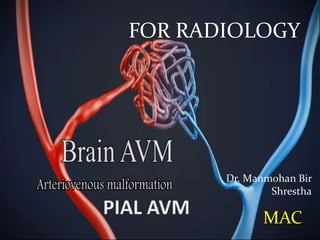
- 1. Dr. Manmohan Bir Shrestha FOR RADIOLOGY MAC
- 2. Vascular Malformations in Brain OVERVIEW Cerebrovascular malformations (CVMs) are a heterogenous group of disorders that represent morphogenetic errors affecting arteries, capillaries, veins or various combinations of vessels.
- 3. Using accurate terminology 2 major groups: A. Vascular malformations Includes AVM & Fistula B. Hemangiomas These are benign vascular neoplasms, not malformations Proliferating, mesenchymal, nonmeningothelial tumors Can be capillary or cavernous
- 4. Classification of Cerebrovascular malformations A. Histopathologic classification B. Functional classification
- 5. A.Histopathologic classification I. Arteriovenous malformation II. Venous angioma III. Capillary telangiectasia IV. Cavernous malformation.
- 6. B. Functional classification Endovascular radiologists have proposed a functional, highly practical system, and divides all CVMs in 2 categories CVMs that display shunting AVM Fistula CVMs without AV shunting Everything else i.e. venous, capillary, cavernous malformations. The 1st group are amenable to intervention & latter are either left alone or treated surgically.
- 8. Pial AVM • Also called Cerebral AVM/ Classic AVM • Definition • It is a vascular malformation with direct artery to vein (AV) shunting, no intervening capillary bed • 3 components • Enlarged feeding arteries, 1 or more • Nidus of tightly packed, enlarged tangled vascular channels • Dilated draining veins, 1 or more **No normal brain parenchyma in between
- 11. Demographics Age Peak presentation: 20-40 years Gender M=F Epidemiology Prevalence: 0.040 - 0.52%
- 12. Location Supratentorial – 85% Posterior fossa – 15% Number Solitary Multiple AVMs usually syndromic (Hereditary hemorrhagic telangiectasia, Wyburn-Mason syndrome). Size Varies from microscopic to giant Most symptomatic AVMs are 3-6 cm.
- 13. Pathology Etiology Origin of AVMs remain uncertain However, thought to occur congenitally, due to dysregulated angiogenesis. Genetic Sporadic AVMs have up/down-regulated genes Homeobox genes, such as HOXD3 AND HOXB3 Pleomorphisms on p21 locus of chromosome 9 Syndromic Hereditary hemorrhagic telangiectasia Wyburn-Mason syndrome
- 14. Clinical presentation Headache Seizure Focal neurological deficit Hemorrhage Parenchymal/subarachnoid/intraventricular Ischaemic events due to vascular steal from normal brain Incidental finding
- 15. Imaging CT MRI Angiography
- 16. CT NECT Normal, if AVM is very small Iso/hyperdense serpentine vessels Calcification in 25-30% AVM bleed = parenchymal/intraventricular/subarachnoid hemorrhage Post-embolization – embolics appear hyperdense within nidus CECT Strong enhancement of arterial feeders, nidus and draining veins, giving appearance of “bag of worms” CTA Depicts enlarged arteries, draining veins
- 17. Case of an adult with complaints of left sided pulsatile tinnitus NECT
- 18. MIP CTA
- 19. Case of 40 yrs. Female with complaints of severe headache
- 20. NECT
- 21. CECT
- 22. MR findings T1WI Tightly packed mass, “honeycomb” of flow voids Signal varies with flow rate, presence/age of hemorrhage T2WI Tangle of serpiginous, ‘honeycomb’ of flow voids Little/no brain inside nidus Some gliotic high signal may be present FLAIR Flow voids with surrounding high signal (gliosis) T2*GRE Blooming if hemorrhage Post Contrast T1 Strong enhancement of nidus, draining veins Rapid flow may not enhance arteries, and seen as flow voids MRA Helpful for gross depiction of flow Does note depict detailed angioarchitecture
- 23. T1WI
- 24. T2WI
- 25. FLAIR
- 26. T1 POST CONTRAST
- 28. Angiographic findings Digital Substraction Angiography (DSA) best delineates internal angioarchitecture Depicts 3 components of AVM
- 29. Case of 50 yrs. Male with complaints of sudden severe headache and left sided weakness
- 30. NECT
- 31. DSA
- 34. Associated abnormalities Flow-related aneurysm on feeding artery= 10-15% Intranidal aneurysm = >50% Vascular steal may cause ischemia in adjacent brain
- 35. Staging/grading Spetzler-Martin scale Score correlates with operative outcome Score = 1-5 Size Small (<3cm) =1 Medium (3-6 cm)= 2 Large (>6 cm) = 3 Location Non-eloquent area = 0 Eloquent area = 1 (sensorimotor cortex, visual cortex, thalamus, hypothalamus, internal capsule, brainstem, cerebellar peduncles, deep nuclei) Venous drainage Superficial only = 0 Deep = 1
- 41. Differentials for Pial AVM Dural AV fistula Glioblastoma with AV shunting
- 42. Dural AV fistula AV shunts within wall of patent+- partially thrombosed dural venous sinus, parallel venous channel, or adjacent cortical vein. Most common location = transverse/sigmoid sinus (35-40%) Differentiate from pial AVM by Nidus intimately related to dural venous sinus Predominant blood supply is from dural(meningeal) arteries>> pial artery Flow-related aneurysm are rare.
- 43. Case of AV fistula 60 yrs. Old female with complaints of sudden severe headache
- 44. NECT
- 45. MIP CTA
- 46. DSA
- 49. Glioblastoma with AV shunting GBM enhances Has mass effect Some brain parenchyma between vessels.
- 50. Key points Hemangiomas are benign vascular neoplasms, not vascular malformations Pial AVMs have direct artery to vein shunting, no intervening capillary bed AVM has 3 components – feeding artery, nidus & draining veins Calcification in 25-30% cases No brain parenchyma No/minimal mass effect DSA best delineates angioarchitecture Flow-related aneurysm in feeding artery & nidus should be looked carefully DD’s – Dural AV fistula Glioblastoma with AV shunting
- 51. THANK YOU