Sesame leaves intake improve and increase epididymal spermatocytes reserve in adult male Sprague Dawley rat

Increasing concern has been expressed about the potential effects of both synthetic and natural estrogenic endocrine disruptors (EEDs) on human reproductive health in our environment in the last decade. However, little attention is paid to histomorphometric structural changes of the epididymis. We aim to evaluate the chronic exposure effects of phytoestrogens found in aqueous extract of Sesame radiatum leaves on the male Sprague Dawley (SD) rats’ epididymes. Thirty adult male SD rats were randomly divided into three groups (2 treated and 1 control groups respectively). In the treated groups, a single daily dose of aqueous leaves extract of S. radiatum (14.0 mg/kg and 28.0 mg/kg body weight) were administered via gastric garvage, while, equal volume of normal saline was administered in control group for six weeks. Histomorphometric study of the epididymal tissues and hormonal assay were analyzed using SPSS software and P < 0.05 was considered statistically significant. Significant (P < 0.05) body weight gain in a dose dependent was observed in all the animals. Also, there was significant weight gain in both raw weight and relative organo-somatic weight of the epididymis per 100 g body weight. However, the weight gain was more in the high dose than the low dose group. The epididymal lumen appeared wider and fuller with spermatocytes when compared to the control. There is significant (P > 0.05) increases in testosterone level compared to control, however, the low dose was also significantly lower than the control. Sesame improves the storage capacity for the spermatozoa in the epididymis in a dose related manner.

Recommended
Recommended
More Related Content
What's hot
What's hot (19)
Similar to Sesame leaves intake improve and increase epididymal spermatocytes reserve in adult male Sprague Dawley rat
Similar to Sesame leaves intake improve and increase epididymal spermatocytes reserve in adult male Sprague Dawley rat (20)
More from lukeman Joseph Ade shittu
More from lukeman Joseph Ade shittu (12)
Recently uploaded
Recently uploaded (20)
Sesame leaves intake improve and increase epididymal spermatocytes reserve in adult male Sprague Dawley rat
- 1. Scientific Research and Essay Vol. 2 (8), pp. 319-324, August 2007 Available online at http://www.academicjournals.org/SRE ISSN 1992-2248 © 2007 Academic Journals Full Length Research Paper Sesame leaves intake improve and increase epididymal spermatocytes reserve in adult male Sprague Dawley rat Shittu, L. A. J.1 *, Bankole, M. A.2 , Oguntola, J. A.1 , Ajala, O.3 , Shittu R. K.4 , Ogundipe, O. A.5 , Bankole M. N.6 , Ahmed, T. 7 and Ashiru, O. A.1 1 Department of Anatomy, Lagos State University College of Medicine, Ikeja, Lagos, Nigeria. 2 Department of Medical Microbiology and Parasitological, College of Medicine, University of Lagos/Lagos University Teaching Hospital, Idi-araba, Lagos, Nigeria. 3 Department of Chemical Pathology, College of Medicine/ Lagos State University Teaching Hospital, Ikeja, Lagos Nigeria. 4 Medical Microbiology Unit, Bolomedics Laboratories, Egbeda, Lagos, Nigeria. 5 Department of Biochemistry, Obafemi Awolowo University, Ile-Ife, Oshun State, Nigeria. 6 Diarrhea Unit, Microbiology Division, Nigerian Institute of Medical Research, Yaba, Lagos, Nigeria. 7 Department of Microbiology, Drug Quality Control Laboratory/Lagos State University Teaching Hospital, Ikeja, Lagos, Nigeria. Accepted 29 June, 2007 Increasing concern has been expressed about the potential effects of both synthetic and natural estrogenic endocrine disruptors (EEDs) on human reproductive health in our environment in the last decade. However, little attention is paid to histomorphometric structural changes of the epididymis. We aim to evaluate the chronic exposure effects of phytoestrogens found in aqueous extract of Sesame radiatum leaves on the male Sprague Dawley (SD) rats’ epididymes. Thirty adult male SD rats were randomly divided into three groups (2 treated and 1 control groups respectively). In the treated groups, a single daily dose of aqueous leaves extract of S. radiatum (14.0 mg/kg and 28.0 mg/kg body weight) were administered via gastric garvage, while, equal volume of normal saline was administered in control group for six weeks. Histomorphometric study of the epididymal tissues and hormonal assay were analyzed using SPSS software and P < 0.05 was considered statistically significant. Significant (P < 0.05) body weight gain in a dose dependent was observed in all the animals. Also, there was significant weight gain in both raw weight and relative organo-somatic weight of the epididymis per 100 g body weight. However, the weight gain was more in the high dose than the low dose group. The epididymal lumen appeared wider and fuller with spermatocytes when compared to the control. There is significant (P > 0.05) increases in testosterone level compared to control, however, the low dose was also significantly lower than the control. Sesame improves the storage capacity for the spermatozoa in the epididymis in a dose related manner. Key words: Epididymis, sesame leaves, histomorphometric study. INTRODUCTION Increasing concern has been expressed about the potential effects of both synthetic and natural estrogenic endocrine disruptors (EEDs) on human reproductive *Corresponding author. E-mail: drlukemanjoseph@yahoo.com. health as evidence by its adverse effect on both the male reproductive tract and sperm quality in wildlife species and humans in our environment in the last decade (Vos et al., 2000). More recently, it has been hypothesized that both testicular cancers and sub-fertility may be caused by the exposure of the developing male embryo to agents that disrupt normal hormonal balance (Sharpe and Shak-
- 2. 320 Sci. Res. Essays kebeak,1993; Sharpe, 2003; Izegbu et al., 2005; Shittu, 2006). Sesame plant is one of the richest food source of lig- nans, a major type of phytoestrogens known to man sin- ce the dawn of civilization (Thompson et al., 1991) and is increasingly being incorporated into human diets because of its reported health benefits. However, sesame lignans such as sesamin, episesamin, sesamolin, and -tocophe- rol isolated from Sesamum indicum and Sesamum radia- tum seeds among other species have been implicated as having anti-tumorigenic (Hirose et al., 1992), estrogenic and/or anti-estrogenic (Collins et al., 1997; Shittu, 2006) and antioxidant (Kang et al., 1999; Hou et al., 2004; Shit- tu, 2006) properties. Moreover, the ROS scavenging moiety of sesame lignans may contribute important com- ponents which prevent body cells from the free radical injury (Jeng et al., 2005). Sesame is reputed in folk medicine in Africa and Asia. All parts of the plant are useful. Sesame plant especially the seed, oil and leaves are consumed locally as a staple food by subsistence far- mers in South-West and Middle Belt areas of Nigeria (Ak- pan –Iwo et al., 2006) and this may account for the high fecundity among the adult male population in these areas (Shittu, 2006). In the South-Western Nigeria, decoction of the leaves is used for the treatment of bruised or erupted skins, catarrh and eye pains. However, leaves decoction has been found to have antimicrobial effects (Shittu et al., 2006; Bankole et al., 2007). The spermatocytes released from the testis, upon rea- ching the epididymis will undergo additional physiological maturation (capacitation) and gain fertilizing capacity and motility. However, sperm maturation in the epididymis involves various morphological and biochemical changes with the initiation of progressive motility and acquisition of fertilizing ability. The acquisition of sperm motility is a key element of epididymal sperm maturation (Orgebin-Crist, 1967), which occurs in the microenvironment provided by the epididymal secretory products such as Sialic acid, acetyl carnitine, Glyceryl phosphoryl choline (GPC) amo- ng others, to help maintain the osmolarity of epididymal luminal fluid (Wales et al., 1966), thereby serving as sta- bilizer of the spermatozoal membrane (Scott et al., 1963), and playing a vital role in the metabolism of spermatozoa after capacitation (Mitra and Chowdhury, 1994). However, estrogen (E2) has been implicated to regula- te ions transport and luminal fluid reabsorption in the efferent ductules of the male. Since, most of the testicular fluid (about 96%) is reabsorbed by the non-ciliated cells in the efferent ductules (Clulow et al., 1998) and without this re-absorption, the sperm will remain diluted and inca- pable of maturation in the epididymis and as such any blockage in the estrogen receptor’s function may result in infertility. Moreover, estrogen is responsible also for ma- intaining the differentiated epithelial morphology through an unknown mechanism. Thus, estrogen or its receptor is important for normal functioning of the male reproductive tract in numerous species (Hess and Carnes, 2004). As a result of paucity of knowledge, we aim to carry out this study on the chronic toxicity effect of aqueous extract of S. radiatum leaves on the adult male Sprague Dawley (SD) rat epididymis using histomorphometric study and hormonal assay. MATERIALS AND METHODS Collection of plant materials Sesame plants (S. radiatum, Schum and Thonn - Pedaliacaea fami- ly) were bought from a vendor in Agege market, Lagos. The plant was authenticated by the herbarium section of Forestry Institute of Research (FRIN) with FHI # 107513 on the 5th of August, 2005 (Shi- ttu et al., 2006). Voucher specimens were deposited in Botany Dep- artments of University of Ibadan and Lagos State University, res- pectively. Preparation of extracts The leaves of the plants were air dried for 2 weeks and powdered. For the preparation of the aqueous extraction of sesame leaves, 100 g of the powdered leaves was added to 1.0 litre of distilled wat- er at a ratio of 1:10 in a beaker and allowed to boil to boiling temp- erature after intermittent stirring on a hotplate for one hour. The decoction was filtered into another clean beaker using a white sieve clothing material and the filtrate evaporated at 50o C to dryness in a desiccator to produce a black shinning crystal residue form with a yield of 83% w/w of the extract. The crude extract was kept in the refrigerator (4o C) before being reconstituted and used for the in vivo study. To prepare the ether extracts, 100 g of the powdered leaves were extracted with 500 ml of 40% diethyl ether for 72 h with Sox- hlet equipment using modified method of Alade and Irobi (1993). Phytochemical screening using gas chromatography-mass spectral Gas chromatography of crude ether extract of sesame leaves was performed using a Hewlett Packard GCD system (model 6890), equipped with a flame ionization detector and injector MS transfer line temperature maintained at 230°C respectively as described in our previous study (Shittu et al., 2006). Compound identification was accomplished by comparing the GC relative retention times and mass spectra to those of authentic substances analyzed under the same conditions, by their retention indices (RI) and by com- parison to reference compounds (Shittu et al., 2006). Animal experiment Thirty mature and healthy adult male Sprague Dawley rats weighing 120 to 216 g were procured from Ladoke Akintola University, Col- lege of Medicine, Ogbomosho and housed in well ventilated wire- wooden cages in the departmental animal house. They were main- tained under controlled light schedule (12 h light and 12 h dark- ness) at room temperature (28o C) and with constant humidity (40 - 50%). The animals were allowed to acclimatize for a period of 7 days before treatment during the experiments. During this period they were fed with standard rat chows/pellets supplied by Pfizer Nigeria Limited and water ad libitum. Individual identification of the animal was made by ear tags.
- 3. Shittu et al 321 Table 1. Average weekly body weight of animal. Weight A (Control) B (High dose) C (Low dose) Initial (pre-experiment) (g) 127.3 ± 5.55 206.2 ± 6.45 186.3 ± 1.99 Final (post- experiment) (g) 185.2 ± 11.05* 248.2 ± 14.40* 219.8± 4.47* weight gain (g) 58.5 ± 5.50* 42.0 ± 7.95* 33.4± 2. 48* Values are mean ± S.D. *P < 0.05 was considered significant. Table 2. Summary of weight (g) of epididymis. Group Raw weight (g) Epididymo-somatic weight (wt/100 g bwt) Group A ( control) 0.55 ± 0.03 - Group B ( high dose) 0.75 ± 0.01* 0.30 ± 0.00* Group C ( low dose) 0.57 ± 0.01* 0.26 ± 0.01* Values are mean ± S.D. *P < 0.05 was considered significant. The rats were randomly divided into three groups (A to C) compri- sing of ten rats each. The group A served as the control while B and C constituted the treated groups. The animals in group A recei- ved equal volume of 0.9% (w/v) normal saline daily while group B received aqueous extract of sesame leaves at 14.0 mg/kg body weigh /day(half the group B dose). The animals in group C were given aqueous extract of sesame at 28.0 mg/kg body weight/day (twice the group B dose). All the doses were given via gastric gava- ge (oro-gastric intubation) daily for a period of 6 weeks. All proce- dures involving animals in this study conformed to the guiding prin- ciples for research involving animals as recommended by the decal- ration of Helsinki and the guiding principles in the care and use of animals (American Physiological Society, 2002) and were approved by the departmental committee on the use and care of animals. All animals were observed for clinical signs of drug toxicity (such as tremors, weakness, lethargy, refusal of feeds, weight loss, hair-loss, coma and death) throughout the duration of the experiment. The rats were anaesthetized at the time of sacrifice by being pla- ced in a sealed cotton wool soaked chloroform inhalation jar bet- ween 0900 and 1100 h done the following day after the termination of the experiment following over night fasting of the animals. The weights of the animals were taken weekly and before the sacrifice. The epididymes were carefully dissected out, trimmed of all fat and blotted dry to remove any blood. Their weights were noted and volume measured by water displacement with the aid of a 10 ml measuring cylinder. Later, the sizes (length and width) were recor- ded by use of a sliding gauge (d = 0.1) before being fixed in freshly prepared 10% formol saline solution. The fixed tissues were trans- ferred into graded alcohol and then processed for 17.5 h in an automated Shandon processor after which was passed through a mixture of equal concentration of xylene. Following clearance in xylene the sections were then infiltrated and embedded in molten paraffin wax. Prior to embedding, it was ensured that the mounted sections to be cut by the rotary microtone were orientated perpen- dicular to the long axes of the epididymes. These sections were designated “vertical sections”. Serial sections of 5 µm thickness were cut, floated onto clean slides coated with Mayer’s egg albu- min for proper cementing of the sections to the slides and were then stained with haematoxylin and eosin as previously described (Shittu, 2006). Hormonal assay The estimation of testosterone was carried out using the procedure enclosed with the kit purchased from Amersham International Plc. (UK) by ELISA technique as previously described (Shittu, 2006). The inter- and intra-assay coefficients of variation for the testoste- rone were <15%. Statistical analysis The weight data were expressed in mean ± S.D. (standard devia- tion) while other data were expressed as Mean ± S.E.M (standard error of mean). Statistical analyses were done by using the student t-test and ANOVA as the case may be with input into SPSS 12 software Microsoft computer (SPSS, Chicago, Illinois). Statistical significance was considered at P 0.05. RESULTS AND DISCUSSION The GC/MS showed the presence of essential oils mainly the carboxylic phenolic groups such as (sesamol, sesa- min) and other compounds such as thiazole, pyrroles, disulphide, ketones and aldehyde (Shittu et al., 2006). No obvious toxicity signs such as weakness, lethargy, tremors, refusal of feeds, weight loss, hair loss, coma and death were seen in any of the animals. However, most of the animals exhibited calmness; improve appetite for food and water and general sense of well-being, all through the duration of the study. Evidence of significant (P < 0.05) weight gain was observed in all the animals. The weight gain observed in the treated groups were dose dependent such that weight gain in the high dose was more than that in the low dose as observed in Table 1. The epididymal weight increased significantly (P < 0.05)
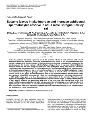
- 4. 322 Sci. Res. Essays X400 X100 A B C Figure 1. Showing the micrographs of the animals. A. The top micrographs showed the control group at X400 and X100 magnification. B. The middle micrographs showed evidence of spermatocytes fullness in the varying tubular lumen of the low dose epididymes observed at X400 and X100 magnification. C. The lower micrographs showed similar features like the low dose groups but with a fuller tubular lumen seen when compared to the control groups (at the top) at both X400 and X100 magnifications. Table 3. Summary of hormonal profile of the animals. Hormone (ng/ml) Group A (Control) Group B (High dose) Group C (Low dose) TEST 0.8 ± 0.03 0.9 ± 0.2* 0.1 ± 0.0* in the high dose as compared to the low dose with no significant weight difference in the relative epididymo- somatic weight (weight/100 g body weight) of the high dose compared to control. However, the low dose weight was significantly lower as observed in Table 2. The epidi- dymal lumens in the sesame treated were wider, fewer with irregular tubular formation and significantly filled with spermatocytes when compared to the control group whi- ch are relatively numerous with smaller luminal sizes as shown in the Figure 1. There is significant (P > 0.05) increases in testosterone level compared to control, however, the low dose was also significantly different from the control as shown in Table 3. This is the first study, to our knowledge, to look at the role of sesame lignans on the male reproductive tracts after extensive literature review. There is increasing role of sesame lignans research contribution to medicine. However sesame, being rich in trace elements or mine- rals, vitamins, antioxidant lignans (phytoestrogens), have the ability of improving fertility potential of the male repro- ductive tract but its mechanism of action need to be elucidated in this study. The beneficial effects derived from the high intake of fruits and vegetables on the various metabolic disease conditions (such as diabetes mellitus, obesity, heart dis- eases and cancer), that may impact on one’s reproduc- tive life may not be due to the effect of their well charact-
- 5. erized antioxidants, such as -carotene, vitamins C and E only, but rather to some other antioxidants or non-antioxi- dant phytochemicals or by an additive actions of the diff- erent compounds present in foods (fruits and vegetables) such as alpha-linolenic acid (poly unsaturated fatty acids), various phenolic compounds (sesamin, sesamol) and fibres, which are present in sesame leaves for exam- ple (Shittu et al., 2006). All the animals that were used in this study, irrespective of their aggressive nature, exhibited calmness (non-aggr- essive state) during treatment. More so, there was a general state of well-being observed in all the animals during the whole experimental period. This may reflect the positive effect of some of the active agents present in the plant on their neuropsychological and neuro-endo- crine pathways. The sesame-treated rats showed signifi- cant weight gain (P < 0.05, using ANOVA), unlike in other studies where no significant difference in the animal wei- ghts was observed (Awoniyi et al., 1997). The relative epididymal weight difference is a reflection of the site of action of the sesame estrogenic lignans whi- ch may also be hormonally influenced. Since the efferent ductile and epididymis of rats are rich in estrogens recap- tors; α and sub-types (Hess et al., 1997; Hess and Car- nes, 2004) just like any other part of the body such as uterus and mammary glands among others (Williams et al., 2000; Nie et al., 2002), any disruption of these receptors as a result of abnormalities in the efferent ductules or epididymis have been hypothesized to lead to impairments of male fertility in mice (Hess, 1997; Hess and Carnes, 2004). However, efferent ductile or epididy- mis is also richer in androgen receptor, the site for action of the testosterone (TT), dihydrotestosterone (DHT) and probably estradiol (E2) (Oliveira et al., 2003). Testosterone level in the high dose sesame was signi- ficantly higher than the control and the low testosterone level observed in the low dose group could be due to the fact that some of the testosterone were aromatized to estradiol and/or converted to dihydrotestosterone by the aromatase and reductase enzymes present within the epididymis. Moreover, Huang et al. (1987) demonstrated that as little as 25% of normal testicular testosterone con- centration is sufficient to support all stages of spermato- genesis and that consistency in testosterone concentra- tion is important for normal spermatogenesis to occur as evidence by the low dose epididymal lumen with fuller spermatocytes compared to control in the present study. Moreover, sesame phytoestrogenic lignans will tend to promote aromatization of testosterone to estradiol, such that, the low dose sesame will make available less endo- genous estradiol and compete less, although there is sy- nergism at this level between the testosterone and estra- diol to favour spermatogenesis. However, reverse is the case for the high dose, which will cause more estradiol production and compete more with dihydrotestosterone for aromatization to occur in its favour if possible. The low testosterone observed here is not as a result of destruct- Shittu et al 323 tion of the Leydig cells but a reflection of the complex hormonal interplay at the level of the hypothalamic- pituitary-testicular axis. Brown and Chakraborty (1992) have suggested that agent like clomiphene will decrease the synthesis and/or release of gonadotrophins such that decreased serum LH (Luitenising Hormone) and testosterone concentration (ng/ml) were found in male rats. However, low dose est- radiol is also needed for spermatogenesis than high dose as evidence in the present study. Little is known about the mechanism of action of sesa- me phytoestrogen lignans with antioxidative properties. But, contrary to speculation that phytoestrogen disrupt male reproductive development through mechanisms wh- ich are independent of the estrogens receptors, in which there is estradiol (E2)-induced transactivation of the and- rogen receptor (AR) (Degen and Metzler, 1987). We are able to demonstrate that sesame lignans has no negative impact on the male reproductive tract through its binding on the estrogen receptors but also modulate the activity of the androgen receptor in the epididymis, thereby ultim- ately influencing the hypothalamic-pituitary-testicular pa- thway as evidence in this study. In general, aromatase has not been found in rat testis, efferent ductules, epididymis or vas deferens (Hess and Carnes, 2004). However, various reports are found on the epididymal presence of aromatase in human efferent ductules and proximal epididymis (Carpino et al., 2004) and cultured rat cells, (Wiszniewska, 2002). We also hypothesized that the action of sesame on the -estrogens receptors as an agonist is more pronounced than that of the α-estrogens receptors with transactivation of the androgen receptor together with its antioxidative property have all contributed to enhancing spermato- genesis with improve male fertility (through production of quality spermatocytes) found in this study. However, stu- dy is on to confirm this hypothesis further. Conclusion Sesame improve epididymal sperm reserve with larger spermatocytes being produced in a dose related manner as evidence in this study through a complex hormonal interplay at the level of the male hypothalamic-pituitary- testicular axis and estrogens receptors. ACKNOWLEDGEMENTS We wish to appreciate the staff of the department of Ana- tomy, LASUCOM, especially Mr. Solomon Adepoju; staff of Help Laboratory, Maryland, Lagos and Drug Quality Laboratories, LASUTH, Ikeja, Lagos. REFERENCES Akpan-Iwo G, Idowu AA , Misari, SM. (2006). Collection and evaluation of sesame (Sesamum spp.) germplasm in Nigeria. IGPR/FAO. 142: 59-62.
- 6. 324 Sci. Res. Essays Alade PI, Irobi ON (1993). Antimicrobial activities of crude leaf extract of Acalypha wilkensiana. J. Ethanopharmacol 39:171-174. American Physiological Society. (2002).Guiding principles for research involving animals and human beings. Am. J. Physiol. Regul. Integr. Comp. Physiol. 283: R281-R283. Awoniyi CA, Santulli R, Chandrashekar V, Schanbacher BD, Zirkin BR. (1997). Neonatal exposure to coumestrol, a phytoestrogen, does not alter spermatogenic potential in rats. Endocrinol. 125: 1303–1309. Bankole Munir A, Shittu Lukeman AJ, Ahmed Titilade A, Bankole Marian N, Shittu Remilekun K, Kpela Terkula, Ashiru Oladapo A (2007). Synergistic Antimicrobial Activities of Phytoestrogens In Crude Extracts Of Two Sesame Species Against Some Common Pathogenic Microorganisms, Afr. J. Trad. CAM 4 (4): 427-433. Brown JL, Chakraborty PK (1988). Characterization of the effects of clomiphene citrate on reproductive physiology in male rats of various ages. Acta Endocrinol. 118(3): 437-443. Carpino A, Romeo F, Rago V (2004). Aromatase immunolocalization in human ductuli efferentes and proximal ductus epididymis. J. Anat. 204: 217-220. Clulow J, Jones RC, Hansen LA, Man SY (1998). Fluid and electrolyte reabsorption in the ductuli efferentes testis. J. Reprod. Fertil. Suppl. 53:1-14. Collins BM, Mclachlan JA, Arnold S (1997). The estrogenic and antiestrogenic activities of phytochemicals with the human receptor expressed in yeast. Steroids 62: 365–372. Degen GH, Metzler M (1987). Sex hormones and neoplasia: genotoxic effects in short term assays. Arch. Toxicol. Suppl. 10: 264–278. Hess RA, Gist DH, Bunick D, Lubahn DB, Farrell A, Bahr J, Cooke PS and Greene GL (1997). Estrogen receptor (a & b) expression in the excurrent ducts of the adult male rat reproductive tract. J. Androl. 18: 602-611. Hess RA, Carnes K ( 2004). The role of estrogen in testis and the male reproductive tract: a review and species comparison. Anim. Reprod. 1: 5-30. Hirose N, Doi F, Ueki T, Akazawa K, Chijiiwa K, Sugano M, Akimoto K, Shimizu S, Yamada H (1992). Anticancer Res., 12: 1259. In: Home Cooking1998. Sesame seeds. www.homecooking.about.com/libra- ry/weekly/aa060898.htm. accessed July 2006.Hou RC, Wu CC, Yang CH, Jeng KC ( 2004). Neurosci. Lett., 367: 10. Jeng KCG, Hou RCW. Sesamin and Sesamolin: Nature`s therapeutic lignans. Current Enzymes Inhibition, 1: 11-20. Huang HFS, Marshall GR, Rosenberg R, Nieschlag E. (1987). Restoration of spermatogenesis by high levels of testosterone in hypophysectomized rats after long-term regression. Acta Endocrinol (Copenh) 116:433– 444. Izegbu MC, Ojo MO, Shittu LAJ. (2005). Clinico-pathological patterns of testicular malignancies in Ilorin, Nigeria-a report of 8 cases. Journal of Cancer Research and Therapeutics, Vol. 1(4): 229-231 Jeng KCG, Hou RCW. ( 2005). Sesamin and Sesamolin: Nature`s therapeutic lignans. Current Enzymes Inhibition, 1: 11-20. In: Jeng KCG, Hou RCW. Sesamin and Sesamolin: Nature`s therapeutic lig- nans. Current Enzymes Inhibition, 1: 11-20, 2005. Kang M.H, Kawai Y, Naito M, Osawa, T. J. (1999). Nutr. 129:1885. In: Jeng KCG, Hou RCW. Sesamin and Sesamolin: Nature`s therapeutic lignans. Current Enzymes Inhibition, 1: 11-20, 2005. Mitra J and Chowdhury M. (1994). Association of glycerylphosphoryl choline with human sperm and effect of capacitation on their metabolism. Reprod Fertil Dev. 6:679–685. Nie R, Zhou Q, Jassim E, Saunders PT and Hess RA (2002). Differential expression of estrogen receptors a and b in the repro- ductive tracts of adult male dogs and cats. Biol. Reprod. 66: 1161- 1168. Oliveira C, Nie R, Carnes K, Franca LR, Prins GS, Saunders PTK, Hess RA. (2003). The antiestrogen ICI 182,780 decreases the expression of estrogen receptor alpha but has no effect on estrogen receptor- beta and androgen receptor in rat efferent ductules. Reprod. Biol. Endocrinol. 1: 75. Orgebin-Crist MC (1967). Sperm maturation in rabbit epididymis .Nature 216: 816-818. Scott TW, Wales RG, Wallace KC, White IG (1963). Composition of ram epididymal and testicular fluid and the biosynthesis of glycerylphosphorylcholine by the rabbit epididymis. J. Reprod. Fertil. 6: 49–59. Sharpe RM. (2003). The 'oestrogen hypothesis'- where do we stand now? Int. J. Androl. 26: 2-15. Sharpe RM, Shakkebeak NE (1993). Are oestrogens involved in falling sperm counts and disorders of the male reproductive tract? Lancet pp. 1392-1395. Shittu Lukeman AJ (2006). The effect of the aqueous crude leaves extract of Sesamum radiatum compared to Mesterolone (proviron) on the adult male Sprague Dawley rats testis and epididymis. MSc Dissertation. Lagos State University, College of Medicine, Ikeja, Nigeria. Shittu LAJ, Bankole MA, Ahmed T, Aile K, Akinsanya MA, Bankole MN, Shittu RK, Ashiru OA (2006). Differential antimicrobial activity of the various crude leaves extracts of Sesame radiatum against some common pathogenic micro-organisms. Sci. Res. Essay 1(3):108-111. Thompson LU, Robb P, Serraino M, Cheung F (1991). Mammalian lignan production from various foods. Nutr. Cancer 16: 43–52. Vos MD, Ellis CA, Bell A, Birrer MJ, Clark GJ (2000). J. Biol. Chem.). 275, 35669-35672. Reinhard Dammann, Takashi Takahashi, Gerd P Pfeifer. (2001). The CpG island of the novel tumor suppressor gene RASSF1A is intensely methylated in primary small cell lung carcinomas. Oncogene, 20 (27): 3563-3567. Wales RG, Wallace JC, White IG (1966). Composition of bull epididymal and testicular fluid. J. Reprod. Fertil. 12: 139–152. Williams K, Saunders PTK, Atanassova N, Fisher JS, Turner KJ, Millar MR, Mckinnell C, Sharpe M (2000). Induction of progesterone receptor immunoexpression in stromal tissue throughout the male reproductive tract after neonatal oestrogen treatment of rats. Mol cell Endocrinol. 16: 117-131. Wiszniewska B (2002). Primary culture of the rat epididymal epithelial cells as a source of oestrogen. Andrologia 34: 180-187.