chaitra-1.pptx fake news detection using machine learning
Termal stab
1. Chemical Papers 68 (1) 121–129 (2014)
DOI: 10.2478/s11696-013-0417-6
ORIGINAL PAPER
Thermal stability, antioxidant activity, and photo-oxidation
of natural polyphenols
Irina Volf, Ioana Ignat*, Mariana Neamtu*, Valentin I. Popa
Faculty of Chemical Engineering and Environmental Protection, “Gheorghe Asachi” Technical University of Iasi,
73 Prof. dr. docent Dimitrie Mangeron Bd., 700050 Ia¸si, Romania
Received 12 January 2013; Revised 25 March 2013; Accepted 5 April 2013
The thermal stability (60◦
C, 80◦
C, 100◦
C), antioxidant activity, and ultraviolet C light (UV-C)
stability of standard polyphenols solutions (catechin, gallic acid, and vanillic acid) and of vegetal
extracts from spruce bark and grape seeds were investigated. Exposure of the standard solutions
and vegetal extracts to high temperatures revealed that phenolic compounds were also relatively
stable (degradations ranged from 15 % to 30 % after 4 h of exposure). The highest antioxidant
activity was obtained for ascorbic acid and gallic acid followed by catechin and caffeic acid and the
grape seeds. The results show that, after 3 h of UV-C exposure, approximately 40 % of vanillic acid,
50 % of gallic acid, and 83 % of catechin were removed. Similar degradation rates were observed
for vegetal extracts, with the exception of the degradation of catechin (40 %) from grape seeds. In
addition, the photo-oxidation of polyphenols in the presence of food constituents such as citric acid,
ascorbic acid, sodium chloride, and sodium nitrate was assessed.
c 2013 Institute of Chemistry, Slovak Academy of Sciences
Keywords: polyphenols, photo-oxidation, thermal stability, radical scavenging activity, food addi-
tives
Introduction
There has recently been a considerable increase in
interest in finding naturally occurring antioxidants to
replace synthetic antioxidants, some of which are be-
ing restricted due to their carcinogenicity. The use
of natural antioxidants also has great potential as a
result of consumers demanding additive-free, fresher,
and more natural-tasting food (Muanda et al., 2011;
Díaz-García et al., 2013).
Antioxidants are compounds that can delay or in-
hibit the oxidation of lipids or other molecules by
inhibiting the initiation or propagation of oxidising
chain reactions. These properties can play an impor-
tant role in adsorbing and neutralising free radicals,
quenching oxygen, or decomposing peroxides (Karou
et al., 2005). Phenolic compounds, one of the most
widely occurring groups of phytochemicals, are of con-
siderable physiological and morphological importance
in plants. Phenolics may act, among others, as phy-
toalexins, anti-feedants, contributors to plant pigmen-
tation, antioxidants, and protective agents against ul-
traviolet (UV) light (Ignat et al., 2011a). Polyphe-
nols have many industrial applications in fields such
as medicine, cosmetics, and the food industry. These
compounds may be used as natural colorants and
preservatives for foods, or as additives in the pro-
duction of paints, paper, cosmetics, and pharmaceu-
tical products (Naczk & Shahidi, 2006; Giusti &
Wrolstad, 2003). Most notably, the antioxidant ac-
tivities of polyphenols are presumed to exert vari-
ous pharmacological effects such as anti-carcinogenic,
anti-mutagenic, and cardio-protective effects, linked to
their free radical scavenging (Parr & Bolwell, 2000;
Casta˜neda-Ovando et al., 2009; Díaz-García et al.,
2013).
In recent years, special attention has been focused
on the isolation of phenolics from different raw materi-
als (medicinal plants, fruits, vegetables, industrial by-
products, and beverages) and on exploration of their
*Corresponding author, e-mail: mariana.neamtu1@yahoo.de, ioana.ignat@gmail.com
A
uthorcopy
2. 122 I. Volf et al./Chemical Papers 68 (1) 121–129 (2014)
potential benefits for human health.
Although these raw materials have been exten-
sively investigated for the isolation of polyphenols,
special attention continues to focus on their extrac-
tion from inexpensive or residual sources. It is well
known that the by-products from industrial processes
still contain a considerable amount of phenolic com-
pounds. Moreover, the large amounts of by-products
resulting annually from vineyards and pulp and paper
mills, along with the use of grape seeds and spruce
bark extracts as food supplements, and alternative
medical products, warrant evaluation of their prop-
erties.
Polyphenols are widely seen as very unstable and
highly susceptible to degradation (B˛akowska et al.,
2003). The stability of polyphenols under different
conditions is a very important aspect which has to be
taken into account to ensure that phenolic compounds
have the desired properties and maintain their activity
and structure during the different stages of processing,
which can involve high temperatures, light, oxygen,
solvents, the presence of enzymes, proteins, metal-
lic ions, or association with other food constituents
(Casta˜neda-Ovando et al., 2009).
UV holds considerable promise in relation to food
processing as an alternative to traditional thermal
processing; however, UV treatment has received less
attention than other non-thermal processing methods.
Applications include pasteurisation of juices, post-
lethality treatment for meats, treatment of food con-
tact surface, and ways to extend the shelf-life of fresh
produce (Koutchma, 2008, 2009; Tikekar et al., 2011a,
2012). Any approach to evaluating UV entails con-
sideration of the properties and composition of the
food product to be treated, the source of the UV ra-
diation, microbial effects, as well as the modelling,
commercial, and economic aspects (Koutchma, 2009).
The use of UV is well established for air, disinfection
of water, and wastewater treatment. The wavelength
of 253.7 nm used in this study is the most efficient
in terms of its germicidal effect, since photons are
most readily absorbed by the DNA of microorgan-
isms at this specific wavelength (Oppenlaender, 2003;
Koutchma, 2009). A significant reduction in the num-
ber of spoilage and human pathogenic microorganisms
has been demonstrated through the use of UV process-
ing (Koutchma, 2009; Tikekar et al., 2011a). Ultravi-
olet C (UV-C) light processing is relatively less costly,
has minimal effects on product flavour, and is adapt-
able to continuous processing methods (Tikekar et al.,
2011a). The Food and Drug Administration (2000)
regulations approved the use of low pressure mercury
(LPM) lamps for juice-processing (Koutchma, 2009).
Little is known about the interaction of UV with com-
plex food matrices. Moreover, the effect of some essen-
tial food additives such as vitamin C, nitrate ions and
salts on the absorption effects of antioxidants needs to
be taken into account during UV treatment.
To date, less attention has been paid to the stabil-
ity of polyphenolic compounds and their degradation
under different conditions. However, these aspects can
influence their potential applications substantially and
might elicit substantial interest in studying the photo-
oxidation and thermal degradation of phenolics.
In the present work, the antioxidant activity, ther-
mal stability, and photo-oxidation of standard solu-
tions of polyphenols (gallic acid, catechin, vanillic
acid) and natural polyphenols (spruce bark and grape
seeds extracts) were investigated. The effects of food
constituents such as citric acid, ascorbic acid, sodium
chloride, and sodium nitrate on the photo-oxidation
of polyphenols were also evaluated.
Experimental
General
Spruce wood bark was provided by a Romanian
pulp and paper company and Merlot grape seeds were
obtained from Panciu vineyard (Vrancea, Romania).
Standard polyphenols (gallic acid, catechin, vanil-
lic acid, siringic acid, p-cumaric acid, ferulic acid, and
sinapic acid) and methanol (HPLC grade) were pur-
chased from Sigma–Aldrich (UK). The other chem-
icals and solvents used were of analytical grade ob-
tained from Merck (Germany) and Sigma–Aldrich
and used without further purification. The mobile
phases for HPLC and aqueous solutions containing
100 mg L−1
standards were prepared with ultrapure
water (conductivity of 0.056 ΩS cm−1
) from a Milli-
pore Waters Milli Q purification unit (France).
Methods
Vegetal extracts were obtained from grape seeds
and spruce bark as raw materials. 50 g of dried ground
material was extracted using distilled water as extrac-
tion solvent, at 70◦
C in a water bath. The extrac-
tion was repeated three times, each time for approxi-
mately 2.5 h, and the extracts were combined and sub-
jected to UV degradation. The initial concentration of
polyphenols was determined by HPLC analysis.
The alcoholic extractions of the two raw materials
were carried out in a Soxhlet installation, over 8 h
at 70◦
C, using ethanol/water (ϕr = 7 : 3) as solvent.
Prior to the HPLC determinations, all the extracts
were concentrated under vacuum and fractioned by
successive liquid–liquid extractions with ethyl acetate.
The organic phases were evaporated to dryness and
diluted in methanol, prior to HPLC determination.
A previously developed reverse-phase high-perfor-
mance liquid chromatographic method (Ignat et al.,
2011b) was used to identify and quantify the phenolic
compounds. The HPLC analysis was performed using
a DionexUltiMate 3000 chromatograph (USA) cou-
pled to a PDA detector. Separations were carried out
A
uthorcopy
3. I. Volf et al./Chemical Papers 68 (1) 121–129 (2014) 123
on a Zorbax RX C18 (USA) (4.6 × 250 mm, particle
size 5 µm) column, operating at 30◦
C with a flow-rate
of 1.2 mL min−1
. The injection volume was 5 µL. The
mobile phase used was 1 vol. % acetic acid in water (A)
vs. methanol (B) for a total run-time of 40 min. The
elution conditions were as follows: the vol. % of sol-
vent B was increased linearly from 10 to 40 in 40 min
and then decreased to 10 and maintained for 10 min.
For quantification, standards for external calibration
were used.
Total phenolic content (TPC) was determined us-
ing the Folin–Ciocalteau reagent, with a protocol de-
veloped previously (Hainal et al., 2011). Gallic acid
was employed as a calibration standard and the re-
sults were expressed as gallic acid equivalents (mg of
GAE per 100 g of dried material).
The radical scavenging activity of the natural
extracts was evaluated using the reduction of the
di(phenyl)-(2,4,6-trinitrophenyl)iminoazanium (2,2-
diphenyl-1-picrylhydrazyl, DPPH) radical. The an-
tioxidant activity of the extracts was expressed as
EC50, an equivalent amount of an extract that neu-
tralises 50 % of the radical. The colorimetric assay was
performed using a modification of the method devel-
oped by Almela et al. (2006).
For the DPPH assay, the alcoholic and aqueous
extracts were concentrated and freeze-dried to avoid
interference from the solvents. Thereafter, different
dosages of a 0.5 mg mL−1
methanolic solution of ei-
ther aqueous or ethanolic freeze-dried extracts (25 µL,
50 µL, 100 µL, 200 µL, 300 µL, 400 µL, 500 µL
each) were added to screw-capped glass vials contain-
ing 2 mL of the DPPH. All the volumes were adjusted
to 3.1 mL with MeOH. After a reaction time of 30 min,
the absorbance was measured at 517 nm. The inhi-
bition percentage of the free radical DPPH (I ) was
calculated according to the following equation:
I =
A0 − A
A0
× 100 % (1)
Methanolic solutions of ascorbic acid, gallic acid,
caffeic acid, and catechin were tested as reference an-
tioxidants. The different quantities of the extracts
tested, expressed in micrograms, were plotted on a
dose-inhibition curve.
The photo-degradation experiments were carried
out in a stirred-batch photo-reactor (volume of irradi-
ated solution = 500 mL, optical path length = 4 cm) at
298 K. The lamp was located on the central axis of the
reactor, in a quartz sleeve. The aqueous solutions, pre-
pared as above, were irradiated with a UV-immersed
low-pressure Heraeus mercury lamp TN 15/32 (Her-
aeus Nobelight, Germany) with a nominal output of
15 W, attaining its spectrum at the 254 nm line. The
incident photonic flux (P0 = 1.013 × 10⁻⁵
E s−1
) was
measured by hydrogen peroxide actinometry.
The stability of standard solutions of polyphenols
and vegetal extracts was evaluated at three different
Fig. 1. Typical chromatogram at 280 nm of standard polyphe-
nols (a), of spruce bark ethanolic extract (b), of grape
seeds ethanolic extract (c). Identified compounds: 1 –
gallic acid; 2 – catechin; 3 – vanillic acid; 4 – syringic
acid; 5 – p-coumaric acid; 6 – ferulic acid; 7 – sinapic
acid.
temperatures (60◦
C, 80◦
C, and 100◦
C) for 4 h. Sam-
ples were taken first after 30 min and every hour sub-
sequently and analysed by HPLC in order to establish
the concentration of polyphenols previously exposed
to temperature treatment.
Analysis of variance or R2
values was used to ver-
ify the statistical significance between the treatments
using Microsoft Excel (version 2007). Each data point
represents the average of three measurements ± stan-
dard deviation (5 %).
Results and discussion
Identification and quantification of polyphe-
nols by HPLC
The chromatographic profiles of the standards and
vegetal extracts are shown in Fig. 1.
The analytical polyphenolic composition of the
samples is given in Table 1; it should be noted that
the major compounds identified in the aqueous and
ethanolic fractions for both vegetal materials were gal-
lic acid and catechin. Furthermore, in the spruce bark
extract, vanillic acid was also identified in relatively
A
uthorcopy
4. 124 I. Volf et al./Chemical Papers 68 (1) 121–129 (2014)
Table 1. Concentration of phenolic compounds (mg per 100 g of dried plant) in samplesa investigated
Raw material Type of extract Gallic acid Catechin Vanillic acid TPC/(mg of GAE per 100 g−1)
Grape seeds Aqueous extract 6.12 ± 0.20 44.36 ± 0.10 – 506 ± 5
Ethanolic extract 12.54 ± 0.80 63.60 ± 1.70 – 1368 ± 14
Spruce bark Aqueous extract – 31.00 ± 1.90 39.40 ± 0.20 517 ± 5
Ethanolic extract 10.20 ± 0.30 71.90 ± 2.70 71.90 ± 0.80 1355 ± 12
a) Results represent average values of triplicate determination (n = 3) ± standard deviation. Caffeic acid, siringic acid, p-cumaric
acid, ferulic acid, and sinapic acid were not detected in the studied samples.
high concentrations. As expected, the concentrations
of the compounds were considerably higher in the al-
coholic extracts. Table 1 also shows the total phenolic
content; the high concentration values (up to 1368 mg
GAE 100 g−1
) indicate that the identified compounds
represent only a small percentage (from 9.6 % to 11 %)
of the total phenolic compounds detected. Similar con-
centrations were reported by Neo et al. (2008) and
Kirca and Arslan (2008).
Radical scavenging activity
The radical scavenging activity of natural extracts
and pure compounds was evaluated by DPPH assay.
The antioxidants react with the stable free radical, i.e.,
1,1-diphenyl-2-picrylhydrazyl (deep violet colour) and
convert it to 1,1-diphenyl-2-picrylhydrazine accompa-
nied with discoloration. The degree of discoloration
indicates the free radical scavenging potentials of the
sample/antioxidant (Sarikurkcu et al., 2008).
Determination of an absolute value for the antioxi-
dant activity of an extract is problematic because it is
dependent on the actual concentration of the radical,
its degradation during the analysis or the matrix inter-
ference. Accordingly, the EC50 parameter was used,
which represents the equivalent amount of an extract
that neutralises 50 % of the radical.
The radical scavenging activity of alcoholic and
aqueous extracts was compared with that of some
standard polyphenols (gallic acid, catechin, caffeic
acid), as well as with ascorbic acid, one of the synthetic
antioxidants commonly used in the food industry.
The reduction in absorbance was measured at
517 nm for different quantities of standards and
extracts, and the results were plotted on a dose-
inhibition curve. The resulting linear calibration
curves were used to derive the EC50 value. The equiv-
alent amount of an extract that neutralises 50 % of the
radical is reported in Table 2.
Table 2 shows that the lowest values of EC50,
which indicates the highest antioxidant activity, were
obtained for ascorbic acid and gallic acid, followed by
catechin and caffeic acid. The linear regression shows
a good accord with the experimental data; high R2
values result for almost all the samples.
In the natural extracts, significant differences in
scavenging activity against the DPPH radical were
Table 2. EC50 values obtained for standard compounds and
vegetal extractsa
Samples EC50/µg R2
Ascorbic acid 4.60 ± 0.87 0.998
Gallic acid 1.70 ± 0.32 0.907
Caffeic acid 42.10 ± 1.02 0.993
Catechin 12.70 ± 0.98 0.999
Grape seeds alcoholic extract 45.75 ± 1.09 0.994
Spruce bark alcoholic extract 159.15 ± 3.21 0.983
Grape seeds aqueous extract 76.25 ± 1.12 0.995
Spruce bark aqueous extract 246.00 ± 5.56 0.997
a) Values are expressed as means ± SD of three replicate anal-
yses.
recorded. The antioxidant activity of the plant ex-
tracts is correlated with their phenolic content (Mon-
toro et al., 2006). The DPPH assay indicated that the
grape seeds’ alcoholic extracts had high radical scav-
enging activities, which could be attributed to high
levels of polyphenols (considerable amounts of gallic
acid and catechin). The results are strongly correlated
with the total phenolic content and the HPLC deter-
minations. The alcoholic extracts show a higher rad-
ical scavenging activity than the aqueous ones, but a
slightly lower one than the standard compounds.
Photo-oxidation
The experiments were carried out with solutions of
polyphenols (gallic acid, catechin, and vanillic acid) in
ultrapure water and also with spruce bark and grape
seeds extracts. The selected standards of polyphenols
used in this study correspond to the compounds iden-
tified and quantified in the plant extracts.
The results of the degradation of polyphenols in
the presence of citric acid, ascorbic acid, sodium ni-
trate, and sodium chloride are presented in Table 3
and Fig. 2. The UV-C exposure of standard polyphe-
nols (100 mg L−1
concentration) for 480 min led to
the complete degradation of catechin, whereas a re-
moval of 85 % of gallic acid and 50 % of vanillic
acid was achieved after the same irradiation time. The
degradation of catechin was seen to be complete after
8 h of irradiation, whereas a removal of 85 % of gal-
lic acid and only 50 % of vanillic acid was achieved
A
uthorcopy
5. I. Volf et al./Chemical Papers 68 (1) 121–129 (2014) 125
Table 3. Kinetic parameters for photo-degradation of polyphenols in standard solution and vegetal extracts using a low-pressure
mercury lamp
Gallic acid Catechin Vanillic acid
Experience 10−3 k 10−5 Pabs 10−3 k 10−6 Pabs 10−3 k 10−5 Pabs
min−1 E s−1 min−1 E s−1 min−1 E s−1
Standard solutions
Without additives 5.70 1.007 1.17 7.960 3.70 1.010
In presence of NaCl 4.10 1.007 8.40 8.830 2.60 1.001
In presence of NaNO3 5.80 1.007 9.60 9.496 2.00 1.007
In presence of ascorbic acid 3.40 1.010 1.01 8.400 3.60 1.010
In presence of citric acid 1.50 1.010 4.80 9.510 1.00 1.010
Vegetal Extracts
Grape seeds aqueous extract 5.40 – 2.10 – – –
Spruce bark aqueous extract – – – – 3.70 –
k – Pseudo-first order rate constant, −
d[M]
dt
= k1[M]; Pabs – photonic flux absorbed by components, Pabs = P0(1 − 10−Aλ ).
Fig. 2. Influence of different additives on catechin (a), gallic
acid (b), and vanillic acid (c) upon exposure to UV. Ini-
tial conditions: 100 mg L−1 of compound; × – 0.5 g L−1
NaCl; – 0.5 g L−1 NaNO−
3 ; – 0.5 g L−1 citric acid;
• – 0.5 g L−1 ascorbic acid; – without additives.
after the same irradiation time. The gallic and vanil-
lic acids presented a higher stability under UV ex-
posure, which is in agreement with their highest an-
tioxidant activity. The first order rate constant of
5.9 × 10⁻³
min−1
of gallic acid photolysis was reported
by Benitez et al. (2005), comparable with our result
(5.7 × 10⁻³
min−1
).
In order to evaluate the impact of preservatives
usually used in food products on the antioxidant com-
pounds under UV exposure for 90 min, citric acid,
ascorbic acid, sodium nitrate, and sodium chloride, in
ratios similar to those used in food, were employed as
additives to the polyphenol standard solutions.
After 90 min of irradiation, the removals of gallic
acid amounted to 27 % and 28 % in the presence of
sodium chloride and ascorbic acid and, respectively,
to 40 % in the presence of sodium nitrate and in the
absence of additives. The use of citric acid as additive
led to a lower degradation of the gallic acid (16 %).
A similar tendency was also observed in the case of
catechin and vanillic acid. The corresponding catechin
removals after 90 min of irradiation were 58 % in the
presence of sodium nitrate and ascorbic acid, 53 % in
the presence of sodium chloride and less than 40 %
when citric acid was used as an additive. In compari-
son, the removal rate constituted approximately 65 %
without additives. Vanillic acid presented a higher sta-
bility against UV exposure, even in the presence of
other compounds.
The effect of food additives on the UV stability
of polyphenols was rather low. Nitrate had a low ab-
sorption rate in the UV-C range (200–280 nm), while
the nitrate molar absorption coefficient at 254 nm
was only 4 L−1
M−1
cm−1
. The photolysis of nitrates
leads, in an overall reaction, to the formation of ni-
trite and oxygen. The photolysis of nitrite in the 200–
A
uthorcopy
6. 126 I. Volf et al./Chemical Papers 68 (1) 121–129 (2014)
400 nm region results in the generation of NO. and O..
At pH < 12 O.−
protonates, to form the OH radical
(Mack & Bolton, 1999). The photolysis proceeds via
complex reaction sequences, involving the formation
of intermediary hydroxyl radicals (Mack & Bolton,
1999; Neamtu & Frimmel, 2006; Schindelin & Frim-
mel, 2000; Warneck & Wurzinger, 1988). The yield of
hydroxyl radicals generated upon exposure to UV ra-
diation of nitrate at 253.7 nm is low.
In theory, dissolved chloride ions react with OH
radicals and lead to the generation of ClOH.−
. This
species can decompose to yield chlorine atoms. The
couple Cl./Cl−
has a reduction potential E from 2.2 V
to 2.6 V. Chlorine atoms can add to the C——C dou-
ble bonds of the compounds present, thus generating
chlorinated hydrocarbons (Openlaender, 2003). Sajiki
and Yonekubo (2004), reported chemical degradation
of bisphenol A (BPA) in the presence of chloride ions
by reactive oxygen species (ROS) such as hydroxyl
radicals. They also suggested that NaCl could enhance
the degradation in the presence of ROS.
Ascorbic acid, also known as vitamin C, is natu-
rally present in some fruit juices or is added as an
antioxidant to minimise losses in colour, flavour, and
nutrients during processing and storage (Koutchma,
2009; Tikekar et al., 2011b, 2012). It is a unique radical
scavenger and is known to enhance the oxygen uptake
(Φ−O2). When exposed to UV radiation, molecular ex-
citation is induced and the photochemical degrada-
tion reactions occurring include multiple free radical
reactions through the formation of ascorbyl radicals.
The ascorbyl radicals generated upon exposure to UV
radiation have a long half-life (≈ 50 s) and persist
even during storage in the dark for a certain period,
continuing to degrade the compounds (Tikekar et al.,
2011b, 2012). The present results are consistent with
this. The presence of ascorbic acid has been shown to
slightly accelerate the oxidation of catechin.
Fig. 3 shows the results obtained for the UV-C ex-
posure of vegetal extracts versus the standard com-
pounds aqueous solutions. Over 180 min of irradia-
tion, approximately 60 % of the gallic acid and more
than 50 % of the vanillic acid was degraded.
Some differences were observed for catechin. Af-
ter 3 h of irradiation, the removal of catechin from
the grape seeds extract was 40 % whereas, in the
case of the standard compound, it reached 90 %.
The results may be explained by the high ini-
tial concentrations of catechin in the analysed ex-
tract. The initial concentration of catechin in the
analysed grape seeds extract was 275 mg L−1
,
while the concentration in the standard solution was
100 mg L−1
.
The results show that the polyphenols’ standard
solutions degraded faster under UV than the vege-
tal extracts. This can be explained by the presence
of complex matrices in the plant extracts, such as
co-pigments. Similar results have been reported by
Fig. 3. Stability against UV irradiation of catechin (a), gallic
acid (b), and vanillic acid (c) from vegetal aqueous ex-
tracts: Initial conditions: 161.69 mg L−1 of gallic acid;
275.55 mg L−1 of catechin; and 149.11 mg L−1 of vanil-
lic acid; – spruce bark extract; – standard.
other authors. B˛akowska et al. (2003) previously in-
vestigated the influence of UV irradiation on the sta-
bility of cyanidin and anthocyanin (AC) – natural
polyphenol co-pigments responsible for some colours
of fruit, vegetables, and other plant tissues. They
found that the presence of co-pigments inhibited the
degradation effect of UV. The presence of co-pigments
in natural extracts prevented the UV degradation
and stabilised the polyphenols in the vegetal ex-
tracts.
A
uthorcopy
7. I. Volf et al./Chemical Papers 68 (1) 121–129 (2014) 127
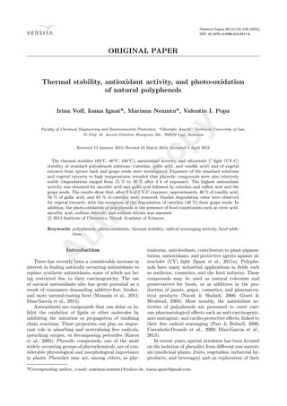
Fig. 4. Thermal degradation of standard polyphenols; gallic
acid (•), catechin ( ), and vanillic acid ( ) at 60◦C (a),
80◦C (b), and 100◦C (c). Initial conditions: 100 mg L−1
of gallic acid; catechin; and vanillic acid.
Thermal degradation
The thermal stability of polyphenols is crucial and
may be correlated with both the extraction and char-
acterisation methods, and the recommendations on
their fields of use. Figs. 4 and 5 show the stability
of gallic acid, catechin, and vanillic acid at different
temperatures.
The data obtained showed a good correlation with
the UV stability. Thus, catechin was the most un-
Fig. 5. Thermal degradation of natural polyphenols; gallic
acid (•), catechin ( ), and vanillic acid ( ) at 60◦C
(a), 80◦C (b), and 100◦C (c). Initial conditions:
161.69 mg L−1 of gallic acid and 275.55 mg L−1 of
catechin from grape seeds extract; 149.11 mg L−1 of
vanillic acid from spruce bark extract.
stable compound, its degradation rate being approx-
imately 20 % at 60◦
C, increasing to 32 % at 100◦
C.
Gallic acid and vanillic acid exhibited a higher sta-
bility, their degradation rates being almost similar at
60◦
C and 80◦
C, with degradations of 15 % and 25 %,
respectively. On the other hand, at 100◦
C over 4 h of
exposure, the removal of gallic acid (30 %), catechin
(32 %), and vanillic acid (37 %) was more visible.
In the case of vegetal extracts, the degradation of
the phenolic compounds surveyed was lower for all the
A
uthorcopy
8. 128 I. Volf et al./Chemical Papers 68 (1) 121–129 (2014)
temperatures used. Similar results were reported by
Fischer et al. (2013) in investigation of the thermal
stability of ACs in three pomegranate juices. The re-
sult may be explained by the higher concentrations of
phenolics in the natural samples. However, their com-
plex chemical composition should not be disregarded.
Conclusions
The antioxidant activity, thermal and UV-C stabil-
ities of standard polyphenols solutions and of vegetal
extracts were investigated. The radical scavenging ac-
tivity of the alcoholic and aqueous extracts indicates
the highest antioxidant activity for the ascorbic and
gallic acids, followed by catechin and caffeic acid. The
stability of standard polyphenols and vegetal extracts
against UV irradiation is relatively high. The pure
compounds exhibited a good stability rate, even in
the presence of various common additives. The highest
stability under UV light was observed for gallic acid
and vanillic acid. These data correspond with similar
conclusions relating to their radicals-scavenging activ-
ity and thermal degradation.
The response to UV irradiation of the extracts in-
vestigated was analogous to that of the standard com-
pounds, which confirms the stability of polyphenols
against UV irradiation (and temperature treatment).
The effect on UV-C exposure of food additives such as
citric acid, ascorbic acid, sodium chloride, and sodium
nitrate was rather low.
The results reveal that the selected by-products re-
sulting from vineyards and pulp and paper industries
are rich sources of phenolic compounds with a high
radical scavenging activity. This outcome favours the
application of natural extracts as natural additives in
different fields. Further studies are needed to evaluate
the effect of UV light on complex liquid food matrices
and on the quality of foods.
References
Almela, L., Sánchez-Mu˜noz, B., Fernández-López, J. A., Roca,
M. J., & Rabe, V. (2006). Liquid chromatograpic–mass spec-
trometric analysis of phenolics and free radical scaveng-
ing activity of rosemary extract from different raw mate-
rial. Journal of Chromatography A, 1120, 221–229. DOI:
10.1016/j.chroma.2006.02.056.
B˛akowska, A., Kucharska, Z. A., & Oszmia´nski, J. (2003). The
effects of heating, UV irradiation, and storage on stabil-
ity of the anthocyanin–polyphenol copigment complex. Food
Chemistry, 81, 349–355. DOI: 10.1016/s0308-8146(02)00429-
6.
Benitez, F. J., Real, F. J., Acero, J. L., Leal, A. I., & Garcia,
C. (2005). Gallic acid degradation in aqueous solutions by
UV/H2O2 treatment, Fenton’s reagent and the photo-Fenton
system. Journal of Hazardous Materials, 126, 31–39. DOI:
10.1016/j.jhazmat.2005.04.040.
Casta˜neda-Ovando, A., Pacheco-Hernández, M. L., Páez-Her-
nández, M. E., Rodríguez, J. A., & Galán-Vidal, C. A. (2009).
Chemical studies of anthocyanins: A review. Food Chemistry,
113, 859–871. DOI: 10.1016/j.foodchem.2008.09.001.
Díaz-García, M. C., Obón, J. M., Castellar, M. R., Collado,
J., & Alacid, M. (2013). Quantification by UHPLC of total
individual polyphenols in fruit juices. Food Chemistry, 138,
938–949. DOI: 10.1016/j.foodchem.2012.11.061.
Fischer, U. A., Carle, R., & Kammerer, D. R. (2013).
Thermal stability of anthocyanins and colourless pheno-
lics in pomegranate (Punica granatum L.) juices and
model solutions. Food Chemistry, 138, 1800–1809. DOI:
10.1016/j.foodchem.2012.10.072.
Food and Drug Administration (2000). Irradiation in the pro-
duction, processing and handling of food. Federal Register,
65, 71056–71058.
Giusti, M. M., & Wrolstad, R. E. (2003). Acylated antho-
cyanins from edible sources and their applications in food sys-
tems. Biochemical Engineering Journal, 14, 217–225. DOI:
10.1016/s1369-703x(02)00221-8.
Hainal, A. R., Ignat, I., Volf, I., & Popa, V. I. (2011). Transfor-
mation of polyphenols from biomass by some yeast species.
Cellulose Chemistry and Technology, 45, 211–219.
Ignat, I., Volf, I., & Popa, V. I. (2011a). A critical review of
methods for characterization of polyphenolic compounds in
fruits and vegetables. Food Chemistry, 126, 1821–1835. DOI:
10.1016/j.foodchem.2010.12.026.
Ignat, I., Stingu, A., Volf, I., & Popa, V. I. (2011b). Character-
ization of grape seeds aqueous extract and possible applica-
tion in biological systems. Cellulose Chemistry and Technol-
ogy, 45, 205–209.
Karou, D., Dicko, M. H., Simpore, J., & Traore, A. S. (2005).
Antioxidant and antibacterial activities of Polyphenols from
ethnomedicinal plants of Burkina Faso. African Journal of
Biotechnology, 4, 823–828.
Kırca, A., & Arslan, E. (2008). Antioxidant capacity and to-
tal phenolic content of selected plants from Turkey. Interna-
tional Journal of Food Science and Technology, 43, 2038–
2046. DOI: 10.1111/j.1365-2621.2008.01818.x.
Koutchma, T. (2008). UV-light for processing foods. Ozone:
Science and Engineering: The Journal of the International
Ozone Association, 30, 93–98. DOI: 10.1080/0191951070181
6346.
Koutchma, T. (2009). Advances in ultraviolet light technology
for non-thermal processing of liquid foods. Food Bioprocess
Technology, 2, 138–155. DOI: 10.1007/s11947-008-0178-3.
Mack, J., & Bolton, J. R. (1999). Photochemistry of nitrite
and nitrate in aqueous solution: a review. Journal of Photo-
chemistry and Photobiology A: Chemistry, 128, 1–13. DOI:
10.1016/s1010-6030(99)00155-0.
Montoro, P., Tuberoso, C. I. G., Piacente, S., Perrone, A., De
Feo, V., Cabras, P., & Pizza, C. (2006). Stability and antioxi-
dant activity of polyphenols in extracts of Myrtus communis
L. berries used for the preparation of myrtle liqueur. Journal
of Pharmaceutical and Biomedical Analysis, 41, 1614–1619.
DOI: 10.1016/j.jpba.2006.02.018.
Muanda, F. N., Soulimani, R., Diop, B., & Dicko, A. (2011).
Study on chemical composition and biological activities of
essential oil and extracts from Stevia rebaudiana Bertoni
leaves. LWT – Food Science and Technology, 44, 1865–1872.
DOI: 10.1016/j.lwt.2010.12.002.
Naczk, M., & Shahidi, F. (2006). Phenolics in cereals, fruits
and vegetables: Occurrence, extraction and analysis. Journal
of Pharmaceutical and Biomedical Analysis, 41, 1523–1542.
DOI: 10.1016/j.jpba.2006.04.002.
Neamtu, M., & Frimmel, F. H. (2006). Degradation of endocrine
disrupting bisphenol A by 254 nm irradiation in different
water matrices and effect on yeast cells. Water Research, 40,
3745–3750. DOI: 10.1016/j.watres.2006.08.019.
Neo, Y. P., Ariffin, A., Tan, C. P., & Tan, Y. A. (2008). De-
termination of oil palm fruit phenolic compounds and their
A
uthorcopy
9. I. Volf et al./Chemical Papers 68 (1) 121–129 (2014) 129
antioxidant activities using spectrophotometric methods. In-
ternational Journal of Food Science & Technology, 43, 1832–
1837. DOI: 10.1111/j.1365-2621.2008.01717.x.
Oppenlaender, T. (2003). Photochemical purification of water
and air. New York, NY, USA: Wiley-VCH.
Parr, A. J., & Bolwell, G. P. (2000). Phenols in plant and
in man. The potential for possible nutritional enhancement
of the diet by modifying the phenols content or profile.
Journal of the Science of Food and Agriculture, 80, 985–
1012. DOI: 10.1002/(sici)1097-0010(20000515)80:7<985::aid-
jsfa572>3.0.co;2-7.
Sajiki, J., & Yonekubo, J. (2003). Leaching of bisphenol A
(BPA) to seawater from polycarbonate plastic and its degra-
dation by reactive oxygen species. Chemosphere, 51, 55–62.
DOI: 10.1016/s0045-6535(02)00789-0.
Sarikurkcu, C., Tepe, B., Daferera, D., Polissiou, M., & Har-
mandar, M. (2008). Studies on the antioxidant activity of
the essential oil and methanol extract of Marrubium globo-
sum subsp. globosum (lamiaceae) by three different chem-
ical assays. Bioresource Technology, 99, 4239–4246. DOI:
10.1016/j.biortech.2007.08.058.
Schindelin, A. J., & Frimmel, F. H. (2000). Nitrate and natural
organic matter in aqueous solutions irradiated by simulated
sunlight. Environmental Science and Pollution Research, 7,
205–210. DOI: 10.1007/bf02987349.
Tikekar, R. V., Anantheswaran, R. C., & LaBorde, L. F.
(2011a). Ascorbic acid degradation in a model apple juice
system and in apple juice during ultraviolet processing
and storage. Journal of Food Science, 76, H62–H71. DOI:
10.1111/j.1750-3841.2010.02015.x.
Tikekar, R. V., Anantheswaran, R. C., Elias, R. J., & LaBorde,
L. F. (2011b). Ultraviolet-induced oxidation of ascorbic acid
in a model juice system: Identification of degradation prod-
ucts. Journal of Agricultural and Food Chemistry, 59, 8244–
8248. DOI: 10.1021/jf201000x.
Tikekar, R. V., Anantheswaran, R. C., & LaBorde, L. F. (2012).
Patulin degradation in a model apple juice system and in
apple juice during ultraviolet processing. Journal of Food
Processing and Preservation. DOI: 10.1111/jfpp.12047. (in
press)
Warneck, P., & Wurzinger, C. (1988). Product quantum yields
for the 305-nm photodecomposition of nitrate in aqueous so-
lution. Journal of Physical Chemistry, 92, 6278–6283. DOI:
10.1021/j100333a022.
A
uthorcopy