Call Girls Kochi Just Call 9907093804 Top Class Call Girl Service Available
j.1476-4431.2011.00628.x.pdf
1. State-of-the-Art-Review
Ureteral obstructions in dogs and cats: a review
of traditional and new interventional diagnostic
and therapeutic options
Allyson C. Berent, DVM, DACVIM
Abstract
Objective – To describe and review both traditional and newer diagnostic and therapeutic options for canine
and feline ureteral obstructions currently being performed clinically in veterinary medicine.
Data Sources – A Medline search with no date restrictions was used for this review.
Human Data Synthesis – The human literature would support the use of minimally invasive endourological
techniques for the treatment of nearly all causes of ureteral obstructions, whenever possible. This typically
includes extracorporeal shockwave lithotripsy, intracorporeal lithotripsy via retrograde ureteroscopy or ante-
grade percutaneous nephroureterolithotomy, ureteral stenting, percutaneous nephrostomy tube placement,
and laparoscopic endopyelotomy. Typically open surgery is only suggested in cases of ureteral or gyne-
cological malignancy when en bloc resection is considered a good option, or when various methods of
endourological techniques have failed.
Veterinary Data Synthesis – The veterinary literature is scarce on the use of interventional endourological
techniques for the treatment of ureteral obstructions and has been growing over the last 5 years. The current
literature reports the use of extracorporeal shockwave lithotripsy for ureteral stones, as well as the use of ureteral
stents for the treatment of trigonal obstructive transitional cell carcinoma, ureterolithiasis, and ureteral strictures.
Traditional surgical interventions, like ureterotomy, ureteronephrectomy, and ureteral reimplantation is more vastly
reported and accepted. This review will focus on new clinical data using interventional endourological techniques
for ureteral obstructions.
Conclusions – Various treatment options for ureteral obstructions are now available for veterinary patients,
and the trend away from traditional surgical techniques will hopefully be followed now that they are
technically and clinically available for dogs and cats.
(J Vet Emerg Crit Care 2011; 21(2): 86–103) doi: 10.1111/j.1476-4431.2011.00628.x
Keywords: interventional endoscopy, interventional radiology, lithotripsy, nephrostomy tubes, ureteral stents,
ureteral stricture
Introduction
Ureteral obstructions can be difficult to diagnose and
treat in veterinary medicine. The increasing incidence of
ureteral obstructions in veterinary practices, combined
with the invasiveness and morbidity associated with
traditional surgical techniques, makes the use of newer
interventional options appealing. Interventional radio-
logic (IR) and interventional endoscopic (IE) techniques
have enabled clinicians to simultaneously diagnose and
treat ureteral disease in an expedited and minimally in-
vasive manner. The technology involved in these tech-
niques includes the use of fluoroscopy, often with flexible
and rigid endoscopy, for ureteral visualization and to
access various parts of the body. This review will focus
on the application of both IR and IE as tools to diagnose
and manage ureteral obstructions and aid in an algorith-
mic approach to these types of cases. It is important to
understand that most of the data pertaining to IR/IE
treatment of ureteral obstructions are new and still con-
sidered investigational in nature. A large proportion of
this review article is based on the author’s experience,
opinions, and data which have only been published
and presented in abstract form. Traditional therapeutic
options will be reviewed in addition to new alternatives
in dealing with these challenging cases.
The author declares no conflict of interest.
Address correspondence and reprint requests to
Dr. Allyson C. Berent, The Animal Medical Center, 510 E. 62nd St,
New York, NY 10065, USA.
Email: allyson.berent@amcny.org
Submitted August 16, 2010; Accepted January 27, 2011.
From The Animal Medical Center, 510 E. 62nd St, New York, NY 10065.
Journal of Veterinary Emergency and Critical Care 21(2) 2011, pp 86–103
doi:10.1111/j.1476-4431.2011.00628.x
& Veterinary Emergency and Critical Care Society 2011
86
2. Equipment
Various flexible and rigid endoscopes are needed for
the following endourologic ureteral procedures. Rigid
cystoscopy is used for female (dog or cat) ureteral in-
tervention. Ureteral access is possible in a retrograde
fashion in most female patients via IR techniques. Rigid
endoscope diameters range from 1.9 to 7.5 mm. Flexible
ureteroscopes (7.5–8.2-Fr) are used for cystoscopic ac-
cess in male dogs and ureteroscopy in both male and
female dogs, when possible. Different types of intra-
corporeal lithotrites and lasers are available including:
holmium:YAG, diode, ultrasonic, pneumatic, and elec-
trohydraulic. These are typically used for stone disease
or tumor ablation when treating ureteral obstructions.
In addition, extracorporeal shock wave lithotripsy
(ESWL) has great application for ureteroliths in dogs
and sometimes, but less commonly, cats.1–3
A traditional fluoroscopic C-arm is sufficient for visu-
alization and ureteral intervention. A mobile C-arm has
the ability to move the image intensifier and gain various
tangential views of the renal pelvis and ureter. Ultra-
sonography is useful for percutaneous renal access in
order to cannulate the ureter in an antegrade manner, or
for the placement of a nephrostomy tube. This can also be
done under direct fluoroscopic guidance when the renal
pelvis is already opacified with contrast. Various guide-
wires and catheters are also needed for each procedure,
and a discussion of these can be found elsewhere.4
Uret-
eral stents, which are available in numerous dimensions,
are soft polyurethane-type catheters that have a double
pigtail multifenestrated design. They can be easily re-
moved after resolution of ureteral disease if necessary,
but are often considered long-term treatment options.
Additional interventional techniques that will be dis-
cussed for the management of ureteral disease include
the placement of nephrostomy tubes and the use of a
subcutaneous ureteral bypass (SUB).
Etiology
Ureterolithiasis is the most common cause of ureteral ob-
structions in both dogs and cats,5,6
though trigonal ne-
oplasia,a,7
ureteral strictures (congenital or acquired),b
dried solidified blood clots/calculi8
or other tumor types
have also been reported.9
Greater than 98% of feline, and
over 50% of canine ureteroliths were recently docu-
mented to be composed of calcium oxalate.5,6,10–12
These
types of stones do not dissolve medically and either need
to pass spontaneously, be removed, or managed, to per-
mit urine passage. Once medical management fails (tra-
ditionally this involves, eg, IV fluid therapy, mannitol
continuous rate infusions [CRI], and a-adrenergic block-
ade, amitriptyline, glucagon), partial obstructions were
traditionally tolerated and often left untreated (benign
neglect) due to the risk:benefit ratio of attempted surgical
removal. If there is a complete ureteral obstruction, de-
compression of the renal pelvis was typically encouraged.
This approach to management has more recently changed
in the author’s practice and immediate treatment of par-
tial ureteral obstructions is typically recommended.
The physiologic response to a ureteral obstruction is
very complex both before and following its relief. Fol-
lowing a ureteral obstruction studies in normal dogs have
demonstrated that ureteral pressures increase immedi-
ately and can take over 24 hours after obstruction relief
for the pressure to decrease.13
After this increase in pres-
sure renal blood flow diminishes to 40% of normal over
the first 24 hours and drops to 20% of normal by 2
weeks.14,15
The excessive pressure is transmitted to the
entire nephron and a decrease in glomerular filtration
rate (GFR) occurs via concurrent vasoactive mediator re-
lease, leukocyte influx, and subsequent fibrosis.14
The
contralateral kidney will have an increase in the GFR in
response. The longer the ureter remains obstructed, the
more damage occurs, which may be irreversible depend-
ing on severity and duration of the obstruction. In a study
of normal dogs, it was found that after 7 days of ob-
struction the GFR was permanently diminished by 35%,
and when the obstruction lasted for 14 days the GFR was
diminished by 54%.13–18
These numbers were obtained in
a canine model with complete obstruction, without pre-
existing azotemia, chronic interstitial nephritis, or chronic
obstructions, so extrapolation of a worse outcome might
be expected in dogs and cats that are obstructed, making
aggressive and timely intervention imperative. It was also
documented in these studies that it took over 4 months to
get the maximal return to function.16–18
Interestingly, in
contrast to the irreversibility of a complete obstruction,
partial obstructions resulted in less severe destruction
with greater return of function after the obstruction is
relieved. In 1 dog model it was found that the GFR re-
turned to normal after a partial obstruction was present
for 4 weeks.13
Knowing that many patients are partially
obstructed with concurrent renal compromise, eg, chronic
renal disease (CKD), aggressive management and reso-
lution is recommended to improve overall outcome.
The normal diameter of the canine ureter based on
computed tomography (CT) measurements ranges from
1.3 to 2.7mm (3.9–8.1-Fr).19
The normal internal diam-
eter of the feline ureter is approximately 0.4 mm (1.2-Fr),
whereas the outside diameter reaches 1 mm (3-Fr).20–24
The ureter sits in the retroperitoneal space connecting
the renal pelvis to the urinary bladder. It is lined with
transitional cell epithelium and composed of several
layers of smooth muscle surrounding the mucosal layer,
allowing the peristalsis of the ureter and urine propul-
sion from the kidney to the urinary bladder.20,21
In the
dog and cat the ureteropelvic junction is covered by re-
nal parenchyma. Once the ureter exits the kidney it
& Veterinary Emergency and Critical Care Society 2011, doi: 10.1111/j.1476-4431.2011.00628.x 87
Image-guided interventions in ureteral obstructions
3. passes dorsally. The right ureter typically passes lateral
to the caudal vena cava, whereas the left ureter is usually
lateral to the aorta. In the case where the ureter is seen to
pass dorsal to the caudal vena cava, this is termed a
circumcaval (or retrocaval) ureter. In a normal cat, as the
ureter passes caudally, it passes ventrally, enters the lat-
eral ligament of the bladder and then the distal end
curves resulting in a ‘J-shaped’ hook, entering the dorsal
aspect of the bladder neck at the ureterovesicular junc-
tion (UVJ).22
In cats the UVJ is typically found in the
proximal urethra at the junction of the trigone.
Ureteral interventions in people
In human urology the development and improvements in
ureteroscopy, ureteral stenting, ESWL, laser lithotripsy,
laparoscopy, and percutaneous nephroureterolithotomy
(PCNUL) have almost eradicated the need for open uret-
eral surgery for stone disease, strictures, trauma, neoplasia,
and congenital anomalies.25–35
Currently, ureteroscopy is
the first line modality for the evaluation and treatment of
ureteral neoplasia, upper tract essential hematuria, ureteral
calculi 45mm, and evaluation for ureteral obstructions.
Ureteroliths o5mm have a 98% chance of spontaneous
passage with medical management alone (eg, a-blockade).
For larger stones, or those that do not pass spontaneously,
ESWL is effective in 50–81% of cases, though most of
the literature suggests this number is closer to 50–
67%.25–27,33,35
Ureteroscopy has a near 100% success rate
when a holmium:YAG laser lithotripsy is used.25,26,34
PCNUL has been successful for large proximal impacted
ureteral stones.7
Ureteral stenting was first introduced in
1967 for management of people with malignant ureteral
obstructions.30
They are still widely used to treat both be-
nign and malignant obstructive disease and this is consid-
ered standard-of-care in many instances. There has been
documented success in stent placement for distal malig-
nant obstructions of 496% when placed in an antegrade
manner when nephrostomy access is obtained through the
kidney, versus only 50% when placed in a retrograde
manner.30
Ureteral stenting for stone disease is typically
done after ureteroscopy for the management of postscop-
ing spasm and edema, and in children it has been per-
formed before ureteroscopy to allow for passive ureteral
dilation, immediate ureteral bypass, future ureteroscopy,
and spontaneous stone passage.33
With this understanding
a similar approach is starting to be used in veterinary
medicine over the past 5 years in a few veterinary facilities.
History and clinical presentation
Feline patients with ureteral obstruction(s) typically pres-
ent with vague signs associated with vomiting, lethargy,
a decreased appetite, and acute or chronic weight loss.5
If
the patient is severely azotemic then signs of uremia may
be present such as polyuria, polydipsia, vomiting, ano-
rexia, oral ulcerations, weakness. Unless there are con-
current bladder or urethral stones, or a trigonal mass,
signs of dysuria are not commonly observed. Concurrent
urinary tract infections are not as common in cats (ap-
prox 33%)4
as in dogs (77%),12
and ureteral colic can be
associated with signs of dysuria or stranguria, but this
appears to be less common in cats than dogs. Pain on
palpation of the affected kidney is more commonly seen
in acute obstructions.
Clinical presentation in dogs with a ureteral obstruc-
tion is typically associated with dysuria (incontinence,
stranguria, hematuria, polyuria, pollakiuria) and signs of
systemic illness (eg, vomiting, inappetance, depression,
lethargy). Most dogs (77%) with a ureteral obstruction
have associated pyelonephritis and cystitis. This is per-
haps why more dogs have generalized signs of systemic
illness.12
Dramatic signs of lower urinary tract disease are
more commonly associated with ureteral colic, cystitis,
or concurrent lower urinary tract disease (eg, trigonal
tumor, urethral or bladder stones, polyps, masses).
Diagnosis
On physical examination it is common to palpate 1 en-
larged kidney and 1 small kidney in cats. In dogs renal
pain is more common and is typically associated with
the concurrent pyelonephritis and capsular inflamma-
tion. Pallor and associated anemia is common in cats,
and evidence of a heart murmur is often auscultated.
Biochemical parameters: Cats are often anemic (48%)
at diagnosis and this is either due to concurrent CKD or
from excessive blood sampling during previous hospi-
talizations.5
Dogs often have a moderate to severe neu-
trophilia associated with concurrent pyelonephritis and
44% of dogs with ureterolithiasis-induced obstructions
were reported to have some degree of thrombocytopenia,
which can be quite severe (o40,000 platelets). The
thrombocytopenia may be secondary to sepsis or a
secondary immune-mediated thrombocytopenia.12
Azo-
temia is common at the time of diagnosis (83% of cats and
50% of dogs were azotemic with a unilateral obstruc-
tion).5,6,12
The degree of azotemia does not appear to be
associated with outcome if early decompression is un-
dertaken. Hyperphosphatemia was documented in 54%,
hyperkalemia in 35%, hypercalcemia in 14%, and hypo-
calcemia in 22% of cats with ureteral obstructions.5,6
On
urinalysis crystals were observed in the urine of 29% of
cats (amorphous crystals and calcium oxalate being most
common). Urinary tract infections were documented in
8% of cats in 1 study6
and over 30% of cats in another
study,c
whereas 77% of dogs were documented to have
positive stone or urine bacterial cultures.12
Imaging: Bilateral ureteral obstructions were docu-
mented in 19% of cats5,6
and 12.5% of dogs.12
& Veterinary Emergency and Critical Care Society 2011, doi: 10.1111/j.1476-4431.2011.00628.x
88
A.C. Berent
4. Radiopaque calculi (Figure 1) are typically visible in
patients with calculi-induced obstructions. In some pa-
tients an enema is necessary in order to better visualize
the entire ureter and associated renal pelvis. The com-
bination of radiographs and ultrasound is preferred in
the diagnosis of ureteral obstructions. The benefit of the
radiographs is to document stone size, number, loca-
tion, and the presence of concurrent nephrolithiasis.
These are often underestimated with ultrasound.
Ultrasound is ideal for the documentation of hydro-
ureter, hydronephrosis, and the exact location of the
obstructive lesion. If a trigonal tumor is presently caus-
ing the ureteral obstruction then a hydroureter would
be expected to extend to the level of the UVJ. If hydro-
ureter is present and very proximal, with no evidence
of a shadowing stone at the junction of normal and
abnormal ureter, then one might expect a ureteral stric-
ture to be present. In a recent study 60% of cats with a
ureteral stricture had evidence of peri-ureteral hyper-
echoic tissue at the stricture site on ultrasound.b
Iden-
tifying concurrent nephroliths and other ureteroliths is
vital in therapeutic decision making when considering
traditional surgery, ureteral stenting, or ESWL. One
study documented 62% of cats had concurrent neph-
roliths and 9% cystic calculi, whereas 50% of canine
patients had concurrent nephroliths.5,6,12
Another key
piece of information is knowing the exact diameter of
the dilated renal pelvis based on the ultrasound. It is
important to know which interventional option is best
for each patient and ensure that the loop of a locking-
loop pigtail nephrostomy tube or ureteral stent fits
inside the renal pelvis if that modality is being consid-
ered. The sensitivity for abdominal radiographs for fe-
line ureteral calculi was 81% (versus 88% in dogs) and
for abdominal ultrasound was 77% (versus 100% in
dogs).12
In combination the sensitivity of both radiog-
raphy and ultrasound was 90% in cats.5,6
Percutaneous antegrade pyelography: Percutaneous
antegrade pyelography provides good visualization of
the renal pelvis and ureter, and allows localization of
the ureteral obstruction and aids in determining
whether a complete or partial obstruction is present.36
In brief, the animal is anesthetized, or heavily sedated,
and the area over the kidney is clipped and aseptically
prepared. Using ultrasound guidance and a 22-G IV
catheter the renal pelvis is approached through the
greater curvature of the kidney. The needle is attached
to a 3-way stopcock and extension set. Once inside the
renal pelvis, urine is collected for bacterial culture and
urinalysis and iohexola
is injected (comprising 50% of
the urine volume that was removed). Images should be
obtained immediately and again at 5 and 15 minutes.
Ideally, this procedure should be performed under
fluoroscopic guidance, which allows visualization of
the filling of the ureter and the point of obstruction in
real time. In a study of 11 cats the sensitivity and spec-
ificity was excellent in the cases where the study could
be interpreted.36
Forty-four percent of the studies had
evidence of leakage and 30% was not diagnostic, mak-
ing its use somewhat more limited.36
With the im-
provement in ultrasound imaging, the confidence of
obstruction based on ultrasound and radiographs, the
more aggressive treatment of partial obstructions, as
well as the need for pyelography during treatment
(ureteral stenting, nephrostomy tube placement, or SUB
placement), the use of purely diagnostic antegrade
pyelography has declined tremendously in the author’s
practice, and is instead performed intraoperatively
during obstruction relief (Figure 1).
Retrograde ureteropyelography is performed via cystos-
copy and fluoroscopy by cannulating the UVJ and in-
jecting contrast.d
This procedure allows for irrigation of
contrast in a retrograde manner in order to document
ureteral patency, space occupying lesions, stone dis-
ease, tortuosity of the ureter, and the ureteral diameter.
This is seemingly more accurate than IV pyelography
(IVP), allowing for ureteral distension and higher con-
trast concentrations during ureteral irrigation without
the dilutional effect observed with an IVP and the po-
tential risk of contrast-induced nephropathy. The con-
trast agent remains in the renal collection system and is
not injected intravascularly, having no ill effect on the
nephrons. This procedure is also less invasive than an-
tegrade pyelography, eliminating the need for needle
access and the risk of subsequent bleeding or urinary
leakage through the renal parenchyma if a ureteral ob-
structive lesion persists (Figure 1).
CT: Computed tomography can be performed pre-
operatively when traditional surgical options are
anticipated if the stone number and location is not
clear based on radiographs and ultrasound. IV contrast
administered during the CT scan can aid in concurrent
differentiation of partial or complete obstructions. IVP
is often not useful in animals with ureteral obstructions
due to the poor filling of an obstructed kidney and the
nephrotoxicity risk of the contrast material that is being
filtered by the kidneys during the nephrogram phase,
particularly considering a significant number of both
dogs and cats are azotemic at the time of diagnosis. The
risk is theoretical at this point but should be considered.
In certain instances an IVP might be indicated. With the
advent of ureteral stenting, the location of the stones is
slightly less important if the entire ureter will be by-
passed by a stent, and most of the stones will not ul-
timately be removed. This is not the case when
traditional surgical therapy is being performed.
& Veterinary Emergency and Critical Care Society 2011, doi: 10.1111/j.1476-4431.2011.00628.x 89
Image-guided interventions in ureteral obstructions
5. GFR studies/scintigraphy: The GFR of individual
kidneys can be measured by technetium Tc 99 dieth-
ylenetriamine pentacetic acid scintigraphy. The GFR of
an obstructed kidney is most often reduced, and the
predictability of return to function based on scinti-
graphy is often unclear, potentially significantly under-
estimating postrelief renal function. The measurement
of the GFR of the contralateral kidney may assist in the
decision whether to treat unilateral or bilateral obstruc-
tions, or potentially perform a nephrectomy if abso-
lutely necessary (not typically recommended).22
Treatment
There is a paucity of information reported in the literature
on the treatment of canine or feline ureteral obstructions
aside from a large surgical case series in cats (153 cases)6
and 1 small surgical case series in dogs (16 cases).12
Other
reports are often based on anecdotal experience or a sub-
set of patients from this larger case series.
Medical management: Medical management should
be initiated immediately following diagnosis of a uret-
eral obstruction, as stabilization is often required and a
majority of cats will present with concurrent kidney
disease and azotemia. Medical management alone has
been shown to be effective in a minority of cats with
stone disease in 1 study,5
and there are no reports in
dogs. Only 17% of patients were reported to have
movement of their ureteral stones to either complete or
partial passage with aggressive medical management.6
It is important to remember that movement to a partial
obstruction is not considered resolution of the condi-
tion, and progressive renal damage will likely still en-
sue, albeit at a slower rate. Because there is a small
chance of complete stone passage (typically under
10%), medical management should always be consid-
ered before any more invasive intervention.6
Dogs may
not always present azotemic, but because over 75% of
dogs with a ureteral obstruction had concurrent urinary
tract infections broad spectrum antimicrobial therapy is
indicated.12
It is very important that if medical man-
agement is not successful in relieving the obstruction
within 48–72 hours, as documented via serial radio-
graphs, blood work, and ultrasound examinations,
more aggressive interventions should be considered
in order to avoid excessive loss of renal function.
Medical management should consist of aggressive IV
fluid therapy, being careful to monitor central venous
pressures, body weight, electrolyte concentrations, and
hydration status. It is not uncommon for patients, par-
ticularly cats, to become fluid overloaded both before
and after any ureteral intervention. This is typically
seen preoperatively during a diuresis period or post-
operatively during high IV fluid therapy rates. Care
should be taken to prevent excessive fluid volumes and
monitor hydration status very carefully. The fluid ther-
apy protocol typically recommended by the author
includes administering a maintenance rate of fluids (eg,
Figure 1: Imaging in animals with ureteral obstructions. (A) Lat-
eral abdominal radiograph showing multiple stones (white arrow)
in the ureter of a cat. (B) Ultrasound image of a canine proximal
ureter and renal pelvis showing multiple stones in the proximal
ureter just past the ureteropelvic junction (UPJ). (C) Aventrodorsal
radiograph in a feline patient after a percutaneous antegrade
pyelogram with a proximal ureteral obstruction (stricture-black
arrow). (D) A ventrodorsal projection during a retrograde ureter-
opyelogram using fluoroscopic and cystoscopic guidance in a cat.
White arrow is the open-ended ureteral catheter.
& Veterinary Emergency and Critical Care Society 2011, doi: 10.1111/j.1476-4431.2011.00628.x
90
A.C. Berent
6. 50–60 mL/kg/d) using 0.45% saline mixed with 2.5%
dextrose and then a replacement fluid (typically avoid-
ing saline if possible due to sodium load) to correct
hydration status and promote diuresis (eg, 45–75 mL/
kg/d). Again, careful monitoring of fluid therapy is
very important. In addition to IV fluid therapy the use
of an osmotic diuretic is often recommended. For pa-
tients that do not have any cardiac compromise, a CRI
of mannitol is typically chosen by the author. The
author starts with a bolus at 0.25–0.5 g/kg over 20–30
minutes followed by a CRI at 1 mg/kg/min for 24
hours. If after 24 hours there is no evidence of im-
provement based on imaging this is discontinued.
Other medical considerations include amitriptyline,37
a-adrenergic blockade (prazosin or tamsulosin),38
or
glucagon therapy.e
In cats only anecdotal evidence ex-
ists regarding the medical management strategies dis-
cussed. One study evaluated the response of normal
canine ureters and compared various spasmolytics.38
In
people, a-1 adrenergic antagonists, eg, tamsulosin (a-
1a/1d adrenergic antagonist), was the treatment of
choice for the expulsion of small ureteral stones. It is a
very potent spasmolytic and is very effective in reliev-
ing a ureteral obstruction when stones are distal and
o5 mm in diameter.39
A study in dogs comparing var-
ious antispasmotics including a-adrenergic antagonists
(tamsulosin, prazosin [a-1 non-selective–adrenergic an-
tagonist], an experimental b-2/b-3 adrenergic agonist,
verapamil [a calcium channel blocker], and papaverine
[a phosphodiesterase inhibitor]), showed that the b-
adrenergic agonist had the most efficacious ureteral
relaxant abilities, and tamsulosin was the second most
effective.39
Prazosin had little effect, and was actually
shown that in high concentrations it enhanced sponta-
neous contractions.38
This suggests that further inves-
tigation into the use of a b-adrenergic agonist or
tamsulosin in dogs and cats with ureteral obstructions
is warranted. Amitriptyline is a potent urinary smooth
muscle relaxer mediated by the opening of voltage-
gated potassium channels. One study evaluating
amitriptyline in cats with urethral obstructions demon-
strated that amitriptyline was effective in relieving ure-
thral obstructions and relaxing the muscle of normal
human and porcine ureteral segments.37
This has since
been extrapolated for use in canine and feline ureteral
obstructions. The recommended dose is 1 mg/kg, PO,
per day but no documented clinical evidence has been
reported for its use in veterinary ureteral disease.37
Glucagon is another medical option which theoretically
causes relaxation of the ureteral smooth muscle, pro-
moting passage of ureteral calculi. An abstract evalu-
ating the use of glucagon for ureteral obstructions in 25
catse
demonstrated that glucagon improved urine out-
put in previously oliguric cats but there was no doc-
umented short- or long-term benefit for the ureteral
obstruction.e
Major side effects associated with glu-
cagon include vomiting, diarrhea, dyspnea, and tachy-
pnea. The dose of glucagon reportedly used is 0.1 mg/
cat, IV, every 12 hours for up to 4 doses; however, many
clinicians would recommend against its use, or recom-
mend its use with caution. Another study evaluated
glucagon and ritodrine in a canine model of ureteral
obstruction.40
This study demonstrated a reduction in
intraluminal pressure of the ureter and a reduction in
the peristalsis rate, but the effect was not particularly
prolonged, and was considered clinically irrelevant.40
When medical management fails or the patient is un-
stable (eg, hyperkalemic, excessively overhydrated, or
becoming oliguric) then immediate intervention should
be considered. If immediate resolution (via ureterotomy,
ureteral reimplantation, ureteral stenting, or SUB) is not
possible then the 2 best options are either to place a
nephrostomy tubef
or initiate intermittent hemodialysis
or continuous renal replacement therapy (CRRT). The
author typically recommends immediate renal pelvis
decompression over hemodialysis or CRRT if the patient is
stable to undergo anesthesia and the operator is comfort-
able placing the draining tube. By relieving the obstruc-
tion several goals are accomplished: (1) improvement of
azotemia and electrolyte status, (2) prevention of further
nephron damage from the increased hydrostatic pressure,
(3) alleviation of the ureteral colic caused by the obstruc-
tion, (4) enabling the potential for retrograde migration of
the obstructing stone after ureteral decompression, and
(5) allowing time for a postobstructive diuresis to occur
through the nephrostomy catheter (which is a larger di-
ameter) rather than a smaller diameter ureteral stent or
edematous ureteral surgical site.
Nephrostomy tube placement: A nephrostomy tube
(Figure 2) will rapidly and effectively relieve a ureteral
obstruction, as well as enable the determination of
whether adequate renal function remains before sub-
jecting a patient to a prolonged anesthesia for definitive
ureteral surgery. The catheter recommended for this
procedure is a locking-loop pigtail catheter.g
A study
using a dog model for ureteral obstruction demon-
strated that after a nephrostomy tube was placed 75%
of the dogs passed an implanted artificial steel ball,
which was the stone model, into the urinary bladder.41
A smooth metal ball should pass much easier than an
irregular calculus, but these data may suggest that by
relieving the increased ureteral and renal pelvic pres-
sure the excessive ureteral spasm and edema created by
the stone may be relieved and encourage stone passage.
This is speculative, but possible. In a recent series of 19
dogs and cats, locking-loop nephrostomy tube place-
ment was reported.f
No leakage occurred in any of
& Veterinary Emergency and Critical Care Society 2011, doi: 10.1111/j.1476-4431.2011.00628.x 91
Image-guided interventions in ureteral obstructions
7. these patients, but 1 dog accidentally removed the tube
at home while running out of the crate 5 days after
placement.f
This dog did not have a nephropexy as his
tube was placed percutaneously, and no ill effects were
noted from this incident.f
It is recommended that nephrostomy tubes should be
placed percutaneously in dogs and surgically in cats
due to the mobility of the feline kidney and greater risk
for leakage without a surgical nephropexy. The loop on
the catheter is approximately 10 mm in diameter so this
procedure is reserved for dogs and cats that have a
renal pelvis 410 mm. If the entire loop is not securely
situated inside the renal pelvis then leakage can occur.
The author typically use a 6-Fr catheterg
in dogs and a
5-Fr catheterh
in cats. Percutaneously, this procedure is
performed with ultrasound, fluoroscopic guidance, or
Figure 2: Nephrosotmy tube placement. Seldinger-technique displaying the placement of a locking-loop pigtail catheter over a
guidewire inside the renal pelvis of a feline patient. (A) A pigtail catheter over a guidewire showing it is straight to start. (B) Once the
wire is removed the loop starts to form. (C) Once the string is locked in the loop tightens. (D) Representing the catheter over the
guidewire inside the renal pelvis after a pyelogram is performed. (E) The loop of the pigtail is advanced over the wire as depicted in
(B). (F) The lock is formed inside the renal pelvis, as depicted in (C). (G) One-stab technique showing the sharp trocar inside the
stylette of the locking-loop pigtail catheter. (H) A percutaneous pyelogram in a cat with a proximal ureteral stricture. (I) A locking-
loop pigtail catheter being advanced into the renal pelvis with the sharp trocar (white arrow) through the greater curvature of the
kidney. (J) The locking-loop pigtail catheter in place and locked inside the renal pelvis.
& Veterinary Emergency and Critical Care Society 2011, doi: 10.1111/j.1476-4431.2011.00628.x
92
A.C. Berent
8. both. With ultrasound alone a ‘one-stab’ technique can
be performed. A stab incision is made through the skin
in the location of puncture. The locking-loop pigtail
catheter is straightened by the hollow stylette. Then the
sharp trocar is placed within the hollow stylette (which
come together as a set). This sharp trocar is used to
puncture the greater curvature of the kidney with care-
ful ultrasound guidance. Large vessels should be
avoided with color-flow Doppler when possible. Once
the catheter is seen inside the renal pelvis the sharp
trocar is removed. Now the hollow stylette is carefully
withdrawn from the catheter as the pigtail catheter is
advanced into the renal pelvis. Once the catheter is seen
to start forming a curl the locking string is gently pulled
tight to encourage the pigtail to form and lock inside
the renal pelvis. Once the catheter is in place urine
should be removed from the catheter and emptying of
the renal pelvis should be documented on ultrasound.
At this time a pyelogram can be performed to confirm
that no leakage has occurred. The catheter is securely
sutured to the skin by purse-string suture followed by
finger trap suture pattern. The tube should be tacked to
the skin in 3–4 separate locations using surgical tape or
serial finger traps to secure the tube from any possible
traction to the kidney. The tube should then be wrapped
and secured carefully around the abdomen. This should
remain in place for 2–4 weeks for tract formation, or
the hole can be closed surgically if a laparotomy for
definitive resolution is subsequently performed. If the
tube is placed surgically, which is recommended in cats,
a surgical nephropexy is concurrently performed and the
tube can be removed as soon as the ureteral obstruction is
permanently relieved.
The ‘Seldinger-technique’ for nephrostomy tube
placement requires both ultrasound and fluoroscopic
guidance. For this procedure a pyelocentesis and uretero-
pyelogram is performed using a renal access needlei
un-
der ultrasound guidance. Under fluoroscopic guidance,
an angled guidewirej
is passed through the needle and
coiled into the dilated renal pelvis. The locking-loop pig-
tail catheter is then straightened out with the hollow tro-
car. The renal access needle is removed, and the
nephrostomy tube set is advanced over the guidewire
into the renal pelvis. Once the catheter is observed via
fluoroscopy to be inside the renal pelvis, the catheter is
advanced off the hollow stiff stylette and the curl is
formed over the wire inside the renal pelvis. Once the
curl is in the renal pelvis entirely the pigtail is locked
and the catheter and hollow stylette are removed. The
catheter is secured to the body wall as described above.
The tube should be tested with a pyelogram to ensure
that no leakage is present or that there is no resistance to
drainage. The entire system is then attached to a closed
gravity drainage collection system. This allows for exter-
nal renal drainage, elimination of excessive hydrostatic
pressure, and patient stabilization before a more perma-
nent fixation (eg, ureterotomy, ureteral reimplantation,
ureteral stent placement, ureteral bypass). During ne-
phrostomy tube placement the guidewire maybe be able
to be advanced down the entire ureter around the ob-
struction. If it bypasses the obstruction, and through and
through access is obtained, a ureteral stent can be placed
as detailed later in this review.
Dialysis: Dialysis, including initiate intermittent
hemodialysis and CRRT, may be useful to stabilize pa-
tients with ureteral obstruction. In particular, patients
with severe hyperkalemia or life-threatening volume
overload (ie, pulmonary edema) may benefit from these
types of treatment. Dialysis can make the patient more
stable for anesthesia for a definitive procedure (eg, ure-
terotomy or ureteral stent placement). While placing a
dual lumen catheter for dialysis may require sedation, it
is generally a quick procedure with less morbidity than
surgery or percutaneous nephrostomy tube placement.
The optimal treatment protocol will depend on the spe-
cifics of the case. Because many of these patients suffer
from volume overload and marginal hypotension, slow
gradual removal of the fluid over several hours (eg,
6–24 h) may be desirable. If definitive care is not im-
mediately available, dialysis can be performed daily or
continuously until the procedure can be scheduled.42
Surgical management: Traditional intervention has
been accomplished surgically via ureterotomy, ureteral
reimplantation, ureteronephrectomy, and at times renal
transplantation. Kyles et al5,6
reported 2 retrospective
studies involving over 150 cats.5,6
There were various
procedure-associated complications (over 30%) and the
mortality rates ranged from 18% to over 30%, depend-
ing on the type of procedure performed or management
necessary (eg, concurrent nephrostomy tubes or hemo-
dialysis).5,6
These studies not only included cats that
had a ureterotomy or ureteral reimplantation, but also
those that had renal transplantation or ureteronephrec-
tomy procedures. Considering most cats with a ureteral
obstruction are not considered candidates for uretero-
nephrectomy (with the majority [480%] being azote-
mic), and a renal transplantation should be reserved for
patients with irreversible renal azotemia, not necessar-
ily postrenal azotemia, the outcome for the type of pa-
tients we see clinically is likely different than the larger
series reported previously.5,6
The studies published by
Kyle and colleagues were collaborations from 2 uni-
versities with extensive experience in ureteral surgery
when compared with most surgical practices or insti-
tutions, as active renal transplantation programs ex-
isted and microsurgical expertise was available at that
time. The morbidity and mortality may be higher in
& Veterinary Emergency and Critical Care Society 2011, doi: 10.1111/j.1476-4431.2011.00628.x 93
Image-guided interventions in ureteral obstructions
9. environments where operating microscopes and micro-
surgical expertise is not as readily available. These were
also cases that were considered ‘surgical candidates,’
where more recently the large number of stones com-
monly seen in a feline ureter and renal pelvisc
make
surgical intervention alone challenging, if not some-
times impossible (Figure 1).
Depending on the site of obstruction, the number of
stones obstructing, and the reason for the obstruction, the
type of surgery considered may vary. A ureteronephrec-
tomy would be the least complicated procedure with the
least number of procedure-associated complications
when compared with other ureteral surgical options. Pa-
tients who are nonazotemic and have a normal GFR to
the contralateral kidney would be the only candidates for
this procedure. Knowing that over 30% of older cats will
develop chronic kidney disease43,44
with time, and many
cats will eventually develop a stone in the contralateral
kidney/ureter, removing 1 of the kidneys rather than
treating the underlying ureteral disease is less than ideal
and cannot not be recommended. There is also evidence
from the limited literature that over 50% of cats and
nearly 40% of dogs will remain azotemic after treatment
of a ureteral obstruction, further supporting the need to
preserve all renal function when possible and avoiding
ureteronephrectomy.c,6,12
A ureterotomy or ureteral reimplantation are the 2 most
commonly performed traditional surgical techniques for
the treatment of ureteral obstructions in dogs and cats. It
is very important to be sure that all stones are removed
during this surgery as stones o1mm in diameter may be
difficult to palpate digitally at the time of surgery and can
result in surgical site obstruction. Finally, if the obstruction
is very proximal then a nephrocystopexy and ureteral re-
implantation or renal descensus and psoas cystopexy may
be considered.22
Many of the associated complications with surgery are
due to site edema, recurrence of a ureteral obstruction
from stones that pass from the renal pelvis to the surgery
site, stricture formation, and ureterotomy-associated or
nephrostomy tube-associated urine leakage.b,5,6
In the
study by Kyles et al5
over 10% of cats that survived the
these surgical complications required a second surgical
procedure during the same visit and 30% were subse-
quently euthanized or died for serial complications.6
Of
the large number of cats in that study, a relatively small
number had long-term imaging follow-up, and 40% of
those that were followed had evidence of a recurrence of a
ureteral obstruction, from either further stone formation
or passage of a previous nephrolith. Eighty-five percent of
the cats that had stone recurrence had evidence of
nephrolithiasis at the time of the first ureteral surgery.6
The number of animals that did not have stone recurrence
with prior nephrolithiasis was not evident in that study. In
spite of all of the surgical concerns, the survival rates were
dramatically higher for cats that had intervention per-
formed when compared with those treated with medical
management alone.
Finding a less invasive alternative that results in im-
mediate decompression and stabilization of associated
azotemia, addresses all stones in both the kidney and
ureter to prevent future obstructions, preventing reob-
struction, stricture or leakage, and concurrently allows
patency to be established would be ideal. In people, min-
imally invasive treatments have largely replaced open
surgery.25–35,45,46
The placement of a double pigtail ureteral
stent, either minimally invasively (IR technique) or surgi-
cally assisted, could potentially circumvent the complica-
tions of surgery alone (eg, leakage, stricture, reobstruction),
prevent nephroliths from causing future obstructions, and
quickly and efficiently stabilize the patient while decreas-
ing renal pelvic pressure and stopping the cycle of pres-
sure-induced nephron death and renal fibrosis.
Interventional management: As already described,
nephrostomy tube placement is considered an interven-
tional procedure, requiring fluoroscopy, ultrasound, or
both. Ureteral stenting (Figures 3–5) has been performed
for a variety of disorders in both dogs and cats.a,b,c
This
procedure has been performed successfully in over 150
cases in the author’s practice to date. The goals of uret-
eral stenting are 5-fold: (1) to divert urine from the renal
pelvis into the urinary bladder to bypass a ureteral
obstruction, (2) to encourage passive ureteral dilation
(for ureteral stenosis/strictures, multiple ureteral stones,
prevent reobstruction, encourage stone passage, or future
ureteroscopy), (3) to decrease surgical tension on the
ureter after/during surgery (especially resection and
anastomosis) and prevent postoperative leakage and
edema, (4) to aid in extracorporeal shockwave lithotripsy
for large obstructive ureteroliths or nephroliths that
could result in serial ureteral obstructions if the stones
do not completely pass down the ureter passively, a term
called Steinstrasse,45
and (5) to prevent the migration of
nephroliths resulting in future ureteral obstruction. The
main type of ureteral stent used in veterinary medicine is
an indwelling double pigtail ureteral stentk
(Figure 3).
The double pigtail stent is completely intracorporeal and
can remain in place for numerous months-years if nec-
essary (recommended for o3–6 mo in people but has
been left in place for over 4 y in dogs and cats). In many
circumstances in the author’s practice this is currently
considered a long-term treatment option for various
causes of ureteral obstructions in both dogs and cats.a,b,c,7
It is important to realize that ureteral stenting in dogs
and cats should still be considered investigational and
that the data below are solely based on the experience of
1 group of investigators.
& Veterinary Emergency and Critical Care Society 2011, doi: 10.1111/j.1476-4431.2011.00628.x
94
A.C. Berent
10. The first report of ureteral stent placement was in 1967
in a person suffering from a malignant obstruction.28
Be-
cause of the presence of the tumor at the UVJ, access
through an antegrade percutaneous approach is typically
performed, in order to gain access down the ureter. There
are few veterinary studies or clinical cases reported in
dogs or cats with ureteral stents.a,b,c,7
Stents have been placed successfully as a long-term
treatment option in veterinary patients, contrary to our
human counterparts. Ureteral stents are most often
placed cystoscopically in dogs and, when possible, in fe-
male cats. This is done in a retrograde manner through
the ureteral orifice at the UVJ using cystoscopic and flu-
oroscopic guidance. They can also be placed antegrade,
through the renal pelvis percutaneously or surgically.
Surgical stent placement is most common in cats and this
is accomplished via pyelocentesis (antegrade), a cysto-
tomy to access the UVJ (retrograde), or through an ure-
terotomy (antegrade or retrograde).
The retrograde technique (Figure 4) typically uses
cystoscopy concurrently with fluoroscopy. An angled
hydrophilic guidewirej
is advanced into the distal ureter
from the UVJ. The wire is advanced up the distal ureter
and an open-ended ureteral catheterl
is advanced over
this wire to the distal ureter for a retrograde uretero-
pyelogram to aid in identifying any lesions, stones, or
filling defects in the ureter or renal pelvis. Once the ure-
terogram is performed the wire is readvanced up the
ureter and care is taken to bypass the obstruction and
gain access into the renal pelvis. The catheter is then
withdrawn and an indwelling double pigtail ureteral
stentk
is placed over the guidewire with 1 curl remaining
in the renal pelvis in front of the obstruction and the
other curl is pushed into the urinary bladder with the
entire shaft sitting in the ureteral lumen.
The antegrade technique (Figure 5) requires percuta-
neous or surgical pyelocentesis with a renal access nee-
dlei
or over-the needle IV catheter (18-G in dogs; 22-G in
cats). This can be performed using ultrasound, flu-
oroscopy, or via surgical palpation for guidance. The
guidewire is passed down the ureter guided by an ure-
teropyelogram, into the urinary bladder and out the
Figure 3: Feline double pigtail ureteral stents. (A) The double pigtail ureteral stent. Notice that 1 pigtail curls inside the renal pelvis and
the shaft courses down the ureteral lumen with the distal pigtail coiling inside the urinary bladder. The black catheter between the stents
is a tapered ureteral dilator that is used to aid in stent placement. (B) Notice the multiple fenestrations on the loop or shaft of the stent.
& Veterinary Emergency and Critical Care Society 2011, doi: 10.1111/j.1476-4431.2011.00628.x 95
Image-guided interventions in ureteral obstructions
11. urethra to have through-and-through access (flossed). This
is the typical approach for a trigonal-induced malignant
ureteral obstruction when the ureteral orifice cannot be
identified cystoscopically, or for small male dogs and male
or female cats where cystoscopy for retrograde ureteral
access is not possible. The stent is then placed in a ret-
rograde fashion over the wire, as described above, to keep
the hole in the kidney as small as possible. These proce-
dures can also be done intraoperatively when surgical
success is in question, leakage is a concern, or obstructive
neoplasia is found. Indwelling drainage is an ideal, long
term, and safe option.
At this time the success rate for ureteral stent place-
ment is 98% in dogs (n584) and 94% in cats
(n562). This has improved tremendously since the
development of a smaller diameter and different material
feline ureteral stent. This can be typically accomplished
noninvasively with fluoroscopy and cystoscopy with or
without ultrasound in most dogs, and with surgical
assistance and fluoroscopy in most cats.
One abstract on ureteral stenting in cats, where the first
47 obstructed feline ureteral units were reported (with
various types of stents)c
approximately 20% of the
obstructions were due to ureteral strictures (half due to a
previous ureteral surgery and the other half due to a
circumcaval ureter), and approximately 80% were due to
ureterolithiasis (calcium oxalate or dried solidified blood
clots). Approximately 75% of patients had evidence of
nephrolithiasis at the time of stent placement, the median
number of stones in the obstructed ureter were 6, and
approximately 10% were bilaterally obstructed. Over 80%
of patients in this population would have required 2 or
more ureterotomies to correct the ureteral obstruction,
and the majority were considered nonsurgical candidates.
Over 90% of the cats were azotemic at presentation (me-
dian creatinine 398mmol/L [ 4.5mg/dL]), while ap-
proximately 40% remained azotemic after stent placement
(median creatinine 220mmol/L [ 2.5mg/dL]).
Complications were separated into 4 categories includ-
ing procedural (during the ureteral stent placement),
peri-operative (within the first week typically during
hospitalization), short term (1wk to 1 mo) and long term
(41 mo). There were relatively few identified procedure
related complications. In patients requiring a concurrent
ureterotomy, when leakage of urine postoperatively was
anticipated, a closed-suction abdominal drain was typ-
ically placed. Leakage was rare, and when occurred typ-
ically resolved within 24 hours. No patient required a
second surgery for leakage once a stent was in place.
Peri-operative reobstruction was not seen. The peri-
operative mortality rate was under 10%, and the cause of
death or euthanasia in this patient population was due to
nonurinary causes, eg, congestive heart failure, pan-
creatitis. No patient was deemed to have died from the
Figure 4: Retrograde ureteral stent placement in a dog. (A) Cystoscopy being performed under fluoroscopic guidance as a guidewire
(black arrow) is being advanced up the ureter at the ureterovesicular junction through the working channel of the cystoscope. An
open-ended ureteral catheter is advanced over the guidewire (white arrow) for a retrograde ureteropyelogram to aid in stent
placement. (B) Guidewire being advanced up the ureter to the level of the renal pelvis (white asterisk). (C) The guidewire (black thin
arrow) being coiled inside the renal pelvis (white asterisk) as the ureteral stent (thick black arrows) is being advanced over the
guidewire through the cystoscope. (D) The double pigtail stent is pushed into the urinary bladder through the cystoscope so that each
loop is indwelling inside the patient.
Veterinary Emergency and Critical Care Society 2011, doi: 10.1111/j.1476-4431.2011.00628.x
96
A.C. Berent
12. procedure or as a complication from ureteral disease.
Only approximately 5% of cats did not have a significant
improvement in the serum creatinine concentration after
ureteral decompression with a stent. Those few patients
went on to either succumb to renal azotemia or receive a
renal transplantation.
Figure 5: Antegrade ureteral stent placement in a cat with bilateral nephrostomy tubes. (A) Percutaneous nephrostomy access via
an antegrade ureteropyelogram. A guidewire (black arrow) (0.018 in.) being advanced through the nephrostomy catheter and is
advanced down the ureter, around the stones, under fluoroscopic guidance. This patient also has a pigtail nephrostomy tube. (B)
Guidewire (black arrow) being advanced down the ureter. (C) Guidewire being advanced through the ureterovesicular junction and
out the urethra in a male cat. (D) The guidewire is through-and-through and is straight from the renal pelvis out the urethra. (E) The
open-ended ureteral dilator (white arrow heads) is being advanced over the guidewire in a retrograde manner. (F) Double pigtail
ureteral stent (white arrow) being advanced inside the renal pelvis and coiled inside the urinary bladder. (G) A lateral radiograph
after stent placement (white arrows) in feline patient after the nephrostomy tube was removed, bypassing the multiple ureteroliths
and nephroliths.
Veterinary Emergency and Critical Care Society 2011, doi: 10.1111/j.1476-4431.2011.00628.x 97
Image-guided interventions in ureteral obstructions
13. There were few short-term (from 2wk to 1 mo) com-
plications documented including dysuria (in a minority
of cats and this was typically self-limiting within 2–14 d
of onset). Patients that failed to improve received a short
course of glucocorticoids and signs improved in nearly
all of them. The long-term (41 mo postoperative) com-
plications were less serious and included pollakiuria
( 17%), stent migration ( 5%), ureteritis ( 3%), tissue
in-growth on the stent ( 7%), chronic mild hematuria
( 10%), ureterovesicular reflux ( 1%), and urinary tract
infections ( 20%). All of these complications were rare
but clients should be aware of the risks. Nearly all of
these complications were manageable with either med-
ical therapy or a minor outpatient procedure, and none
were deemed related to patient mortality.
Stent encrustation is the major complication observed in
people but this has not been appreciated in the veterinary
patients. Typically, stent mineralization in people is asso-
ciated with encrustation and is indicated on abdominal
radiographs as mineralization of the stent material cover-
ing it with stone debris. This makes it become obstructed
and extremely difficult to remove. This has not been ob-
served in any of the canine or feline stented patients on
routine radiography. Interestingly, the ureters remained
patent long term in nearly all cats with the longest stent in
place for over 4 years. No patient to date is deemed to
have died of ureteral-related disease and to date only 5%
of cats died of progressive CKD within the time span of
this study (median 4380d; as most are still alive).
The complications observed in dogs were few in all
time periods (peri-operative, short- and long term). These
included stent migration (o5%), stent occlusion (o5%),
urinary tract infections (o10%). Dysuria is much less
common in dogs than in cats after stent placement and
both species are typically glucocorticoid responsive
if necessary.
These preliminary data would suggest that ureteral
stenting in both dogs and cats are safe and effective re-
sulting in immediate decompression of the renal collection
system. Few major procedural or peri-operative compli-
cations occurred, particularly in dogs, but the learning
curve was steep. The equipment has dramatically im-
proved over the past 4 years making stent placement less
complicated and faster. In cats long-term stent exchange
or manipulation may be necessary if a ureteral reaction or
stent migration occurs, but this is relatively uncommon.
All owners should be prepared for ‘stent upkeep.’ Readers
should understand that feline ureteral stent placement is
technically challenging and care should be taken before
attempting this procedure in practice.
A new device called SUB (Figure 6) is currently under
investigation for the treatment of feline ureteral obstruc-
tions when stent placement is either not possible or has
failed. The placement of nephrostomy tubes, as described
above, is useful when renal pelvic drainage is required.
The biggest limitation is the externalized drainage,
requiring careful management and hospitalization to pre-
vent infection and dislodgement. The development of an
indwelling ureteral bypass device using a combination of
locking-loop nephrostomy and locking-loop cystostomy
catheters allows a nephrostomy tube to remain indwell-
ing. In people, a different type of ureteral bypass has been
demonstrated to improve the quality of life and reduce the
complication rates when compared with externalized
nephrostomy tubes, and have remained in situ long term
when compared with the chronic encrustations seen with
indwelling ureteral stents.47,48
This device is placed with fluoroscopic and surgical
assistance using a similar Seldinger technique as de-
scribed above for surgical nephrostomy tube placement.
Once a nephrostomy and cystostomy tube are in place a
nephropexy and cystopexy are performed and both cath-
eters are carefully tunneled under the skin and attached
onto a special port. Once both catheters are secured to the
port system the device is tested by flushing it with con-
trast material under fluoroscopic guidance ensuring no
leakage is present. Finally, the port is secured to the ven-
tral body wall to prevent migration or dislodgement. It is
Figure 6: The placement of a subcutaneous ureteral bypass (SUB). (A) A lateral radiograph of a cat with a unilateral SUB and a
contralateral ureteral stent. There is a nephrostomy catheter and cystostomy catheter connected to a vascular access port SC using
a male-to-male adaptor port (white arrow). (B) Surgical picture of the SUB that is being secured SC to the port (yellow arrow).
(C) Injection of the SUB using fluoroscopic guidance through the SC port (white). Notice a Huber needle being used to inject the port
resulting in a nephrogram, cystogram and ureterogram. Because of contralateral stent patency there is contrast filling the contralateral
renal pelvis.
Veterinary Emergency and Critical Care Society 2011, doi: 10.1111/j.1476-4431.2011.00628.x
98
A.C. Berent
14. important to remember that this device can be accessed in
the future for either sampling or flushing the system, and
this should only be done using a Huber needle. Care
should be taken in performing a cystocentesis and that
this should never be performed from the side of the SUB
and should only be performed using ultrasound guid-
ance. The need for SUB flushing through the port is rarely
necessary, but can be performed using a 22-G Huber nee-
dle.m
The skin over the port should be clipped of fur and
asceptically prepared. A Huber needle on an extension set
with a 3-way stopcock is used with 1 empty syringe for
urine sampling and 1 syringe filled with 50% contrast
material. Under fluoroscopic guidance the port can be
flushed and evacuated to ensure no encrustation is pres-
ent and full patency is maintained (Figure 6).
The author has performed this procedure in 20 cats
to date for various reasons, most commonly for prox-
imal ureteral strictures. In a recent abstractn
all patients
were assessed for patency, and with median follow-up
period of 1 year, all were deemed patent and all
patients had a decrease in the creatinine concentration.
No SUB was seen to encrust or obstruct and they were
well tolerated. With this procedure there were a few
peri-operative complications including nephrostomy
site leakage due to the breakdown of the nephropexy,
leakage at the junction of 1 catheter to the port, and
SUB occlusion with a blood clot, which was relieved
with the infusion of 1 mg of tissue plasminogen acti-
vator. Leaking catheters were able to be salvaged by
securing the catheter with stronger suture material (3-0
polydioxonone) and sterile tissue glue.o
With this
adaptation no further leakage has been documented
in cases thus far. To this point there were no short- or
long-term complications in this small patient popula-
tion. Overall, the use of a SUB for cats with a ureteral
obstruction can be considered a functional option
when other traditional therapies have failed or are
contraindicated. The author considers this a salvage
procedure as complications are not common, but can
be severe if they occur and are typically procedure
related. Further investigation into the use of this device
in both cats and dogs is underway.
After placement of any device a 2-week course of a
broad spectrum antimicrobial therapy is typically rec-
ommended. Routine urinary tract ultrasonography and
radiography focusing on the renal pelvis diameter,
stent location, ureteral diameter, and SUB catheters are
performed to assure there is no evidence of migration,
occlusion, or encrustation. Bacterial urine cultures
should be obtained every 3 months for the first year
then every 6 months thereafter. Based on guidelines in
people, ureteral stents are traditionally meant to be re-
moved whenever possible and this has been extrapo-
lated to veterinary medicine as well. This is often the
case for dogs but not cats. Long-term complications
with ureteral stenting in both dogs and cat, if they oc-
cur, are typically minor, and should be anticipated and
monitored for.
Extracorporeal shock-wave lithotripsy is another
minimally invasive alternative for the removal of uret-
eral calculi.1–3
ESWL delivers external shockwaves
through a water medium directed under fluoroscopic
guidance in 2 planes. The stone is shocked anywhere
from 1,000 to 3,500 times at different energy levels to
allow for implosion and powdering. The debris is then
left to pass down the ureter into the urinary bladder
over a 1–2-week period (Figure 7). This procedure can
be performed safely for ureteroliths smaller than 5 mm
in dogs and 3–5 mm in cats. For larger stone burdens
an indwelling double pigtailed ureteral stent is placed
before ESWL to aid in stone debris passage, ureteral
imaging, and immediate relief of the ureteral obstruc-
tion. For stones of larger sizes, or those imbedded in
the ureteral mucosa, other minimally invasive options
like a PCNUL may be necessary.
PCNUL is a minimally invasive procedure where an
antegrade nephroureteroscopy is performed via renal
access.25–27
The endoscope is advanced over a guide-
wire wire, down the ureter and ureteroscopic evalua-
tion is performed. If a stone is present a stone basket
can be used to remove the stone through the access
sheath, or broken with the laser lithotrite. This is rarely
necessary in veterinary medicine.
Ureteroscopy is possible in dogs larger than approx-
imately 20 kg. This procedure is difficult to perform in
dogs through a normal ureteral orifice, as the ureter in
a normal dog is o2 mm and the smallest ureteroscope
is approximately 2.5 mm. Ureteral access is obtained
via cystoscopy, as described above with a guidewire
being advanced into the renal pelvis (Figure 8). The
flexible ureteroscope is then advanced over the guide-
wire into the ureter under fluoroscopic and endoscopic
guidance. Once the cause of the obstruction is identi-
fied it can then be treated appropriately (eg, laser
lithotripsy for stone disease or balloon dilation for a
ureteral stricture).
Postoperative management: Postoperative care is
critically important in patients with ureteral obstruc-
tions. All patients should be monitored carefully. Cats
are at particularly high risk of developing severe post-
obstructive diuresis (sometimes exceeding 4100 mL/h
of urine production) and therefore are at a high risk for
fluid overload. In the author’s experience, the majority
of patients that develop congestive heart failure after
their procedure (usually 2–5 d later) have a normal
echocardiogram before anesthesia. For this reason all
cats are treated as if they have heart disease, or at a high
Veterinary Emergency and Critical Care Society 2011, doi: 10.1111/j.1476-4431.2011.00628.x 99
Image-guided interventions in ureteral obstructions
15. risk of fluid overload, and are monitored with central
venous pressures, serial body weights, and urine out-
put measurements to ensure they are not becoming
fluid overloaded. The fluid rate is kept as conservative
as possible to ensure proper hydration, cardiovascular
stability, and improving azotemia. Care is taken to
maintain an appropriate fluid balance using enteral
hydration when possible (ie, via feeding tubes). Our
recommendation is to place an esophagostomy tube in
all cats with ureteral obstruction during their decom-
pressive procedure. They are maintained on enteral
water, if tolerated, during hospitalization at 60–
120 mL/kg/d. If the urine production is above this rate
then they are given 0.45% saline with 2.5% dextrose at
60 mL/kg/d IV. If the urine production exceeds the
combination of this volume then they are further
matched with a balanced electrolyte replacement fluid.
Electrolyte and creatinine concentrations are followed
carefully to prevent the development of hyponatremia.
As long as the patient is cardiovascularly stable, and
Figure 8: Retrograde ureteroscopy in a female dog. (A) Using cystoscopic guidance the ureter is cannulated with a guidewire and
ureteral catheter for a retrograde ureteropyelogram. (B) A guidewire (black arrows) is up the ureter as a ureteral dilator (white
arrowhead) is advanced over the guidewire to aid in acceptance of the ureteroscope. (C) Ureteral dilator traveling up the ureter
(white arrow). (D) Ureteroscope (white arrows) that is up the ureter and inside the renal pelvis.
Figure 7: Extracorporeal shock wave lithotripsy (ESWL) in a dog with multiple ureteroliths. (A) The ESWL machine with the dog in
lateral recumbency under general anesthesia. The dry lithotrite is sitting above the dog’s ureter. (B) A lateral radiograph of the
calcium oxalate ureteroliths before ESWL. (C) A lateral radiograph after a ureteral stent and ESWL was performed. Notice
the ureteral stent traveling from the renal pelvis, down the ureter, and into the urinary bladder preventing a ureteral obstruction as
the ureteroliths fragment and pass.
Veterinary Emergency and Critical Care Society 2011, doi: 10.1111/j.1476-4431.2011.00628.x
100
A.C. Berent
16. the creatinine is continually declining, then the author’s
recommendation is to try and keep the patient fluid
intake slightly under the estimated or quantified urine
output (3–5%) to prevent overhydration. These recom-
mendations are based on the author’s experience as
currently, there is no evidence-based literature to for-
mulate a treatment protocol.
Follow-up management: Persistent azotemia is a
widespread problem after a successful intervention
(over 40–50% of cats and 25–50% of dogs)c,p,6,12
and
careful monitoring for renal disease progression, reob-
struction, urinary tract infections, hypertension, hyper-
phosphatemia, and device malfunction is necessary.
Fortunately, the persistent azotemia is typically within
the IRIS Stages 1–2 chronic kidney disease, allowing a
relatively long survival time. Initial reevaluation after an
intervention is typically at 1–2 week intervals, then at 1
month, and every 2–3 months thereafter for 1–2 years,
then every 6 months thereafter. Typical evaluation en-
tails a CBC, biochemical profile, thyroid hormone con-
centration (cat), urinalysis, urine bacterial culture, urine
protein:creatinine ratio (cats), blood pressure, abdominal
radiograph, and abdominal sonography focusing on the
urinary tract. Appropriate management of CKD is often
necessary and may involve diet changes, the use of
phosphorus binders, antacids, potassium citrate (for cal-
cium oxalate stone formers), and angiotensin-converting
enzyme inhibitor if proteinuria is present.49,50
Prognosis: The prognosis for renal recovery and
persistence of patency after a ureteral obstruction is re-
lieved is variable depending upon the chronicity of the
obstruction, the species, the cause for the obstruction,
the method of fixation, the degree of obstruction, and
the postoperative care. Considering the data in normal
dogs demonstrate a dramatic loss of renal function
within 7 days16,18
and little recovery is seen after 40
days,18
timing appears to be critically important for re-
nal recovery and a ureteral obstruction should be
treated as an emergency. The data also suggest that re-
covery can take weeks to months to occur, so being
patient and giving the kidney time for recovery is nec-
essary.16,18
The author has noted dramatic improve-
ments in creatinine concentrations 4–6 months after
ureteral decompression. The resolution of hydroureter
and hydronephrosis is typically observed quickly after
resolution of the obstruction and should be followed
routinely.a,22
Interestingly, in the over 75 cats that had
ureteral stents placed in the author’s practice, only 5%
of them did not experience a dramatic return of renal
function. There was no clear documented risk factor
(eg, renal pelvis size, kidney size, renal Doppler flow on
ultrasonography, presence of anemia, degree of azote-
mia, chronicity of obstruction) that was predictive of
renal outcome and ultimate stable creatinine concen-
trations in this large group of cats.c
In the 16 dogs reported after surgical management of
ureteral calculi12
the median survival time was 904
days (range 2–1,876) and the mortality rate was 25%.12
An additional 2 dogs required a second surgery. With
the advent of ESWL for canine ureteroliths the out-
comes are superior in the limited number of cases re-
ported with an extremely low morbidity and mortality
rate. In one study of 32 dogs with renal or ureteral
calculi there was a 90% success rate, although 30% of
dogs required more than 1 ESWL treatment.3
In the
author’s experience, with concurrent ureteral stenting,
o15% of dogs required a second treatment.
In the 153 cats reported,6
of which 89 had a surgical
intervention and 52 had medical management alone,
there was an approximately 30% major postoperative
complication rate (eg, leakage, stricture, reobstruction)
and approximately 20% peri-operative mortality rate.
Thirty-three percent of cats treated medically died within
1 month of hospitalization. The mortality rate was as
high as 39% for those patients requiring hemodialysis or
a nephrostomy tube placement before surgery. Postoper-
atively 40% of cats had a ureteral obstruction recurrence,
and 86% of these cats had nephroliths at the time of the
first surgery, suggesting that nephrolithiasis could pre-
dispose patients to future ureteral obstructions.6
The current recommended treatment in our practice
is ESWL for dogs with ureteral obstructions (with or
without a concurrent ureteral stent) and a ureteral stent
in cats if they have concurrent nephroliths, multiple
ureteroliths, are considered a poor surgical candidate,
or have a history of previous stone surgery. These op-
tions are associated with the lowest morbidity and
mortality rates to date. For cats with ureteral strictures,
particularly those at the ureteropelvic junction, a SUB
is typically recommended if traditional surgical treat-
ment is declined.
Conclusions
The ureter is a frustrating area to gain access to for both
diagnostic and therapeutic purposes. With the recent
advances in veterinary interventional endourologic
techniques,a,b,c,e,f,n,1–3
diagnosis and treatment has
become less invasive and can often take place simul-
taneously. Proper training and the availability of
specialized equipment is required to help these proce-
dures become more available in the future. With these
new modalities in veterinary medicine, it is hoped that
better alternatives for these problematic conditions will
be identified, as has been the case in human medicine.
Ureteral diversion is the current treatment of choice in
human medicine and is becoming similar in our prac-
Veterinary Emergency and Critical Care Society 2011, doi: 10.1111/j.1476-4431.2011.00628.x 101
Image-guided interventions in ureteral obstructions
17. tice as well. It is important to remain cognizant that
much of the data in this review are still largely consid-
ered investigational but the future for these techniques
clearly holds great promise.
Footnotes
a
Berent A, Weisse C, Bagley D, Casale P. Ureteral stenting for benign and
malignant disease in dogs and cats (abstr). Vet Surgery 2007; 36(6): E1–
E29.
b
Zaid M, Berent A, Weisse C, et al. Feline ureteral strictures: 10 cases
(abstr). Am Coll Vet Intern Med 2010; 24:660–795.
c
Berent A, Weisse C, Bagley D, et al. Ureteral stenting for feline ureteral
obstructions: technical and clinical outcomes (abstr). Renal Week 2010.
d
Forman MA, Francey T, Fischer JR, et al. Use of glucagon in the
management of acute ureteral obstruction in 25 cats (abstr). Am Coll Vet
Intern Med 2004; 18(3): 375–460.
e
Berent A, Weisse C, Bagley D, et al. The use of locking-loop
nephrostomy tubes for ureteral obstructions in dogs and cats (abstr).
Am Coll Vet Surgeons 2010; 39:E30.
f
Berent A, Weisse C, Bagley D. The use of a subcutaneous ureteral bypass
for feline ureteral obstructions: 13 cases (abstr). World Congress
Endourol 2010.
g
Omnipaque, Iohexol 240mg/mL, GE Healthcare, Princeton, NJ.
h
Infiniti 6-Fr Locking loop drainage catheter, Infiniti Medical LLC,
Malibu, CA.
i
Dawson Meuller 5-Fr Locking loop drainage catheter, Cook Medical,
Bloomington, IN.
j
Disposable Trocar Needle, 18-G 15 cm, Cook Medical.
k
Angle-tipped hydrophilic 0.035 in. guidewire, Infiniti Medical LLC.
l
Vet Stent, Double pigtail Ureteral stent, Infiniti Medical LLC.
m
Open-ended ureteral catheter, Cook Medical.
n
Huber needle, 22-G, Access Technololgy, Skokie, IL.
o
Sterile cyanoacrylate, Abbott Animal Health, Abbott Park, IL.
p
Berent A, Weisse C, Bagley D, et al. Ureteral stenting for obstructive
ureterolithiasis (abstr). Am Coll Vet Inter Med 2009.
References
1. Lane IF. Lithotripsy: an update on urologic applications in small
animals. Vet Clin North Am Small Anim Pract 2004; 34(4):1011–1025.
2. Block G, Adams LG, Widmer WR, et al. Use of extracorporeal shock
wave lithotripsy for treatment of nephrolithiasis and uretero-
lithiasis in five dogs. J Am Vet Med Assoc 1996; 208(4):531–536.
3. Adams LG, Senior DF. Electrohydraulic and extracorporeal shock-
wave lithotripsy. Vet Clin North Am Small Anim Pract 1999; 29:293–
302.
4. Weisse C. Introduction to interventional radiology for the critical-
ist. J Vet Emerg Crit Care 2011; 21(2):79–85.
5. Kyles A, Hardie EM, Wooden BG, et al. Clinial, clinicopathologic,
radiographic, and ultrasonographic abnormalities in cats with
ureteral calculi: 163 cases (1984–2002). J Am Vet Med Assoc 2005;
226(6):932–936.
6. Kyles A, Hardie E, Wooden E, et al. Management and outcome of
cats with ureteral calculi: 153 cases (1984–2002). J Am Vet Med
Assoc 2005; 226(6):937–944.
7. Berent A. Ureteral stenting in cats: a minimally invasive alterna-
tive. Proceedings presented at American College of Veterinary
Surgery, October 22–25, 2008, San Diego, CA.
8. Westropp JL, Ruby AL, Bailiff NL, et al. Dried solidified blood cal-
culi in the urinary tract of cats. J Vet Intern Med 2006; 20(4):828–832.
9. Steffey M, Rassnick KM, Porter B, et al. Ureteral mast cell tumor in
a dog. J Am Anim Hosp Assoc 2004; 40(1):82–85.
10. Cannon AB, Westropp JL, Ruby AL, et al. Evaluation of trends in
urolith composition in cats: 5,230 cases (1985–2004). J Am Vet Med
Assoc 2007; 231(4):570–576.
11. Low WW, Uhl JM, Kass PH, et al. Evaluation of trends in urolith
composition and characteristic of dogs with urolithiasis: 25,499
cases (1985–2006). J Am Vet Med Assoc 2010; 236(2):193–200.
12. Snyder DM, Steffery MA, Mehler SJ, et al. Diagnosis and surgical
management of ureteral calculi in dogs: 16 cases (1990–2003). NZ
Vet J 2004; 53(1):19–25.
13. Wen JG, Frokiaer J, Jorgensen TM, et al. Obstructive nephropathy:
an update of the experimental research. Urol Res 1999; 27:29–39.
14. Coroneos E, Assouad M, Krishnan B, et al. Urinary obstruction
causes irreversible renal failure by inducing chronic tubulo-
interstitial nephritis. Clin Nephrol 1997; 48:125–128.
15. Wilson DR. Renal function during and following obstruction. Ann
Rev Med 1977; 28:329–339.
16. Fink RW, Caradis DT, Chmiel R, et al. Renal impairment and its
reversibility following variable periods of complete ureteric
obstruction. Aust NZ J Surg 1980; 50:77–83.
17. Kerr WS. Effect of complete ureteral obstruction for one week on
kidney function. J Appl Physiol 1954; 6:762–772.
18. Vaughan DE, Sweet RE, Gillenwater JY. Unilateral ureteral
occlusion: pattern of nephron repair and compensatory response.
J Urol 1973; 109:979–982.
19. Rozear L, Tidwell AS. Evaluation of the ureter and uretero-
vesicular junction using helical computed tomographic excretory
urography in healthy dogs. Vet Radiol Ultrasound 2003; 44:
155–164.
20. Christie BA. Anatomy of the urinary tract, In: Slatter D. ed. Text-
book of Small Animal Surgery, 3rd edn. Philadelphia: WB Saun-
ders; 2003, pp. 1558–1575.
21. Lanz OI, Waldron DR. Renal and ureteral surgery in dogs. Clin
Tech Small Anim Pract 2000; 15:1–10.
22. Hardie EM, Kyles AE. Management of ureteral obstruction. Vet
Clin North Am Small Anim Pract 2004; 34(4):989–1010.
23. Kochin EJ, Gregory CR, Wisner E, et al. Evaluation of a method of
ureteroneocystotomy in cats. J Am Vet Med Assoc 1993; 202:257–260.
24. McLouglin MA, Bjorling DE. Ureters, In: Slatter D. ed. Textbook
of Small Animal Surgery, 3rd edn. Philadelphia: WB Saunders; 2003,
pp. 1619–1628.
25. Bagley DH, Das A. Endourologic Use of the Holmium Laser.
Jackson: Tenton NewMedia; 2001.
26. Denstedt JD, Razvi HA, Sales JL, et al. Preliminary experience with
holmium: YAG laser lithotripsy. J Endourol 1995; 9:255–258.
27. Al-Shammari AM, Al-Otaibi K, Leonard MP, et al. Percutaneous
nephrolithotomy in the pediatric population. J Urol 1999; 162:
1721–1724.
28. Zimskind PD. Clinical use of long-term indwelling silicone rubber
ureteral splints inserted cystoscopically. J Urol 1967; 97:840–844.
29. Yossepowitch O. Predicting the success of retrograde stenting for
managing ureteral obstruction. J Urol 2001; 166:1746–1749.
30. Uthappa MC, Cowan NC. Retrograde or antegrade double-pigtail
stent placement for malignant ureteric obstruction? Clinical Rad
2005; 60:608–612.
31. Goldin AR. Percutaneous ureteral splinting. Urology 1977; 10(2):
165–168.
32. Haleblian G, Kijvikai K, de la Rosette J, et al. Ureteral stenting and
urinary stone management: a systemic review. J Urol 2008; 179(2):
424–430.
33. Mustafa M. The role of stenting in relieving loin pain following
ureteroscopic stone therapy for persisting renal colic with
hydronephrosis. Int Urol Nephrol 2007; 39(1):91–94.
34. Hubert KC, Palmar JS. Passive dilation by ureteral stenting before
ureteroscopy: eliminating the need for active dilation. J Urol 2005;
174(3):1079–1080.
35. Salah MA, Holman E, Munim Khan A, et al. Percutaneous cyst-
olithotomy for pediatric endemic bladder stone: experience with
155 cases from 2 developing countries. J Pediatr Surg 2005;
40:1628–1631.
36. Adin CA, Herrgesell EJ, Nyland TG, et al. Antegrade pyelography
for suspected ureteral obstruction in cats: 11 cases (1995–2001).
J Am Vet Med Assoc 2003; 222(11):1576–1581.
37. Achar E, Achar RA, Paiva TB, et al. Amitriptyline eliminates
calculi through urinary tract smooth muscle relaxation. Kidney
Int 2003; 64(4):1356–1364.
38. Wanajo I, Tomiyama Y, tadachi M, et al. The potentcy of
KUL-7211, a selective ureteral relaxant, in isolated canine ureter:
Veterinary Emergency and Critical Care Society 2011, doi: 10.1111/j.1476-4431.2011.00628.x
102
A.C. Berent
18. comparison with various spasmolytics. Urol Res 2005; 33(6):
409–414.
39. Dellabella M, Milanese G, Muzzonigro G. Efficacy of tamsulosin in
the medical management of juxtavesical ureteral stones. J Urol
2003; 170(6):2202–2205.
40. Stower MJ, Clark AG, Wright JW, et al. The effect of ritodrine and
glucagons on the acutely obstructed canine ureter. Urol Res 1986;
14(1):37–40.
41. Lennon GM, Thornhill JA, Grainger R, et al. Double pigtail uret-
eric stent versus percutaneous nephrostomy: effects on stone tran-
sit and ureteric motility. Eur Urol 2007; 31(1):24–29.
42. Langston CE, Cowgill LD, Spano JA. Applications and outcome of
hemodialysis in cats: a review of 29 cases. J Vet Intern Med 1997;
11(6):348–355.
43. Ross SJ, Osborne CA, Kirk CA, et al. Clinical evaluation of dietary
modification for treatment of spontaneous chronic kidney disease
in cats. J Am Vet Med Assoc 2006; 229(6):949–957.
44. Elliot J, Barber PJ. Feline chronic renal failure: clinical findings in
80 cases diagnosed between 1992–1995. J Small Anim Pract 1998;
39(2):78–85.
45. Al-Awadi KA. Steinstrasse: a comparison of incidence with and
without J stenting and the effect of J stenting on subsequent man-
agement. BJU Int 1999; 84:618–621.
46. Carrafiello G, Lagana D, Lumia D, et al. Direct primary or
secondary percutaneous ureteral stenting: what is the most com-
pliant option in patients with malignant ureteral obstructions?
Cardioveasc Intervent Radiol 2007; 30(5):974–980.
47. Schmidbauer J, Kratzik C, Klingler HC, et al. Nephrovesical
subcutaneous ureteric bypass: long-term results in patients with
advanced metastatic disease-improvement of renal function and
quality of life. Eur Urol 2006; 50(5):1073–1078.
48. Jurczok A, Loertzer H, Wagner S, et al. Subcutaneous nephro-
vesical and nephrocutaneous bypass. Gynecol Obstet Invest 2005;
59(3):144–148.
49. Syme HM. Proteinuria in cats. Prognostic marker or mediator?
J Feline Med Surg 2009; 11(3):211–218.
50. Boyd L, Langston C, Thompson K, et al. Survival in cats with
naturally occurring chronic kidney disease (2000–2002). J Vet
Intern Med 2008; 22(5):1111–1117.
Veterinary Emergency and Critical Care Society 2011, doi: 10.1111/j.1476-4431.2011.00628.x 103
Image-guided interventions in ureteral obstructions
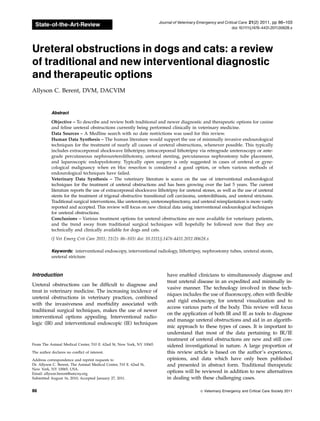
![Equipment
Various flexible and rigid endoscopes are needed for
the following endourologic ureteral procedures. Rigid
cystoscopy is used for female (dog or cat) ureteral in-
tervention. Ureteral access is possible in a retrograde
fashion in most female patients via IR techniques. Rigid
endoscope diameters range from 1.9 to 7.5 mm. Flexible
ureteroscopes (7.5–8.2-Fr) are used for cystoscopic ac-
cess in male dogs and ureteroscopy in both male and
female dogs, when possible. Different types of intra-
corporeal lithotrites and lasers are available including:
holmium:YAG, diode, ultrasonic, pneumatic, and elec-
trohydraulic. These are typically used for stone disease
or tumor ablation when treating ureteral obstructions.
In addition, extracorporeal shock wave lithotripsy
(ESWL) has great application for ureteroliths in dogs
and sometimes, but less commonly, cats.1–3
A traditional fluoroscopic C-arm is sufficient for visu-
alization and ureteral intervention. A mobile C-arm has
the ability to move the image intensifier and gain various
tangential views of the renal pelvis and ureter. Ultra-
sonography is useful for percutaneous renal access in
order to cannulate the ureter in an antegrade manner, or
for the placement of a nephrostomy tube. This can also be
done under direct fluoroscopic guidance when the renal
pelvis is already opacified with contrast. Various guide-
wires and catheters are also needed for each procedure,
and a discussion of these can be found elsewhere.4
Uret-
eral stents, which are available in numerous dimensions,
are soft polyurethane-type catheters that have a double
pigtail multifenestrated design. They can be easily re-
moved after resolution of ureteral disease if necessary,
but are often considered long-term treatment options.
Additional interventional techniques that will be dis-
cussed for the management of ureteral disease include
the placement of nephrostomy tubes and the use of a
subcutaneous ureteral bypass (SUB).
Etiology
Ureterolithiasis is the most common cause of ureteral ob-
structions in both dogs and cats,5,6
though trigonal ne-
oplasia,a,7
ureteral strictures (congenital or acquired),b
dried solidified blood clots/calculi8
or other tumor types
have also been reported.9
Greater than 98% of feline, and
over 50% of canine ureteroliths were recently docu-
mented to be composed of calcium oxalate.5,6,10–12
These
types of stones do not dissolve medically and either need
to pass spontaneously, be removed, or managed, to per-
mit urine passage. Once medical management fails (tra-
ditionally this involves, eg, IV fluid therapy, mannitol
continuous rate infusions [CRI], and a-adrenergic block-
ade, amitriptyline, glucagon), partial obstructions were
traditionally tolerated and often left untreated (benign
neglect) due to the risk:benefit ratio of attempted surgical
removal. If there is a complete ureteral obstruction, de-
compression of the renal pelvis was typically encouraged.
This approach to management has more recently changed
in the author’s practice and immediate treatment of par-
tial ureteral obstructions is typically recommended.
The physiologic response to a ureteral obstruction is
very complex both before and following its relief. Fol-
lowing a ureteral obstruction studies in normal dogs have
demonstrated that ureteral pressures increase immedi-
ately and can take over 24 hours after obstruction relief
for the pressure to decrease.13
After this increase in pres-
sure renal blood flow diminishes to 40% of normal over
the first 24 hours and drops to 20% of normal by 2
weeks.14,15
The excessive pressure is transmitted to the
entire nephron and a decrease in glomerular filtration
rate (GFR) occurs via concurrent vasoactive mediator re-
lease, leukocyte influx, and subsequent fibrosis.14
The
contralateral kidney will have an increase in the GFR in
response. The longer the ureter remains obstructed, the
more damage occurs, which may be irreversible depend-
ing on severity and duration of the obstruction. In a study
of normal dogs, it was found that after 7 days of ob-
struction the GFR was permanently diminished by 35%,
and when the obstruction lasted for 14 days the GFR was
diminished by 54%.13–18
These numbers were obtained in
a canine model with complete obstruction, without pre-
existing azotemia, chronic interstitial nephritis, or chronic
obstructions, so extrapolation of a worse outcome might
be expected in dogs and cats that are obstructed, making
aggressive and timely intervention imperative. It was also
documented in these studies that it took over 4 months to
get the maximal return to function.16–18
Interestingly, in
contrast to the irreversibility of a complete obstruction,
partial obstructions resulted in less severe destruction
with greater return of function after the obstruction is
relieved. In 1 dog model it was found that the GFR re-
turned to normal after a partial obstruction was present
for 4 weeks.13
Knowing that many patients are partially
obstructed with concurrent renal compromise, eg, chronic
renal disease (CKD), aggressive management and reso-
lution is recommended to improve overall outcome.
The normal diameter of the canine ureter based on
computed tomography (CT) measurements ranges from
1.3 to 2.7mm (3.9–8.1-Fr).19
The normal internal diam-
eter of the feline ureter is approximately 0.4 mm (1.2-Fr),
whereas the outside diameter reaches 1 mm (3-Fr).20–24
The ureter sits in the retroperitoneal space connecting
the renal pelvis to the urinary bladder. It is lined with
transitional cell epithelium and composed of several
layers of smooth muscle surrounding the mucosal layer,
allowing the peristalsis of the ureter and urine propul-
sion from the kidney to the urinary bladder.20,21
In the
dog and cat the ureteropelvic junction is covered by re-
nal parenchyma. Once the ureter exits the kidney it
& Veterinary Emergency and Critical Care Society 2011, doi: 10.1111/j.1476-4431.2011.00628.x 87
Image-guided interventions in ureteral obstructions](data:image/gif;base64,R0lGODlhAQABAIAAAAAAAP///yH5BAEAAAAALAAAAAABAAEAAAIBRAA7)