SlideShare a Scribd company logo
1 of 30
Saransh Khandelwal
Technical Superintendent,
Department of Biomedical Engineering
IIT Hyderabad, 502284
PAGE 1
Flow
Cytometryin
Microfluidics
Content
Outline
Introduction
Understanding Flow Cytometry
Concept of Fluorescence in Flowcytometry
State-of-the-art application-based Research Paper
Conclusion and Future Aspects
PAGE 2
Introduction
Basic Concept
Parameters
Fluorescence involved in Flow
Cytometry
Flow Cytometry
PAGE 3
Flow Cytometry
 The measurement (-metry) of cells (cyto-) as they flow
past a detection system.
 First developed for single-cell analysis in the late 1960’s
 Technology to measure one cell/particle at a time in a
moving fluid stream
 By the late 1970s, instruments configured with two
LASERS [1]
 Measure and Quantify cells plus Cell Sorting
 Microfluidic Flow Cytometer: cells can be confined in
micrometer-sized chambers and channels which are
compatible to their intrinsic volume, thus enabling the
analysis of single cell with the minimized dilution or
buffer solution [1]
PAGE 4
Flow Cytometry
Paper Review Conclusion and Discussions Questions
PAGE 5
Flow Cytometry
Paper Review Conclusion and Discussions Questions
Fig.1 Basic schematic of Flow Cytometer
LASER
SAMPLE
FS-Detector
SS-Detector
To PC
Hydrodynamic
Focusing
Dichroic Mirror
Fig. 1: Basic schematic of Flow Cytometer [2]
Hydrodynamic Focusing
PAGE 6
Flow Cytometry
Paper Review Conclusion and Discussions Questions
The sample is injected into an outer sheath stream. The
pressure differential and design of the flow cell creates
laminar flow where the sample core is focussed into a
stream of single particles within the outer sheath layer
Piezoelectric/Acoustic waves can also be used to improve
focusing.
Fig. 2: Hydrodynamic Focusing
Sheath
Stream
Sample
Stream
Scattering
PAGE 7
Flow Cytometry
Paper Review Conclusion and Discussions Questions
Forward Scattering: Magnitude proportional to Size of the sample
Fig. 3: Forward scattering with different size of cells
Scattering
PAGE 8
Flow Cytometry
Paper Review Conclusion and Discussions Questions
Side Scattering: Magnitude proportional to Granularity and
Structural Complexity
Fig. 4: Side scattering featuring the granularity of a cell
Parameters
PAGE 9
Flow Cytometry
Paper Review Conclusion and Discussions Questions
• Small angle (0.5-5°)
• Corresponds to cell size
• Large angle (15-150°)
• Corresponds to internal granularity,
unevenness of the cell surface
Forward Scatter (FSC) Side Scatter (SSC)
Fig. 5: Voltage peak acquired after FSC and SSC
Parameters
PAGE 10
Flow Cytometry
Paper Review Conclusion and Discussions Questions
Forward Scatter (FSC) Side Scatter (SSC)
Fig. 6: Scatter plot obtained after sampling through FSC and SSC setup
Dot plot
PAGE 11
Flow Cytometry
Paper Review Conclusion and Discussions Questions
Fig. 7: Dot plotting from Forward and Side Scatter data
Scatter plot
PAGE 12
Flow Cytometry
Paper Review Conclusion and Discussions Questions
Neutrophils
Monocytes
Lymphocytes
Fluorescence
PAGE 13
Flow Cytometry
Paper Review Conclusion and Discussions Questions
Many fluorescent substances are used in
flow cytometry:
• Fluorochrome antibody conjugates
• Nucleic acid binding dyes
• Metabolic indicators
• Fluorescent protein
Most flow cytometers use filters and
detectors to measure ranges of light
that are associated with specific
fluorochromes
The advanced FCM have array of 16-28
detectors for capturing different range.
Fluorescent tagged
antibody or dye
PAGE 14
Flow Cytometry
Paper Review Conclusion and Discussions Questions
Stokes Shift
British Scientist Sir George G. Stokes who first
described fluorescence in 1852 and was
responsible for coining the term in honour of
the blue-white fluorescent mineral fluorite
(fluorspar)
He discovered the wavelength shift to longer
values in emission spectra that is known as
the Stokes Shift.
However, Plank’s Equation was derived in
1990.
Figure 8: Fluorophore Absorption and Emission Profiles [3]
PAGE 15
Flow Cytometry
Paper Review Conclusion and Discussions Questions
Dichromatic Mirror
 It is positioned at 450angle to both the incident illumination arriving
from the light source, as well as the optical axis of the microscope
 Light from the source passes through the defined bandpass excitation
filter, and is then reflected down into the objective by the DM to be
focused at the specimen plane
 Fluorescence emission is captured by the objective and directed back
through the DM, which in turn reflects most of the contaminating
excitation light back toward the light source
 Emission wavelengths passing through the DM are further purified by
the emission filter, before traveling to the eyepieces or the camera
image plane
Figure 9: Dichromatic Mirror[3]
PAGE 16
Flow Cytometry
Paper Review Conclusion and Discussions Questions
Filters
 Fluorophores that can absorb a quantity of
illumination, the percentage that will emit
secondary fluorescence is even lower.
 The fundamental problem in fluorescence
microscopy is to produce high-efficiency
illumination of the specimen, while
simultaneously capturing weak
fluorescence emission that is effectively
separated from the much more intense
illumination band.
 These conditions are satisfied in modern
fluorescence instruments by a combination
of filters that coordinate excitation and
emission requirements.
Fig. 10: Filters characteristics [3]
PAGE 17
Flow Cytometry
Paper Review Conclusion and Discussions Questions
Advancements and Current Techniques
 Usually, sample pumping, focusing and sorting are achieved on a microfluidic
chip.
 Due to the extremely small size of the microchip, less volume of sample and
reagent is usually required in the microfluidic flow cytometer resulting in
reducing the cost significantly
 Cells can be assayed one at a time at speeds of up to 100,000 cells per second
 High-end cytometers can measure up to 30 parameters (28 colours + FSC/SSC)
 Cell sorting speeds up to 25,000 per second
PAGE 18
Flow Cytometry
Paper Review Conclusion and Discussions Questions
Applications and state of the art equipment's
 Cell sorting – cells are not destroyed
 Pumping, Sampling, Dispensing Sequential Loading etc
 POC diagnostic tests
 Haematology, Oncology, Immunology
 Blood Banking
• LSR II – 5 laser
• LSR Fortessa 4 laser
• 2 x LSR Fortessa X20 4 laser
• LSR Fortessa X50 – 5 laser
• 3 x Attune NxT – 4 laser
• FACSAria II
• FACSAria III
• 2 x Aria Fusion
• Sony MA900
PAGE 19
Flow Cytometry
Paper Review Conclusion and Discussions Questions
Limitations and Future Aspects
With the rapid development of electronics and computer science, the data processing is much
easier to be miniaturized and integrated into the systems. However, compared to the conventional
flow cytometers, the microfluidic flow cytometers also exhibit some limitations
 Lower transfer rates
 Integration and simplification of many functional components on a microchip still is a challenge
 The standardization of advanced microfluidic flow cytometers in clinical and research laboratories
is the key to expand these technologies rapidly
The sensitivity and throughput of such systems need to be further improved. Besides, parallel
detection and processing is used to improve the throughput. [4]
 A highly parallel acoustic flow cytometer that used an acoustic standing wave to focus particles
into 16 parallel analysis points.
 Smartphone-Based Analysis of single RBCs [5]
PAGE 20
Flow Cytometry
Paper Review
Conclusion and Discussions Questions
Paper Review
 Biosensors and
Bioelectronics
 Impact Factor: 12.545
 Volume 222, 15 February
2023, 114916
PAGE 21
Flow Cytometry
Paper Review
Conclusion and Discussions Questions
Introduction
 Conventional immunophenotyping techniques such as flow cytometry or fluorescence microscopy require
immunolabeling of cells, expensive and complex instrumentation, skilled operators, and are therefore
incompatible with field deployment and automated cell manufacturing systems.
 In this work, an autonomous microchip that can electronically quantify the immunophenotypical
composition of a cell suspension.
 Microchip identifies different cell subtypes by capturing each in different microfluidic chambers
functionalized against the markers of the target populations.
 All on-chip activity is electronically monitored by an integrated sensor network, which informs an
algorithm determining subpopulation fractions from chip-wide immunocapture statistics in real time.
 Microchip analyzed a mixture of unlabelled CD4+ and CD8+ T cell sub-populations and validated the
results against flow cytometry measurements.
PAGE 22
Flow Cytometry
Paper Review
Conclusion and Discussions Questions
Theory
 Multicolour flow cytometry has thus far served as the gold standard for immunophenotype
measurements.
 Cells are first pre-labeled with specific antibodies that have been conjugated with fluorochromes and
then interrogated under laser illumination.
 Seeking to create low-cost, portable cell immunophenotyping assays, researchers have
developed microchip-based technologies.
 Typically, such systems drive a cell suspension through an antibody-functionalized microchip
which in turn screens the cell population constituents using immunoaffinity as the discriminatory
mechanism.
 To achieve this capability, this work combines microfluidics, integrated on-chip sensors, computation and
real time feedback control.
PAGE 23
Flow Cytometry
Paper Review
Conclusion and Discussions Questions
Design of
Experiment
 Microchip utilizes a multi-
chamber immunocapture
scheme
 Separates cell
subpopulations by capturing
them based on their surface
markers
 An integrated electrical
sensor network to quantify
the capture statistics
Fig. 11: A schematic illustrating the method for electronic immune cell analysis [6]
PAGE 24
Flow Cytometry
Paper Review
Conclusion and Discussions Questions
Fabrication
Fig. 12: A photograph of the fabricated microchip showing the microfluidic features filled with red dye for visualization and the electrical sensor network
formed by micropatterned gold electrodes. Insets show a close-up microscope image of (top) one of the coded electrical sensors on the device along with
the inter-chamber microfluidic passage it monitors and (bottom) a group of micropillars within the capture chamber that cells interact with as they flow
through the device. [6]
 15 μm thick negative
photoresist SU-8 was spin
coated on Si wafer
 Design Pattern was
transferred
 a mixture of PDMS
elastomer and its crosslinker
was prepared at a 10:1 ratio
(by weight) and poured onto
the mold.
 Cured PDMS was peeled off
PAGE 25
Flow Cytometry
Paper Review
Conclusion and Discussions Questions
Design of
Experiment
 The readout produced by
the sensor network is
analysed by custom-built
deep learning algorithms.
 Algorithms also inform a
runtime feedback control
algorithm that continually
monitors the cell flow
dynamics
 Maintain optimal cell flow
conditions for
immunocapture
Fig. 13: A schematic showing all of the components of the developed autonomous microchip-based immunoassay
system and their interaction [6]
 The readout produced by
the sensor network is
analysed by custom-built
deep learning algorithms.
 Algorithms also inform a
runtime feedback control
algorithm that continually
monitors the cell flow
dynamics
 Maintain optimal cell flow
conditions for
immunocapture
PAGE 26
Flow Cytometry
Paper Review
Conclusion and Discussions Questions
Results
 The complete system
consisting of the developed
hardware and software by
processing heterogenous
samples of mixed T cell
populations.
 The first chamber would
capture exclusively CD4+ T
cells, the second chamber
would capture CD8+ T cells
 To confirm that both
capture chambers were
capturing their intended
target cell populations,
imaging was done with
fluorescence microscopy. Fig. 14: Schematic of the device showing the layout of capture chambers. Insets show images of CD4+ (left) and
CD8+ (right) cells labeled fluorescently after they were captured on the device [6]
PAGE 27
Flow Cytometry
Paper Review
Conclusion and Discussions Questions
Results
Fig. 15: A plot showing the CD4+ and CD8+ T cell subpopulation frequencies as reported by the system (color-filled bars) vs the nominal mix ratio determined
by hemacytometer (unfilled bars). Consecutive image gives the difference between the subpopulation counts determined by flow cytometry vs. our
microchip-based immunoassay [6]
PAGE 28
Flow Cytometry Paper Review
Conclusion and Discussions
Questions
Conclusion and Discussions
 The percentage of CD4+CD8− T cells, CD4−CD8+ T cells, and CD4−CD8−expressors were 48.3%,
46.8%, and 4.1% respectively as per flow cytometry results.
 These results were in good agreement with those from the designed immunoanalysis system,
which reported 49.3%, 43.8% and 6.9% as the frequencies of aforementioned
immunophenotypes.
 Considering the flow cytometry data as the ground truth, these results amounted to an average of
≤6%, effectively representing an accuracy of >94% for the designed system.
 Conclusions are supported by the fact that the used CD4+ T cell and CD8+ T cell samples had
manufacturer-reported purities of 94% and 93%, respectively.
 A proof-of-concept implementation that targets two surface markers, the scalable nature of our
immunocapture scheme enables a straightforward application of our approach to target a
multitude of surface markers by simply adding chambers to the design and functionalizing them
accordingly.
 The measurement throughput can be further increased by revising the fluidic design to lower the
hydraulic resistance.
PAGE 29
Flow Cytometry Paper Review Conclusion and Discussions
Questions
Questions…??
References
PAGE 30
1. New advances in microfluidic flow cytometry, Yanli Gong et.al., Wiley Analytical Science, Electrophoresis, Volume 40, Issue
8, P.P. 1212-1229, 2019
2. Flow Cytometry: Principles and Clinical Applications in Hematology, Brown M and Wittwer C. et.al. ,Clin Chem 46(8) P.P.
1221-29, 2000
3. https://zeiss-campus.magnet.fsu.edu/articles/basics/fluorescence.html
4. Line-Focused Optical Excitation of Parallel Acoustic Focused Sample Streams for High Volumetric and Analytical Rate Flow
Cytometry, Kalb, D. M., Fencl et.al. Anal. Chem., 89, 9967-9975, 2017
5. A smartphone-based optical platform for colorimetric analysis of microfluidic device, Kim, S.C.; Jalal, U.M.; Im, S.B.; Ko, S.;
Shim, J.S.. Sens. Actuators B Chem, 239, 52–59, 2017
6. An autonomous microchip for real-time, label-free immune cell analysis, A.K.M. Arifuzzman et.al. Biosensors and
Bioelectronics Volume 222, 114916, 2023

More Related Content

Similar to Flow Cytometry in Microfluidics

Similar to Flow Cytometry in Microfluidics (20)

VAC Study material VAC
VAC Study material VAC VAC Study material VAC
VAC Study material VAC
 
Flow cytometry
Flow cytometryFlow cytometry
Flow cytometry
 
Presentation on flow cytometry1
Presentation on flow cytometry1Presentation on flow cytometry1
Presentation on flow cytometry1
 
Flow Cytometry
Flow CytometryFlow Cytometry
Flow Cytometry
 
Fluorescence- Activated Cell Sorter (FACS)
Fluorescence- Activated Cell Sorter (FACS)Fluorescence- Activated Cell Sorter (FACS)
Fluorescence- Activated Cell Sorter (FACS)
 
Flow cytometry
Flow cytometry Flow cytometry
Flow cytometry
 
flow cytometry.pptx
flow cytometry.pptxflow cytometry.pptx
flow cytometry.pptx
 
Flow cytometry
Flow cytometryFlow cytometry
Flow cytometry
 
How to become a flow cytometry expert in 4 days
How to become a flow cytometry expert in 4 daysHow to become a flow cytometry expert in 4 days
How to become a flow cytometry expert in 4 days
 
How to become a flow cytometry expert in 4 days
How to become a flow cytometry expert in 4 daysHow to become a flow cytometry expert in 4 days
How to become a flow cytometry expert in 4 days
 
FLOWCYTOMETRY........................pptx
FLOWCYTOMETRY........................pptxFLOWCYTOMETRY........................pptx
FLOWCYTOMETRY........................pptx
 
An Overview of Microfluidics
An Overview of MicrofluidicsAn Overview of Microfluidics
An Overview of Microfluidics
 
Flow Cytometry (FCM)
Flow Cytometry (FCM) Flow Cytometry (FCM)
Flow Cytometry (FCM)
 
Imaging Flow Cytometry
Imaging Flow CytometryImaging Flow Cytometry
Imaging Flow Cytometry
 
Presentation 7
Presentation 7Presentation 7
Presentation 7
 
Flow cytometry
Flow cytometryFlow cytometry
Flow cytometry
 
Flow cytometry
Flow cytometryFlow cytometry
Flow cytometry
 
Flow cytometry in cell biology
Flow cytometry in cell biologyFlow cytometry in cell biology
Flow cytometry in cell biology
 
Flow cytometry
Flow cytometryFlow cytometry
Flow cytometry
 
FlowBasics2[1]
FlowBasics2[1]FlowBasics2[1]
FlowBasics2[1]
 

Recently uploaded

Concept of Vouching. B.Com(Hons) /B.Compdf
Concept of Vouching. B.Com(Hons) /B.CompdfConcept of Vouching. B.Com(Hons) /B.Compdf
Concept of Vouching. B.Com(Hons) /B.CompdfUmakantAnnand
 
Organic Name Reactions for the students and aspirants of Chemistry12th.pptx
Organic Name Reactions  for the students and aspirants of Chemistry12th.pptxOrganic Name Reactions  for the students and aspirants of Chemistry12th.pptx
Organic Name Reactions for the students and aspirants of Chemistry12th.pptxVS Mahajan Coaching Centre
 
How to Make a Pirate ship Primary Education.pptx
How to Make a Pirate ship Primary Education.pptxHow to Make a Pirate ship Primary Education.pptx
How to Make a Pirate ship Primary Education.pptxmanuelaromero2013
 
BASLIQ CURRENT LOOKBOOK LOOKBOOK(1) (1).pdf
BASLIQ CURRENT LOOKBOOK  LOOKBOOK(1) (1).pdfBASLIQ CURRENT LOOKBOOK  LOOKBOOK(1) (1).pdf
BASLIQ CURRENT LOOKBOOK LOOKBOOK(1) (1).pdfSoniaTolstoy
 
Paris 2024 Olympic Geographies - an activity
Paris 2024 Olympic Geographies - an activityParis 2024 Olympic Geographies - an activity
Paris 2024 Olympic Geographies - an activityGeoBlogs
 
Alper Gobel In Media Res Media Component
Alper Gobel In Media Res Media ComponentAlper Gobel In Media Res Media Component
Alper Gobel In Media Res Media ComponentInMediaRes1
 
Introduction to AI in Higher Education_draft.pptx
Introduction to AI in Higher Education_draft.pptxIntroduction to AI in Higher Education_draft.pptx
Introduction to AI in Higher Education_draft.pptxpboyjonauth
 
SOCIAL AND HISTORICAL CONTEXT - LFTVD.pptx
SOCIAL AND HISTORICAL CONTEXT - LFTVD.pptxSOCIAL AND HISTORICAL CONTEXT - LFTVD.pptx
SOCIAL AND HISTORICAL CONTEXT - LFTVD.pptxiammrhaywood
 
Separation of Lanthanides/ Lanthanides and Actinides
Separation of Lanthanides/ Lanthanides and ActinidesSeparation of Lanthanides/ Lanthanides and Actinides
Separation of Lanthanides/ Lanthanides and ActinidesFatimaKhan178732
 
Kisan Call Centre - To harness potential of ICT in Agriculture by answer farm...
Kisan Call Centre - To harness potential of ICT in Agriculture by answer farm...Kisan Call Centre - To harness potential of ICT in Agriculture by answer farm...
Kisan Call Centre - To harness potential of ICT in Agriculture by answer farm...Krashi Coaching
 
microwave assisted reaction. General introduction
microwave assisted reaction. General introductionmicrowave assisted reaction. General introduction
microwave assisted reaction. General introductionMaksud Ahmed
 
Mastering the Unannounced Regulatory Inspection
Mastering the Unannounced Regulatory InspectionMastering the Unannounced Regulatory Inspection
Mastering the Unannounced Regulatory InspectionSafetyChain Software
 
Interactive Powerpoint_How to Master effective communication
Interactive Powerpoint_How to Master effective communicationInteractive Powerpoint_How to Master effective communication
Interactive Powerpoint_How to Master effective communicationnomboosow
 
POINT- BIOCHEMISTRY SEM 2 ENZYMES UNIT 5.pptx
POINT- BIOCHEMISTRY SEM 2 ENZYMES UNIT 5.pptxPOINT- BIOCHEMISTRY SEM 2 ENZYMES UNIT 5.pptx
POINT- BIOCHEMISTRY SEM 2 ENZYMES UNIT 5.pptxSayali Powar
 
Accessible design: Minimum effort, maximum impact
Accessible design: Minimum effort, maximum impactAccessible design: Minimum effort, maximum impact
Accessible design: Minimum effort, maximum impactdawncurless
 
KSHARA STURA .pptx---KSHARA KARMA THERAPY (CAUSTIC THERAPY)————IMP.OF KSHARA ...
KSHARA STURA .pptx---KSHARA KARMA THERAPY (CAUSTIC THERAPY)————IMP.OF KSHARA ...KSHARA STURA .pptx---KSHARA KARMA THERAPY (CAUSTIC THERAPY)————IMP.OF KSHARA ...
KSHARA STURA .pptx---KSHARA KARMA THERAPY (CAUSTIC THERAPY)————IMP.OF KSHARA ...M56BOOKSTORE PRODUCT/SERVICE
 
Incoming and Outgoing Shipments in 1 STEP Using Odoo 17
Incoming and Outgoing Shipments in 1 STEP Using Odoo 17Incoming and Outgoing Shipments in 1 STEP Using Odoo 17
Incoming and Outgoing Shipments in 1 STEP Using Odoo 17Celine George
 
Solving Puzzles Benefits Everyone (English).pptx
Solving Puzzles Benefits Everyone (English).pptxSolving Puzzles Benefits Everyone (English).pptx
Solving Puzzles Benefits Everyone (English).pptxOH TEIK BIN
 
18-04-UA_REPORT_MEDIALITERAСY_INDEX-DM_23-1-final-eng.pdf
18-04-UA_REPORT_MEDIALITERAСY_INDEX-DM_23-1-final-eng.pdf18-04-UA_REPORT_MEDIALITERAСY_INDEX-DM_23-1-final-eng.pdf
18-04-UA_REPORT_MEDIALITERAСY_INDEX-DM_23-1-final-eng.pdfssuser54595a
 

Recently uploaded (20)

Concept of Vouching. B.Com(Hons) /B.Compdf
Concept of Vouching. B.Com(Hons) /B.CompdfConcept of Vouching. B.Com(Hons) /B.Compdf
Concept of Vouching. B.Com(Hons) /B.Compdf
 
Organic Name Reactions for the students and aspirants of Chemistry12th.pptx
Organic Name Reactions  for the students and aspirants of Chemistry12th.pptxOrganic Name Reactions  for the students and aspirants of Chemistry12th.pptx
Organic Name Reactions for the students and aspirants of Chemistry12th.pptx
 
How to Make a Pirate ship Primary Education.pptx
How to Make a Pirate ship Primary Education.pptxHow to Make a Pirate ship Primary Education.pptx
How to Make a Pirate ship Primary Education.pptx
 
BASLIQ CURRENT LOOKBOOK LOOKBOOK(1) (1).pdf
BASLIQ CURRENT LOOKBOOK  LOOKBOOK(1) (1).pdfBASLIQ CURRENT LOOKBOOK  LOOKBOOK(1) (1).pdf
BASLIQ CURRENT LOOKBOOK LOOKBOOK(1) (1).pdf
 
Paris 2024 Olympic Geographies - an activity
Paris 2024 Olympic Geographies - an activityParis 2024 Olympic Geographies - an activity
Paris 2024 Olympic Geographies - an activity
 
Alper Gobel In Media Res Media Component
Alper Gobel In Media Res Media ComponentAlper Gobel In Media Res Media Component
Alper Gobel In Media Res Media Component
 
Introduction to AI in Higher Education_draft.pptx
Introduction to AI in Higher Education_draft.pptxIntroduction to AI in Higher Education_draft.pptx
Introduction to AI in Higher Education_draft.pptx
 
SOCIAL AND HISTORICAL CONTEXT - LFTVD.pptx
SOCIAL AND HISTORICAL CONTEXT - LFTVD.pptxSOCIAL AND HISTORICAL CONTEXT - LFTVD.pptx
SOCIAL AND HISTORICAL CONTEXT - LFTVD.pptx
 
Separation of Lanthanides/ Lanthanides and Actinides
Separation of Lanthanides/ Lanthanides and ActinidesSeparation of Lanthanides/ Lanthanides and Actinides
Separation of Lanthanides/ Lanthanides and Actinides
 
Kisan Call Centre - To harness potential of ICT in Agriculture by answer farm...
Kisan Call Centre - To harness potential of ICT in Agriculture by answer farm...Kisan Call Centre - To harness potential of ICT in Agriculture by answer farm...
Kisan Call Centre - To harness potential of ICT in Agriculture by answer farm...
 
microwave assisted reaction. General introduction
microwave assisted reaction. General introductionmicrowave assisted reaction. General introduction
microwave assisted reaction. General introduction
 
Mastering the Unannounced Regulatory Inspection
Mastering the Unannounced Regulatory InspectionMastering the Unannounced Regulatory Inspection
Mastering the Unannounced Regulatory Inspection
 
Interactive Powerpoint_How to Master effective communication
Interactive Powerpoint_How to Master effective communicationInteractive Powerpoint_How to Master effective communication
Interactive Powerpoint_How to Master effective communication
 
9953330565 Low Rate Call Girls In Rohini Delhi NCR
9953330565 Low Rate Call Girls In Rohini  Delhi NCR9953330565 Low Rate Call Girls In Rohini  Delhi NCR
9953330565 Low Rate Call Girls In Rohini Delhi NCR
 
POINT- BIOCHEMISTRY SEM 2 ENZYMES UNIT 5.pptx
POINT- BIOCHEMISTRY SEM 2 ENZYMES UNIT 5.pptxPOINT- BIOCHEMISTRY SEM 2 ENZYMES UNIT 5.pptx
POINT- BIOCHEMISTRY SEM 2 ENZYMES UNIT 5.pptx
 
Accessible design: Minimum effort, maximum impact
Accessible design: Minimum effort, maximum impactAccessible design: Minimum effort, maximum impact
Accessible design: Minimum effort, maximum impact
 
KSHARA STURA .pptx---KSHARA KARMA THERAPY (CAUSTIC THERAPY)————IMP.OF KSHARA ...
KSHARA STURA .pptx---KSHARA KARMA THERAPY (CAUSTIC THERAPY)————IMP.OF KSHARA ...KSHARA STURA .pptx---KSHARA KARMA THERAPY (CAUSTIC THERAPY)————IMP.OF KSHARA ...
KSHARA STURA .pptx---KSHARA KARMA THERAPY (CAUSTIC THERAPY)————IMP.OF KSHARA ...
 
Incoming and Outgoing Shipments in 1 STEP Using Odoo 17
Incoming and Outgoing Shipments in 1 STEP Using Odoo 17Incoming and Outgoing Shipments in 1 STEP Using Odoo 17
Incoming and Outgoing Shipments in 1 STEP Using Odoo 17
 
Solving Puzzles Benefits Everyone (English).pptx
Solving Puzzles Benefits Everyone (English).pptxSolving Puzzles Benefits Everyone (English).pptx
Solving Puzzles Benefits Everyone (English).pptx
 
18-04-UA_REPORT_MEDIALITERAСY_INDEX-DM_23-1-final-eng.pdf
18-04-UA_REPORT_MEDIALITERAСY_INDEX-DM_23-1-final-eng.pdf18-04-UA_REPORT_MEDIALITERAСY_INDEX-DM_23-1-final-eng.pdf
18-04-UA_REPORT_MEDIALITERAСY_INDEX-DM_23-1-final-eng.pdf
 

Flow Cytometry in Microfluidics

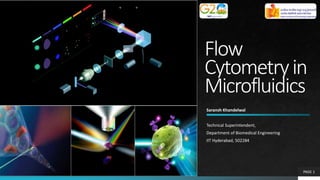
  • 1. Saransh Khandelwal Technical Superintendent, Department of Biomedical Engineering IIT Hyderabad, 502284 PAGE 1 Flow Cytometryin Microfluidics
  • 2. Content Outline Introduction Understanding Flow Cytometry Concept of Fluorescence in Flowcytometry State-of-the-art application-based Research Paper Conclusion and Future Aspects PAGE 2
  • 4. Flow Cytometry  The measurement (-metry) of cells (cyto-) as they flow past a detection system.  First developed for single-cell analysis in the late 1960’s  Technology to measure one cell/particle at a time in a moving fluid stream  By the late 1970s, instruments configured with two LASERS [1]  Measure and Quantify cells plus Cell Sorting  Microfluidic Flow Cytometer: cells can be confined in micrometer-sized chambers and channels which are compatible to their intrinsic volume, thus enabling the analysis of single cell with the minimized dilution or buffer solution [1] PAGE 4 Flow Cytometry Paper Review Conclusion and Discussions Questions
  • 5. PAGE 5 Flow Cytometry Paper Review Conclusion and Discussions Questions Fig.1 Basic schematic of Flow Cytometer LASER SAMPLE FS-Detector SS-Detector To PC Hydrodynamic Focusing Dichroic Mirror Fig. 1: Basic schematic of Flow Cytometer [2]
  • 6. Hydrodynamic Focusing PAGE 6 Flow Cytometry Paper Review Conclusion and Discussions Questions The sample is injected into an outer sheath stream. The pressure differential and design of the flow cell creates laminar flow where the sample core is focussed into a stream of single particles within the outer sheath layer Piezoelectric/Acoustic waves can also be used to improve focusing. Fig. 2: Hydrodynamic Focusing Sheath Stream Sample Stream
  • 7. Scattering PAGE 7 Flow Cytometry Paper Review Conclusion and Discussions Questions Forward Scattering: Magnitude proportional to Size of the sample Fig. 3: Forward scattering with different size of cells
  • 8. Scattering PAGE 8 Flow Cytometry Paper Review Conclusion and Discussions Questions Side Scattering: Magnitude proportional to Granularity and Structural Complexity Fig. 4: Side scattering featuring the granularity of a cell
  • 9. Parameters PAGE 9 Flow Cytometry Paper Review Conclusion and Discussions Questions • Small angle (0.5-5°) • Corresponds to cell size • Large angle (15-150°) • Corresponds to internal granularity, unevenness of the cell surface Forward Scatter (FSC) Side Scatter (SSC) Fig. 5: Voltage peak acquired after FSC and SSC
  • 10. Parameters PAGE 10 Flow Cytometry Paper Review Conclusion and Discussions Questions Forward Scatter (FSC) Side Scatter (SSC) Fig. 6: Scatter plot obtained after sampling through FSC and SSC setup
  • 11. Dot plot PAGE 11 Flow Cytometry Paper Review Conclusion and Discussions Questions Fig. 7: Dot plotting from Forward and Side Scatter data
  • 12. Scatter plot PAGE 12 Flow Cytometry Paper Review Conclusion and Discussions Questions Neutrophils Monocytes Lymphocytes
  • 13. Fluorescence PAGE 13 Flow Cytometry Paper Review Conclusion and Discussions Questions Many fluorescent substances are used in flow cytometry: • Fluorochrome antibody conjugates • Nucleic acid binding dyes • Metabolic indicators • Fluorescent protein Most flow cytometers use filters and detectors to measure ranges of light that are associated with specific fluorochromes The advanced FCM have array of 16-28 detectors for capturing different range. Fluorescent tagged antibody or dye
  • 14. PAGE 14 Flow Cytometry Paper Review Conclusion and Discussions Questions Stokes Shift British Scientist Sir George G. Stokes who first described fluorescence in 1852 and was responsible for coining the term in honour of the blue-white fluorescent mineral fluorite (fluorspar) He discovered the wavelength shift to longer values in emission spectra that is known as the Stokes Shift. However, Plank’s Equation was derived in 1990. Figure 8: Fluorophore Absorption and Emission Profiles [3]
  • 15. PAGE 15 Flow Cytometry Paper Review Conclusion and Discussions Questions Dichromatic Mirror  It is positioned at 450angle to both the incident illumination arriving from the light source, as well as the optical axis of the microscope  Light from the source passes through the defined bandpass excitation filter, and is then reflected down into the objective by the DM to be focused at the specimen plane  Fluorescence emission is captured by the objective and directed back through the DM, which in turn reflects most of the contaminating excitation light back toward the light source  Emission wavelengths passing through the DM are further purified by the emission filter, before traveling to the eyepieces or the camera image plane Figure 9: Dichromatic Mirror[3]
  • 16. PAGE 16 Flow Cytometry Paper Review Conclusion and Discussions Questions Filters  Fluorophores that can absorb a quantity of illumination, the percentage that will emit secondary fluorescence is even lower.  The fundamental problem in fluorescence microscopy is to produce high-efficiency illumination of the specimen, while simultaneously capturing weak fluorescence emission that is effectively separated from the much more intense illumination band.  These conditions are satisfied in modern fluorescence instruments by a combination of filters that coordinate excitation and emission requirements. Fig. 10: Filters characteristics [3]
  • 17. PAGE 17 Flow Cytometry Paper Review Conclusion and Discussions Questions Advancements and Current Techniques  Usually, sample pumping, focusing and sorting are achieved on a microfluidic chip.  Due to the extremely small size of the microchip, less volume of sample and reagent is usually required in the microfluidic flow cytometer resulting in reducing the cost significantly  Cells can be assayed one at a time at speeds of up to 100,000 cells per second  High-end cytometers can measure up to 30 parameters (28 colours + FSC/SSC)  Cell sorting speeds up to 25,000 per second
  • 18. PAGE 18 Flow Cytometry Paper Review Conclusion and Discussions Questions Applications and state of the art equipment's  Cell sorting – cells are not destroyed  Pumping, Sampling, Dispensing Sequential Loading etc  POC diagnostic tests  Haematology, Oncology, Immunology  Blood Banking • LSR II – 5 laser • LSR Fortessa 4 laser • 2 x LSR Fortessa X20 4 laser • LSR Fortessa X50 – 5 laser • 3 x Attune NxT – 4 laser • FACSAria II • FACSAria III • 2 x Aria Fusion • Sony MA900
  • 19. PAGE 19 Flow Cytometry Paper Review Conclusion and Discussions Questions Limitations and Future Aspects With the rapid development of electronics and computer science, the data processing is much easier to be miniaturized and integrated into the systems. However, compared to the conventional flow cytometers, the microfluidic flow cytometers also exhibit some limitations  Lower transfer rates  Integration and simplification of many functional components on a microchip still is a challenge  The standardization of advanced microfluidic flow cytometers in clinical and research laboratories is the key to expand these technologies rapidly The sensitivity and throughput of such systems need to be further improved. Besides, parallel detection and processing is used to improve the throughput. [4]  A highly parallel acoustic flow cytometer that used an acoustic standing wave to focus particles into 16 parallel analysis points.  Smartphone-Based Analysis of single RBCs [5]
  • 20. PAGE 20 Flow Cytometry Paper Review Conclusion and Discussions Questions Paper Review  Biosensors and Bioelectronics  Impact Factor: 12.545  Volume 222, 15 February 2023, 114916
  • 21. PAGE 21 Flow Cytometry Paper Review Conclusion and Discussions Questions Introduction  Conventional immunophenotyping techniques such as flow cytometry or fluorescence microscopy require immunolabeling of cells, expensive and complex instrumentation, skilled operators, and are therefore incompatible with field deployment and automated cell manufacturing systems.  In this work, an autonomous microchip that can electronically quantify the immunophenotypical composition of a cell suspension.  Microchip identifies different cell subtypes by capturing each in different microfluidic chambers functionalized against the markers of the target populations.  All on-chip activity is electronically monitored by an integrated sensor network, which informs an algorithm determining subpopulation fractions from chip-wide immunocapture statistics in real time.  Microchip analyzed a mixture of unlabelled CD4+ and CD8+ T cell sub-populations and validated the results against flow cytometry measurements.
  • 22. PAGE 22 Flow Cytometry Paper Review Conclusion and Discussions Questions Theory  Multicolour flow cytometry has thus far served as the gold standard for immunophenotype measurements.  Cells are first pre-labeled with specific antibodies that have been conjugated with fluorochromes and then interrogated under laser illumination.  Seeking to create low-cost, portable cell immunophenotyping assays, researchers have developed microchip-based technologies.  Typically, such systems drive a cell suspension through an antibody-functionalized microchip which in turn screens the cell population constituents using immunoaffinity as the discriminatory mechanism.  To achieve this capability, this work combines microfluidics, integrated on-chip sensors, computation and real time feedback control.
  • 23. PAGE 23 Flow Cytometry Paper Review Conclusion and Discussions Questions Design of Experiment  Microchip utilizes a multi- chamber immunocapture scheme  Separates cell subpopulations by capturing them based on their surface markers  An integrated electrical sensor network to quantify the capture statistics Fig. 11: A schematic illustrating the method for electronic immune cell analysis [6]
  • 24. PAGE 24 Flow Cytometry Paper Review Conclusion and Discussions Questions Fabrication Fig. 12: A photograph of the fabricated microchip showing the microfluidic features filled with red dye for visualization and the electrical sensor network formed by micropatterned gold electrodes. Insets show a close-up microscope image of (top) one of the coded electrical sensors on the device along with the inter-chamber microfluidic passage it monitors and (bottom) a group of micropillars within the capture chamber that cells interact with as they flow through the device. [6]  15 μm thick negative photoresist SU-8 was spin coated on Si wafer  Design Pattern was transferred  a mixture of PDMS elastomer and its crosslinker was prepared at a 10:1 ratio (by weight) and poured onto the mold.  Cured PDMS was peeled off
  • 25. PAGE 25 Flow Cytometry Paper Review Conclusion and Discussions Questions Design of Experiment  The readout produced by the sensor network is analysed by custom-built deep learning algorithms.  Algorithms also inform a runtime feedback control algorithm that continually monitors the cell flow dynamics  Maintain optimal cell flow conditions for immunocapture Fig. 13: A schematic showing all of the components of the developed autonomous microchip-based immunoassay system and their interaction [6]  The readout produced by the sensor network is analysed by custom-built deep learning algorithms.  Algorithms also inform a runtime feedback control algorithm that continually monitors the cell flow dynamics  Maintain optimal cell flow conditions for immunocapture
  • 26. PAGE 26 Flow Cytometry Paper Review Conclusion and Discussions Questions Results  The complete system consisting of the developed hardware and software by processing heterogenous samples of mixed T cell populations.  The first chamber would capture exclusively CD4+ T cells, the second chamber would capture CD8+ T cells  To confirm that both capture chambers were capturing their intended target cell populations, imaging was done with fluorescence microscopy. Fig. 14: Schematic of the device showing the layout of capture chambers. Insets show images of CD4+ (left) and CD8+ (right) cells labeled fluorescently after they were captured on the device [6]
  • 27. PAGE 27 Flow Cytometry Paper Review Conclusion and Discussions Questions Results Fig. 15: A plot showing the CD4+ and CD8+ T cell subpopulation frequencies as reported by the system (color-filled bars) vs the nominal mix ratio determined by hemacytometer (unfilled bars). Consecutive image gives the difference between the subpopulation counts determined by flow cytometry vs. our microchip-based immunoassay [6]
  • 28. PAGE 28 Flow Cytometry Paper Review Conclusion and Discussions Questions Conclusion and Discussions  The percentage of CD4+CD8− T cells, CD4−CD8+ T cells, and CD4−CD8−expressors were 48.3%, 46.8%, and 4.1% respectively as per flow cytometry results.  These results were in good agreement with those from the designed immunoanalysis system, which reported 49.3%, 43.8% and 6.9% as the frequencies of aforementioned immunophenotypes.  Considering the flow cytometry data as the ground truth, these results amounted to an average of ≤6%, effectively representing an accuracy of >94% for the designed system.  Conclusions are supported by the fact that the used CD4+ T cell and CD8+ T cell samples had manufacturer-reported purities of 94% and 93%, respectively.  A proof-of-concept implementation that targets two surface markers, the scalable nature of our immunocapture scheme enables a straightforward application of our approach to target a multitude of surface markers by simply adding chambers to the design and functionalizing them accordingly.  The measurement throughput can be further increased by revising the fluidic design to lower the hydraulic resistance.
  • 29. PAGE 29 Flow Cytometry Paper Review Conclusion and Discussions Questions Questions…??
  • 30. References PAGE 30 1. New advances in microfluidic flow cytometry, Yanli Gong et.al., Wiley Analytical Science, Electrophoresis, Volume 40, Issue 8, P.P. 1212-1229, 2019 2. Flow Cytometry: Principles and Clinical Applications in Hematology, Brown M and Wittwer C. et.al. ,Clin Chem 46(8) P.P. 1221-29, 2000 3. https://zeiss-campus.magnet.fsu.edu/articles/basics/fluorescence.html 4. Line-Focused Optical Excitation of Parallel Acoustic Focused Sample Streams for High Volumetric and Analytical Rate Flow Cytometry, Kalb, D. M., Fencl et.al. Anal. Chem., 89, 9967-9975, 2017 5. A smartphone-based optical platform for colorimetric analysis of microfluidic device, Kim, S.C.; Jalal, U.M.; Im, S.B.; Ko, S.; Shim, J.S.. Sens. Actuators B Chem, 239, 52–59, 2017 6. An autonomous microchip for real-time, label-free immune cell analysis, A.K.M. Arifuzzman et.al. Biosensors and Bioelectronics Volume 222, 114916, 2023

Editor's Notes

  1. MD Abdullah (BM22RESCH11007)
  2. Principles: Fluid dynamics, Optics, and Electronics. Usually, sample pumping, focusing and sorting are achieved on a microfluidic chip. Due to the extremely small size of the microchip, less volume of sample and reagent is usually required in the microfluidic flow cytometer resulting in reducing the cost significantly
  3. • Becton Dickinson and Company (US) • Danaher Corporation (US) • Thermo Fisher Scientific, Inc. (US) • Agilent Technologies, Inc. (US) • Luminex Corporation (US) • Bio-Rad Laboratories, Inc. (US) • Miltenyi Biotec (Germany) • Sysmex Corporation (Japan) • Stratedigm, Inc. (US) • Apogee Flow Systems Ltd. (UK) • Sony Biotechnology, Inc. (US) • Enzo Life Sciences, Inc (US) • Merck KGaA (Germany)
  4. To fabricate the microfluidic layer, at 15 μm thick negative photoresist SU-8 (SU-8 2015, MicroChem) was spin coated onto a silicon wafer. The design pattern was transferred to the resist layer by exposing the resist layer using a maskless aligner (MLA1500, Heidelberg) and then, the uncured photoresist was developed using SU-8 developer and treated with trichloro(octyl)silane in a desiccator for 8 h. Then, a mixture of PDMS elastomer and its crosslinker (Sylgard 184 kit, Dow Corning) was prepared at a 10:1 ratio (by weight) and poured onto the mold, degassed and cured for 4 h at 65 ◦C. Finally, the cured PDMS was peeled off and cut into individual chips. To fabricate the electrical sensor network, 1.5 μm thick negative photoresist (NR9-1500PY, Futurrex) was spin coated onto a 2-inch by 3-inch glass microscope slide (6101, Premiere). The electrode pattern was transferred to the resist layer using a maskless aligner (MLA-1500, Heidelberg) and developed using photoresist developer (RD6 developer, Futurrex). Then, the glass slides were treated with reactive ion etcher for descumming followed by e-beam deposition of 20 nm-thick Cr and 250 nm-thick Au film stacks. The sacrificial photoresist layer was lifted-off by submerging the glass substrate into an acetone bath under mild sonication. Finally, the PDMS layer and the glass substrate were treated with oxygen plasma for 1 min for surface activation before being aligned under microscope and bonded at 65 ◦C to create the final microchip.
  5. The electrical sensor network is excited with a 2 Vpp sinusoidal wave at 550 kHz through the input pads, and the output currents at the positive and negative pads were amplified via transimpedance amplifiers before being sampled by the lock-in amplifier (HF2LI, Zurich Instruments). The lock-in amplifier output was sampled into a computer using a standalone analog to digital converter (PCIe-6361, National Instruments)
  6. Fluorescence Imaging for labeling captured cell population on the microchip with different-colored fluorophores (FITC anti-CD4 antibody and APC anti-CD8 antibody)
  7. The whole process of analyzing ∼12,000 T cells and computing the capture statistics required ∼30 min Our conclusions are supported by the fact that the used CD4+ T cell and CD8+ T cell samples had manufacturer-reported purities of 94% and 93%, respectively
  8. The whole process of analyzing ∼12,000 T cells and computing the capture statistics required ∼30 min Our conclusions are supported by the fact that the used CD4+ T cell and CD8+ T cell samples had manufacturer-reported purities of 94% and 93%, respectively