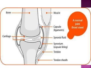
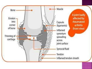
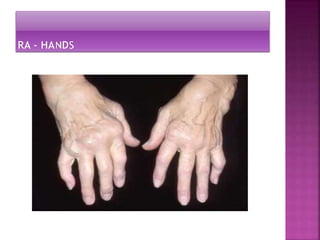
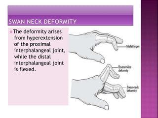
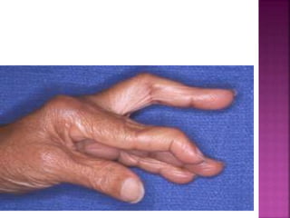
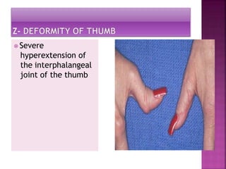
⦿ Rheumatoid arthritis is
autoimmune disorder in
which Immune system
identifies the synovial
membrane as "foreign"
and begins attacking it.
⦿ Synovial membrane shown in
picture
⦿ Rheumatoid Arthritis (RA) is a chronic
inflammatory disorder that may affect many
tissues and organs, but mainly attacks the
joints producing an inflammatory synovitis.
⦿ Idiopathic
⦿ Positive family history
⦿ Inherited tissue type major
histocompatibility complex (MHC) antigen
⦿ Smoking
⦿ Bacterial and Fungal Infection
⦿ Herpes simplex virus infections
⦿ Epstein-Barr virus (EBV)
⦿ Vitamin D deficiency
⦿ RA affects 0.5-1.0% of population in USA
⦿ Females > males 3:1
⦿ but people of any age can be affected
⦿ Peak age 45-65 but onset early from age 20-45
yrs
⦿ About 75% of these are women.
⦿ The disease strikes women three times more
often than men
⦿ Presentation of antigen to T cells
⦿T- and B-cell Proliferation Angiogenesis in
synovial lining
⦿ Neutrophil accumulation in synovial fluid.
⦿ Synovitis.
⦿ Early pannus Formation(The pannus is a
sheet of inflammatory granulation tissue that
spreads from the synovial membrane and
invades the joint)
⦿ Subchondral bone erosion.
⦿ Pannus invasion of cartilage Chondrocyte
proliferation.
⦿ Laxity of ligaments
⦿ Joint instability
, contractures, decreased
ROM, systemic complications
In chronically affected joints, the normally
thin synovium proliferates, thickens, and
develops many villous folds.
The synovial lining cells produce various
materials, including collagenase and
stromelysin(distroys connective tissues).
These contribute to cartilage destruction,
and interleukin-1 (IL-1) and TNF-alpha,
which stimulate cartilage destruction,
osteoclast-mediated bone absorption,
synovial inflammation, and prostaglandins
(which potentiate inflammation).
Sign and Symptoms
⦿The joints of the
hands are often the
very first joints
affected by
rheumatoid arthritis.
These joints are
swollen red and
tender when
squeezed.
⦿ Swelling due to synovitis
⦿The deformity arises
from hyperextension
of the proximal
interphalangeal joint,
while the distal
interphalangeal joint
is flexed.
⦿Mallet finger is a
simple flexion
deformity of the
distal
interphalangeal
joint preventing
extension.
⦿ Severe
hyperextension of
the interphalangeal
joint of the thumb
⦿ Fatigue
⦿ Joint pain
⦿ Joint tenderness
⦿ Joint swelling
⦿ Joint redness
⦿ Joint warmth
⦿ Joint stiffness
⦿ Loss of joint range of motion
⦿ Many joints affected (polyarthritis)
⦿ Limping
⦿ Joint deformity
⦿ Both sides of the body affected (symmetric)
• Extra articular s/s – low grade
fevr,fatigue,anorexia, chest
pain,numbness tingling ,vasculitis,
pericarditis,osteoporosis,neoropathy
etc.
Diagnosis
⦿ X Rays
◾X rays of hands and feet are generally performed in
people with RA.
⦿ Magnetic Resonance Imaging (MRI)
⦿ Ultrasounds
⦿ Blood Tests
◾Rheumatoid Factor (RF)
 RF is a specific antibody in the blood.
 A negative RF does not rule out RA. The arthritis is then called
seronegative, most common during the first year of illness and
converting to seropositive status over time.
◾Anti-citrullinated Protein Antibodies (ACPAs)
 Like RF
, this testing is only positive in a proportion of all RA
cases.
 Unlike RF
, this test is rarely found positive if RA is NOT present,
giving it a specificity of about 95%.
◾Other blood tests performed when RA is suspected:
 Erythrocyte Sedimentation Rate (ESR)
⚫The rate at which red blood cells precipitate in a 1
hour period.
 C-Reactive Protein
⚫A protein found in the blood in response to
inflammation.
 Full Blood Count
⚫Gives information about all blood cells.
 Renal Function
⚫Kidney Function
 Liver Enzymes
⚫Gives information on the state of a patient’s liver
Arthroscopic examination
synovial fluid analysis
ANA Antibodies
⦿ Salicylates
⦿ Acetylated
◾aspirin
⦿ Nonacetylated
◾choline magnesium
◾trisalicylate (Trilisate)
◾choline salicylate (Arthropan)
⦿Nonsteroidal Anti-inflammatory Drugs
(NSAIDs)
◾diclofenac (Voveran)
◾etodolac (Lodine)
◾flurbiprofen (Ansaid)
◾ibuprofen
◾meclofenamate (Meclomen)
◾meloxicam
◾nabumatone (Relafen)
⦿ Disease-Modifying Antirheumatic Drugs (DMARDs)
⦿ Antimalarials
 Action: Anti-inflammatory, inhibits lysosomal enzymes
◾hydroxychloroquine
◾chloroquine
⦿ Gold-containing compounds
 Action: Inhibits T- and B-cell activity, suppresses
synovitis during active stage of rheumatoid
◾aurothioglucose (Solganol)
◾gold sodium thiomalate
◾auranofin (Ridaura)
◾sulfasalazine (Azulfidine)
◾penicillamine (Cuprimine)
 Immunosuppressives
⦿ Action: Immune suppression, effects DNA
synthesis and other cellular effects
 methotrexate (Rheumatrex)
 azathioprine (Imuran)
 cyclophosphamide (Cytoxan)
⦿ Corticosteroids
⦿ Action: Anti-inflammatory, analgesic Used
for shortest duration and at lowest dose
possible to minimize adverse effects
◾prednisone
◾prednisolone
◾hydrocortisone
◾intra-articular injections
⦿ Topical Analgesics
◾capsaicin (Zostrix)
TNF alpha inhibitors
infliximab, adalimumab, etanercept,
golimumab, and certrolizumab.
These agents can be used by themselves or
in combination with other medications such
as prednisone, methotrexate,
hydroxychloroquine, leflunomide or
sulfasalazine.
Surgeries considered for people who
have severe rheumatoid
arthritis include:
⦿ Arthroplasty
, to replace part or all of a joint, such
as the hip or knee.
⦿Arthroscopy, which uses a small lighted instrument
to remove debris or inflamed tissue from a joint.
⦿ Carpal tunnel release, to relieve pressure on the
median nerve in the wrist.
⦿ Cervical spinal fusion, to treat severe neck
pain and nerve problems.
⦿ Finger and hand surgeries, to correct joint
problems in the hand.
⦿ Foot surgery such as phalangeal head
resection.
⦿ Synovectomy
, to remove inflamed joint
tissue.
Arthrodesis
In this process, any diseased cartilage
between the two bones is removed, the
bone ends are cut off, and the two bone
ends are connected to one another using
metal internal fixation, such as screws and
plates.
The two bones eventually grow into one
another, fusing to become one.
Other treatments
physiotyherapy
Exercise
Joint protection splints
Complementary therapies-
acupuncture,meditation,relaxation
Nutrition- omega 3 fatty acids
⦿ NURSING DIAGNOSES
• Acute and chronic pain related to inflammation
and increased disease activity, tissue damage,
fatigue, or lowered tolerance level
• Fatigue related to increased disease activity,
pain, inadequate sleep/rest, deconditioning,
inadequate nutrition, emotional
stress/depression
• Disturbed sleep pattern related to pain,
depression, and medications
• Self-care deficits related to contractures,
fatigue, or loss of motion
⦿ Impaired physical mobility related to
decreased range of motion, muscle
weakness, pain on movement, limited
endurance, lack of or improper use of
ambulatory devices
⦿ Disturbed body image related to physical and
psychological changes and dependency
imposed by chronic illness
• Ineffective coping related to actual or
perceived lifestyle or role changes
Ankylosing spondylitis
Ankylosing spondylitis, also known as axial
spondyloarthritis, is an inflammatory
disease that, over time, can cause some of
the bones in the spine, called vertebrae, to
fuse.
This fusing makes the spine less flexible and
can result in a hunched posture. If ribs are
affected, it can be difficult to breathe
deeply
Family history and genetics. If you have a
family history of ankylosing spondylitis, you
are more likely to develop the disease.
Age. Most people develop symptoms of
ankylosing spondylitis before age
45. However, some people develop the
disease when they are children or teens.
Other conditions. People who have Crohn’s
disease, ulcerative colitis, or psoriasis may
be more likely to develop the disease.
As ankylosing spondylitis worsens, new bone
forms as part of the body's attempt to heal.
The new bone gradually bridges the gaps
between vertebrae and eventually fuses
sections of vertebrae together.
Fused vertebrae can flatten the natural
curves of the spine, which causes an
inflexible, hunched posture.
There is no cure for ankylosing spondylitis,
but treatments can lessen symptoms and
possibly slow progression of the disease.
Early symptoms –
might include back pain and stiffness in the lower back
and hips, especially in the morning and after periods of
inactivity.
Neck pain and fatigue also are common. Over time,
symptoms might worsen, improve or stop at irregular
intervals.
The areas most commonly affected are:
The joint between the base of the spine and the pelvis.
The vertebrae in the lower back.
The places where tendons and ligaments attach to bones,
mainly in the spine, but sometimes along the back of the
heel.
The cartilage between the breastbone and the ribs.
The hip and shoulder joints.
Causes
genetic factors seem to be involved. In
particular, people who have a gene
called HLA-B27 are at a greatly increased
risk of developing ankylosing spondylitis.
Onset generally occurs in late adolescence
or early adulthood.
Pathophysiology
This chronic inflammation involves
infiltrating immune cells such as CD4 and
CD8 T lymphocytes and macrophages.
Cytokines, particularly tumor necrosis
factor-α (TNF-α) and transforming growth
factor-β (TGF-β), are also important in the
inflammatory process.
They contribute to inflammation, fibrosis,
and ossification at sites affected
by enthesitis (Enthesitis is the medical term
for inflammation of one or more entheses,
which are sites where tendons and
ligaments attach to bones.)
Lab studies
erythrocyte sedimentation rate (ESR) and
elevated C-reactive protein (CRP).
Presence of HLA –B 27 gene
• Common treatments for ankylosing
spondylitis include:
Exercise: Regular physical activity can
reduce stiffness and stop AS from getting
worse.
Nonsteroidal anti-inflammatory drugs
(NSAIDs): Over-the-counter (OTC)
NSAIDs, including ibuprofen and
naproxen ease pain and inflammation.
Biologic disease-modifying anti-
rheumatic drugs (DMARDs)
Corticosteroids: Joints
Surgery: joint replacement
GVHD (Graft-versus-host disease)
Graft-versus-host disease (GvHD) is a
systemic disorder that occurs when the
graft's immune cells recognize the host as
foreign and attack the recipient’s body
cells.
“Graft” refers to transplanted, or donated
tissue, and “host” refers to the tissues of
the recipient
GVHD has been classically classified based on the
timing of presentation into acute and chronic using
a cutoff of 100 days post-transplant.
Acute classic GVHD: Presents within 100 days of
transplantation.
Persistent, recurrent, or late-onset acute GVHD:
Manifests after 100 days of transplantation.
Overlap syndrome: May present at any time post-
transplant with features of both acute and chronic
GvHD
GvHD occurs in the following settings:
Following allogeneic bone transplantation
(most common)
Following transplantation of solid organs that
are rich in lymphoid cells (eg. liver)
Following transfusion of un-irradiated blood
Pathophysiology
The ability to recognize "non-self" cells
depends on a set of genes knows as the
histocompatibility genes that provide
instructions for making a group of related
proteins known as major histocompatibility
complex (MHC proteins) or human
leukocyte antigens (HLA).
During transplantation, the donor tissue is
usually obtained from a genetically
different individual known as an "allograft".
Immune cells in the graft recognize
the MHC proteins of the recipient tissue as
"non-self" and triggers an immune response
between the donor and the recipient.
Donor cytotoxic CD-8 t cells recognize host
tissue as foreign and proliferate to cause
severe organ damage (type IV cytotoxic T
cell hypersensitivity reaction).
Treatment / Management
Most treatment options focus on
immunosuppression of donor T cells and
must be balanced to reduce the symptoms
of GVHD while avoiding decreasing the
beneficial graft vs. tumor (GVT) response.
Corticosteroids remain the most commonly
used treatment.
Most commonly methylprednisolone 2
mg/kg/day in divided doses. In cases of GI
involvement, the addition of a
nonabsorbable corticosteroid (budesonide
or beclomethasone) is more effective than
systemic treatment alone.
Steroids should be avoided if a GI infection
is present.
rheumatoid arthritis.pptx

rheumatoid arthritis.pptx

  • 2.
    ⦿ Rheumatoid arthritisis autoimmune disorder in which Immune system identifies the synovial membrane as "foreign" and begins attacking it. ⦿ Synovial membrane shown in picture
  • 3.
    ⦿ Rheumatoid Arthritis(RA) is a chronic inflammatory disorder that may affect many tissues and organs, but mainly attacks the joints producing an inflammatory synovitis.
  • 6.
    ⦿ Idiopathic ⦿ Positivefamily history ⦿ Inherited tissue type major histocompatibility complex (MHC) antigen ⦿ Smoking ⦿ Bacterial and Fungal Infection ⦿ Herpes simplex virus infections ⦿ Epstein-Barr virus (EBV) ⦿ Vitamin D deficiency
  • 7.
    ⦿ RA affects0.5-1.0% of population in USA ⦿ Females > males 3:1 ⦿ but people of any age can be affected ⦿ Peak age 45-65 but onset early from age 20-45 yrs ⦿ About 75% of these are women. ⦿ The disease strikes women three times more often than men
  • 8.
    ⦿ Presentation ofantigen to T cells ⦿T- and B-cell Proliferation Angiogenesis in synovial lining ⦿ Neutrophil accumulation in synovial fluid. ⦿ Synovitis. ⦿ Early pannus Formation(The pannus is a sheet of inflammatory granulation tissue that spreads from the synovial membrane and invades the joint)
  • 9.
    ⦿ Subchondral boneerosion. ⦿ Pannus invasion of cartilage Chondrocyte proliferation. ⦿ Laxity of ligaments ⦿ Joint instability , contractures, decreased ROM, systemic complications
  • 10.
    In chronically affectedjoints, the normally thin synovium proliferates, thickens, and develops many villous folds. The synovial lining cells produce various materials, including collagenase and stromelysin(distroys connective tissues). These contribute to cartilage destruction, and interleukin-1 (IL-1) and TNF-alpha, which stimulate cartilage destruction, osteoclast-mediated bone absorption, synovial inflammation, and prostaglandins (which potentiate inflammation).
  • 11.
  • 12.
    ⦿The joints ofthe hands are often the very first joints affected by rheumatoid arthritis. These joints are swollen red and tender when squeezed. ⦿ Swelling due to synovitis
  • 15.
    ⦿The deformity arises fromhyperextension of the proximal interphalangeal joint, while the distal interphalangeal joint is flexed.
  • 17.
    ⦿Mallet finger isa simple flexion deformity of the distal interphalangeal joint preventing extension.
  • 18.
    ⦿ Severe hyperextension of theinterphalangeal joint of the thumb
  • 19.
    ⦿ Fatigue ⦿ Jointpain ⦿ Joint tenderness ⦿ Joint swelling ⦿ Joint redness ⦿ Joint warmth
  • 20.
    ⦿ Joint stiffness ⦿Loss of joint range of motion ⦿ Many joints affected (polyarthritis) ⦿ Limping ⦿ Joint deformity ⦿ Both sides of the body affected (symmetric)
  • 21.
    • Extra articulars/s – low grade fevr,fatigue,anorexia, chest pain,numbness tingling ,vasculitis, pericarditis,osteoporosis,neoropathy etc.
  • 22.
  • 23.
    ⦿ X Rays ◾Xrays of hands and feet are generally performed in people with RA. ⦿ Magnetic Resonance Imaging (MRI) ⦿ Ultrasounds
  • 24.
    ⦿ Blood Tests ◾RheumatoidFactor (RF)  RF is a specific antibody in the blood.  A negative RF does not rule out RA. The arthritis is then called seronegative, most common during the first year of illness and converting to seropositive status over time. ◾Anti-citrullinated Protein Antibodies (ACPAs)  Like RF , this testing is only positive in a proportion of all RA cases.  Unlike RF , this test is rarely found positive if RA is NOT present, giving it a specificity of about 95%.
  • 25.
    ◾Other blood testsperformed when RA is suspected:  Erythrocyte Sedimentation Rate (ESR) ⚫The rate at which red blood cells precipitate in a 1 hour period.  C-Reactive Protein ⚫A protein found in the blood in response to inflammation.  Full Blood Count ⚫Gives information about all blood cells.  Renal Function ⚫Kidney Function  Liver Enzymes ⚫Gives information on the state of a patient’s liver
  • 26.
  • 27.
    ⦿ Salicylates ⦿ Acetylated ◾aspirin ⦿Nonacetylated ◾choline magnesium ◾trisalicylate (Trilisate) ◾choline salicylate (Arthropan)
  • 28.
    ⦿Nonsteroidal Anti-inflammatory Drugs (NSAIDs) ◾diclofenac(Voveran) ◾etodolac (Lodine) ◾flurbiprofen (Ansaid) ◾ibuprofen ◾meclofenamate (Meclomen) ◾meloxicam ◾nabumatone (Relafen)
  • 29.
    ⦿ Disease-Modifying AntirheumaticDrugs (DMARDs) ⦿ Antimalarials  Action: Anti-inflammatory, inhibits lysosomal enzymes ◾hydroxychloroquine ◾chloroquine
  • 30.
    ⦿ Gold-containing compounds Action: Inhibits T- and B-cell activity, suppresses synovitis during active stage of rheumatoid ◾aurothioglucose (Solganol) ◾gold sodium thiomalate ◾auranofin (Ridaura) ◾sulfasalazine (Azulfidine) ◾penicillamine (Cuprimine)
  • 31.
     Immunosuppressives ⦿ Action:Immune suppression, effects DNA synthesis and other cellular effects  methotrexate (Rheumatrex)  azathioprine (Imuran)  cyclophosphamide (Cytoxan)
  • 32.
    ⦿ Corticosteroids ⦿ Action:Anti-inflammatory, analgesic Used for shortest duration and at lowest dose possible to minimize adverse effects ◾prednisone ◾prednisolone ◾hydrocortisone ◾intra-articular injections
  • 33.
  • 34.
    TNF alpha inhibitors infliximab,adalimumab, etanercept, golimumab, and certrolizumab. These agents can be used by themselves or in combination with other medications such as prednisone, methotrexate, hydroxychloroquine, leflunomide or sulfasalazine.
  • 35.
    Surgeries considered forpeople who have severe rheumatoid arthritis include: ⦿ Arthroplasty , to replace part or all of a joint, such as the hip or knee. ⦿Arthroscopy, which uses a small lighted instrument to remove debris or inflamed tissue from a joint. ⦿ Carpal tunnel release, to relieve pressure on the median nerve in the wrist.
  • 36.
    ⦿ Cervical spinalfusion, to treat severe neck pain and nerve problems. ⦿ Finger and hand surgeries, to correct joint problems in the hand. ⦿ Foot surgery such as phalangeal head resection. ⦿ Synovectomy , to remove inflamed joint tissue.
  • 37.
    Arthrodesis In this process,any diseased cartilage between the two bones is removed, the bone ends are cut off, and the two bone ends are connected to one another using metal internal fixation, such as screws and plates. The two bones eventually grow into one another, fusing to become one.
  • 38.
    Other treatments physiotyherapy Exercise Joint protectionsplints Complementary therapies- acupuncture,meditation,relaxation Nutrition- omega 3 fatty acids
  • 39.
    ⦿ NURSING DIAGNOSES •Acute and chronic pain related to inflammation and increased disease activity, tissue damage, fatigue, or lowered tolerance level • Fatigue related to increased disease activity, pain, inadequate sleep/rest, deconditioning, inadequate nutrition, emotional stress/depression • Disturbed sleep pattern related to pain, depression, and medications • Self-care deficits related to contractures, fatigue, or loss of motion
  • 40.
    ⦿ Impaired physicalmobility related to decreased range of motion, muscle weakness, pain on movement, limited endurance, lack of or improper use of ambulatory devices ⦿ Disturbed body image related to physical and psychological changes and dependency imposed by chronic illness • Ineffective coping related to actual or perceived lifestyle or role changes
  • 42.
  • 43.
    Ankylosing spondylitis, alsoknown as axial spondyloarthritis, is an inflammatory disease that, over time, can cause some of the bones in the spine, called vertebrae, to fuse. This fusing makes the spine less flexible and can result in a hunched posture. If ribs are affected, it can be difficult to breathe deeply
  • 44.
    Family history andgenetics. If you have a family history of ankylosing spondylitis, you are more likely to develop the disease. Age. Most people develop symptoms of ankylosing spondylitis before age 45. However, some people develop the disease when they are children or teens. Other conditions. People who have Crohn’s disease, ulcerative colitis, or psoriasis may be more likely to develop the disease.
  • 45.
    As ankylosing spondylitisworsens, new bone forms as part of the body's attempt to heal. The new bone gradually bridges the gaps between vertebrae and eventually fuses sections of vertebrae together. Fused vertebrae can flatten the natural curves of the spine, which causes an inflexible, hunched posture.
  • 46.
    There is nocure for ankylosing spondylitis, but treatments can lessen symptoms and possibly slow progression of the disease.
  • 47.
    Early symptoms – mightinclude back pain and stiffness in the lower back and hips, especially in the morning and after periods of inactivity. Neck pain and fatigue also are common. Over time, symptoms might worsen, improve or stop at irregular intervals. The areas most commonly affected are: The joint between the base of the spine and the pelvis. The vertebrae in the lower back. The places where tendons and ligaments attach to bones, mainly in the spine, but sometimes along the back of the heel. The cartilage between the breastbone and the ribs. The hip and shoulder joints.
  • 48.
    Causes genetic factors seemto be involved. In particular, people who have a gene called HLA-B27 are at a greatly increased risk of developing ankylosing spondylitis. Onset generally occurs in late adolescence or early adulthood.
  • 49.
    Pathophysiology This chronic inflammationinvolves infiltrating immune cells such as CD4 and CD8 T lymphocytes and macrophages. Cytokines, particularly tumor necrosis factor-α (TNF-α) and transforming growth factor-β (TGF-β), are also important in the inflammatory process. They contribute to inflammation, fibrosis, and ossification at sites affected by enthesitis (Enthesitis is the medical term for inflammation of one or more entheses, which are sites where tendons and ligaments attach to bones.)
  • 50.
    Lab studies erythrocyte sedimentationrate (ESR) and elevated C-reactive protein (CRP). Presence of HLA –B 27 gene
  • 51.
    • Common treatmentsfor ankylosing spondylitis include: Exercise: Regular physical activity can reduce stiffness and stop AS from getting worse. Nonsteroidal anti-inflammatory drugs (NSAIDs): Over-the-counter (OTC) NSAIDs, including ibuprofen and naproxen ease pain and inflammation. Biologic disease-modifying anti- rheumatic drugs (DMARDs) Corticosteroids: Joints Surgery: joint replacement
  • 52.
    GVHD (Graft-versus-host disease) Graft-versus-hostdisease (GvHD) is a systemic disorder that occurs when the graft's immune cells recognize the host as foreign and attack the recipient’s body cells. “Graft” refers to transplanted, or donated tissue, and “host” refers to the tissues of the recipient
  • 53.
    GVHD has beenclassically classified based on the timing of presentation into acute and chronic using a cutoff of 100 days post-transplant. Acute classic GVHD: Presents within 100 days of transplantation. Persistent, recurrent, or late-onset acute GVHD: Manifests after 100 days of transplantation. Overlap syndrome: May present at any time post- transplant with features of both acute and chronic GvHD
  • 54.
    GvHD occurs inthe following settings: Following allogeneic bone transplantation (most common) Following transplantation of solid organs that are rich in lymphoid cells (eg. liver) Following transfusion of un-irradiated blood
  • 55.
    Pathophysiology The ability torecognize "non-self" cells depends on a set of genes knows as the histocompatibility genes that provide instructions for making a group of related proteins known as major histocompatibility complex (MHC proteins) or human leukocyte antigens (HLA).
  • 56.
    During transplantation, thedonor tissue is usually obtained from a genetically different individual known as an "allograft". Immune cells in the graft recognize the MHC proteins of the recipient tissue as "non-self" and triggers an immune response between the donor and the recipient.
  • 57.
    Donor cytotoxic CD-8t cells recognize host tissue as foreign and proliferate to cause severe organ damage (type IV cytotoxic T cell hypersensitivity reaction).
  • 58.
    Treatment / Management Mosttreatment options focus on immunosuppression of donor T cells and must be balanced to reduce the symptoms of GVHD while avoiding decreasing the beneficial graft vs. tumor (GVT) response. Corticosteroids remain the most commonly used treatment.
  • 59.
    Most commonly methylprednisolone2 mg/kg/day in divided doses. In cases of GI involvement, the addition of a nonabsorbable corticosteroid (budesonide or beclomethasone) is more effective than systemic treatment alone. Steroids should be avoided if a GI infection is present.

Editor's Notes

  • #25 The resulting anti-citrullinated protein/peptide antibodies (ACPAs) are distributed through the circulation and may form immune complexes with citrullinated proteins produced in an inflamed synovium, thereby boosting the inflammatory process. This will be associated with the infiltration and activation of neutrophils, macrophages, and lymphocytes; cell death; extracellular DNA trap formation; the activation and release of peptidylarginine deiminases (PADs)
  • #27 The antinuclear antibody (ANA) is a defining feature of autoimmune connective tissue disease. ANAs are a class of antibodies that bind to cellular components in the nucleus, including proteins, DNA, RNA, and nucleic acid-protein complexes