
Recommended
More Related Content
What's hot
What's hot (20)
Similar to Examination of Ear
Similar to Examination of Ear (20)
Recently uploaded
Recently uploaded (20)
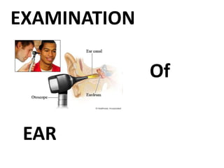
Examination of Ear
- 2. Otorrhoea (Ear discharge) Otalgia (Ear ache) Hearing Loss/impairment Vertigo Tinnitus Ear fullness/blockage Aural polyp Itching in ear Swelling and Deformity Foreign body/ injury
- 3. Discharge from the ear Etiology Infection – • EAC - otitis externa, otomycosis, furunculosis, acute dermatitis, neoplasm, TM joint disease • Middle ear – ASOM, CSOM, Mastoiditis, cholesteatoma • CSF leak Side – B/L, Right , left Onset – sudden – ASOM, gradual/insidious – CSOM, malignancy Amount – scanty –CSOM -AAD, Otitis externa, profuse – CSOM –TTD
- 4. • Duration – long – CSOM-AAD, Otitis externa intermediate – CSOM-TTD short – ASOM, Furuncle • Progress – intermittent – CSOM-TTD, continous – CSOM-AAD, granulations, malignancy • Nature – 1. purulent – furuncle, mastoiditis, malignant otitis externa, CSOM-AAD 2. Mucoid/mucopurulent – granular myringitis, CSOM-TTD, ASOM (late stage) 3. Watery – CSF leak, eczematous/viral otitis externa 4. Bloody – ASOM (initial stage), trauma, granulations, malignancy 5. Pulsatile – ASOM with pin point perforation, glomus tumour, ICA aneurysm
- 5. Colour – green – pseudomonas infection, yellow black – otomycosis, yellow Smell – odourless – allergic otitis externa, CSOM- TTD foul smell – CSOM- AAD, cholesteatoma Aggravating factors – cold, head bath, pharyngitis, tonsillitis – CSOM-TTD Preceding history – trauma – CSF leak, ear surgery, skin disease Associated complaints Ear ache – Acute otitis externa, pruritus – chronic otitis externa, otomycosis, eczema of skin – recurrent otitis externa, retro orbital pain – abscess, hearing loss, vertigo
- 6. Pain in and around the ear Etiology Primary otalgia – local causes – inflammation, trauma, neoplasm affecting external and middle ear, inner ear – no pain • Auricle – perichondritis, trauma • EAC – furuncle, impacted wax, acute otitis externa, FB, otomycosis, neoplasm, myringitis • Middle ear – ASOM, cholesteatoma, mastoiditis, ET obstruction, malignancy, CSOM- no pain unless otitis externa, intra cranial complications of CSOM • Barotrauma- due to flying or scuba diving • Referred otalgia.
- 7. Secondary otalgia Referred pain to ear from other regions of head and neck – common nerve supply V CN – Auriculo temporal branch of mandibular nerve – anterior part of pinna, TM, EAC – referred from dental, oral cavity, salivary glands, nose, PNS, TM joint, face, parotid VII CN – branch of facial nerve –skin of concha, anti helix, lobule, post EAC – referred in bell’s palsy, herpes zoster infection IX CN – Jacobson’s nerve – tympanic branch to middle ear, tympanic plexus, medial part of TM – referred from nasopharynx, oropharynx, tonsil, soft palate, styloid process, ET, mastoid Referred Otalgia
- 8. Duration – short – ASOM, perichondritis long – malignancy Nature – dull – impacted wax, secretory otitis media, eczematous otitis externa, sharp – furuncle throbbing – ASOM Location – front of ear – furuncle, deep in ear – middle ear pathology, behind ear – mastoiditis, lymphadenitis, below ear – ET pathology Aggravating and relieving factors Relieved on discharge from ear – ASOM, increase on swallowing – ASOM, increase on yawning, chewing – furuncle, increase on pulling pinna and pressing tragus – acute otitis externa
- 9. Associated factors Tinnitus present – acoustic neuroma Itching present – otomycosis Association with ear discharge, hearing loss Past history – trauma, ear surgery Psychogenic More on exertion and left side pain – CAD Pain is always more on lying down – increased blood supply- primary otalgia Costen’s syndrome – pain due to TM joint abnormality – defective bite – associated with tinnitus, vertigo, blocked sensation
- 10. Hard of hearing – if hearing loss can improve on treatment Deaf – very severe or profound with little or no residual hearing Laterality - Rt/Lt/bilateral 1. Unilateral – CSOM, Acoustic neuroma, mumps 2. Bilateral – presbycusis, meniere’s disease, otosclerosis, noise induced Onset – sudden – wax, viral deafness, ASOM, traumatic perforation, head injury, blast injury, vascular causes, acoustic trauma, labyrinthitis Gradual/insidious – CSOM, OME, otosclerosis, NIHL, presbycusis, acoustic neuroma
- 11. Type – conductive – defect in external and middle ear, SNHL – defect in inner ear or VIII CN, mixed Progress – stable – CSOM TTD (non discharging), perforated TM Progressive – CSOM AAD,CSOM TTD discharging, otosclerosis, meniere’s disease, acoustic neuroma, presbycusis Fluctuating – meniere’s disease, secretory otitis media Degree – mild – diseases of EAC like wax, FB, mild to moderate – diseases of middle ear, mild to profound – inner ear diseases
- 12. Duration – since birth – genetic, prenatal drugs, maternal infections, prolonged labour, infancy infections like mumps, measles, meningitis Recent – trauma, inflammation, neoplasm, vascular Childhood – ASOM, OME, young adults – otosclerosis, old age – presbycusis Family history – otosclerosis, meniere’s disease Drug history – ototoxic drugs like aminoglycoside, quinine, salicylates, cytotoxic drugs Occupational history – noisy enviroment Trauma, viral fever, psychogenic
- 13. Diplacusis – different pitch in both ears – meniere’s disease Paracusis Willisi – hears better in noisy surroundings – otosclerosis Hears better in quiet place – SNHL Autophony – hears own voice louder – serous otitis media, patulous ET Hyperacusis/ phonophobia – increased or painful sensitivity to everyday sound that wont trouble normal person – stapedius muscle paralysis, congenital syphilis Recruitment – cant hear at normal intensity but slight increase in intensity leads to discomfort – cochlear pathology
- 14. Perception of auditory sensation/sound ringing or noise with no external stimuli 33% population Classification Subjective tinnitus – only perceived by patient, Mainly psychogenic/functional, more common Objective tinnitus – perceived by patient as well as examiner. Seen in chronic contractions of palatal or tympanic muscles, live insects in ear, intracranial vascular tumours, patulous ET,AV malformations, clicking TM joint
- 15. Pulsatile tinnitus – non continous – idiopathic, non vascular causes like myoclonus, neoplasm, TM joint disease, vascular causes like HTN, atherosclerosis, otosclerosis, glomus tumour, anaemia, pregnancy, exercise Non pulsatile tinnitus – continous – with hearing loss seen in wax, FB, otitis media, otosclerosis, noise exposure, presbycusis, meniere’s disease, acoustic neuroma Without hearing loss – psychogenic, idiopathic, migraine
- 16. Site – ear/head Unilateral or bilateral Duration – short – middle ear disease, long – inner ear disease like ototoxicity, meniere’s disease Severity Fluctuant – meniere’s disease Past history – head injury, ear surgery, drug intake, noise exposure Aggravated by smoking – inner ear pathology Aggravated by yawning, blowing – ET dysfunction Relieved by putting pressure on side of neck – vascular cause
- 17. Associated with hearing loss – ear disease Tinnitus is first symptom of salicylate poisoning Auditory hallucination – in psychiatric patients – hear voices and sounds like music
- 18. Sensation of rotation of surrounding enviroment with respect to person or person with respect to surrounding. Disturbance of equilibrium or movements Associated with LOC – central cause, not associated – peripheral cause – inner ear Associated with loss of hearing – labyrinthitis, meniere’s disease, acoustic neuroma (U/L) Associated with discharging ear – labyrinthitis secondary to ASOM, CSOM Sudden onset – ear pathology Associated with posture – BPPV Associated with URTI – viral labyrinthitis
- 19. Duration – 6 weeks or longer – labyrinthitis, 24 minutes to 24 hours – meniere’s disease, few seconds several times a day – BPPV Otological causes – furuncle, wax due to stimulation of vagus nerve, ET catarrh due to negative pressure in middle ear, surgical trauma to inner ear due to mastoidectomy, stapedectomy, labyrinthitis, mumps, measles, meningitis, ototoxic drugs like streptomycin Outside ear causes – cervical pathology,CVS – HTN, hypotension, CNS – tumours, head injury, metabolic – DM, Hypothyroidism, anaemia Functional or idiopathic
- 20. Drugs like sedatives, antibiotics, anti hypertensives, aspirin Tullio’s phenomenon – very loud sound causes vertigo – seen in patients with labyrinthine fistula or those underwent fenestration operation Perilymph fistula- coughing and sneezing causes vertigo – due to rupture of round window (barotrauma) or at oval window due to stapedectomy
- 21. Fungal infection – otomycosis Allergy Wax Dermatitis Wax/ FB ET blockage/dysfunction – due to URTI – aggravated on lying down Patulous ET – disappears on lying down or alters with position of head Meniere’s disease – pressure in ear
- 22. Pedunculated mass in EAC arising from EAC or middle ear, associated with ear discharge, hearing loss and pain in ear Can bleed Etiology EAC – furuncle, trauma, FB, granuloma CSOM TTD/AAD Glomus tumour – red polyp which easily bleeds
- 23. Nasal complaints like nasal obstruction, discharge, post nasal discharge Throat complaints like irritation, dysphagia, change in voice Allergy and bronchial asthma – ET dysfunction, serous otitis media DM – Malignant otitis externa, sudden SNHL HTN – Sudden SNHL Radiation – SNHL Mumps, measles, chicken pox – SNHL Anti thyroid drugs - giddiness
- 24. Treatment for the same illness in the past or any other illness Diabetes, HTN, TB, Asthma and allergies, HIV, HBV, syphilis, radiation exposure Surgeries - ear, hospital admissions, Trauma Deliveries and pregnancies Drug history- at present or past- steroids, insulin, ocp, anti hypertensives, nasal decongestants, ototoxic drugs Allergy history – drugs or diet or allergen FOR DRUG ALLERGY – WRITE IN RED
- 25. Life style – exercise, sedentary, hygiene Food habits – regular-irregular, spicy-non spicy, nonveg- veg, excess tea or coffee Work place – noisy enviroment Home – dampness, pets, hobbies Alcohol, Tobacco – quantity, quality Sexual life Bladder & Bowel habits Menstrual history
- 26. Enquire about parents, siblings and children h/o similar illness in family Familial diseases like Peptic ulcer, cancers, allergies, diabetes and HTN, otosclerosis, deaf mutism, meniere’s disease Consanguinous marriage Infectious diseases- by contact – TB , acute infections
- 27. Children – immunisation schedule OBSTETRIC HISTORY – early deafness Ototoxic drugs to mother during 1st trimester Infections to mother – rubella, mumps Birth trauma Post natal jaundice
- 29. External Ear - Auricle/Pinna - Pre auricular region - Post auricular/Mastoid region EAC TM Middle ear mucosa Eustachian tube Facial nerve and other CN Neck Nose and Throat
- 30. INSPECTION Size – anotia (absence), microtia (small) Shape – cauliflower ear, bat ear (auricle protrudes anteriorly) Colour- red (perichondritis) Position- displacement of auricle forwards, laterally or inferiorly – mastoid abscess Swelling Scar Ulcer
- 32. PALPATION Superficial palpation - Using fingers (digital palpation) of cartilage and soft tissue Soft tissue – mobility of skin (lost in malignancy), thickening, swelling Raised temperature, tenderness Cartilage – defect or loss Deep palpation - Tragal tenderness- inflammation Painful movement of pinna – acute otitis externa
- 33. Sinus – pre auricular sinus Fistula Scar Swelling – cystic, lymphadenitis
- 34. INSPECTION Scar, dermatitis, swelling, fistula Change in mastoid contour Normally – uneven bone, skin over it mobile PALPATION Superficial palpation Skin – mobile Raised temperature Swelling – margins, cystic (sebaceous cyst) Iron out mastoid – smooth surface – coalescent mastoiditis
- 36. Deep palpation Tenderness – mastoiditis 1. Cymba concha – bony landmark for mastoid antrum 2. Midpoint of posterior border of mastoid 3. Tip of mastoid NECK EXAMINATION 1. Bezold’s abscess – pus track along SCM inferiorly 2. Citelli’s abscess – pus track along digastric into submandibular triangle 3. Luc’s abscess – pus around zygoma 4. IJV as hard cord on palpation along SCM – IJV thrombosis
- 37. Direct examination Without speculum • Size of meatus – atresia/wide/narrow • Wide – post op, syphilis, otosclerosis • Narrow – congenital atresia, scar due to trauma, burns, tumour - osteoma • Content of meatus – wax, discharge, FB, Polyp Digital examination – Adults – pinna pulled upwards, backwards and laterally, tragus pulled forwards Children – pinna pulled downwards and laterally
- 38. Look for furuncle, swelling Fungal infection (otomycosis) – black – aspergillus niger, yellow – candida albicans Polyp – probe test – if probe all around – arise from middle ear Impacted wax – whole EAC occluded Tumours With speculum examination – deep meatus to straighten the canal Largest speculum which can enter Black coated Introduce in slow rotatory fashion upto cartilage only
- 39. Cough – vagal irritation Sagging of posterior superior EAC – cholesteatoma, mastoid abscess, mastoiditis Absence of sensation in post sup EAC – Hitselberger’s sign – mass lesion in CP angle
- 40. Figure : Showing method of holding ear speculum during ear examination
- 41. Normal – pearly white colour, obliquely set, anterior and posterior malleolar fold (longer) Handle of malleus – whitish bony landmark Umbo Cone of light/light relex – triangular in shape anterio inferiorly – unreliable landmark Short or lateral process of malleus – small yellowish prominence – at 12 o clock position - always present – last landmark to be destroyed in disease Quadrants – 4 – imaginary line through tip of umbo, 2 nd vertically along handle of malleus – ant sup/inf, post sup/inf
- 43. Colour of TM – normal pearly white/greyish white, red in ASOM, acute myringitis Congestion with yellowish tint – ASOM Diffuse congestion/ localised at handle of malleus – ASOM Dark grey colour/dull appearance – tubal occlusion Dull white/thickened cotton like drum – senile sclerosis, scarring Chalky white appearance – tympanosclerosis after otitis media Dull lusterless/bulging – secretory otitis media Blue drum – transudative otitis media Dark blue drum - haemotympanum
- 44. TM normally inclined downwards and medially Protrude outwards – bulging drum – acute otitis media (pus),Haemotympanum (blood),OME with good ET function (air) – increase length of handle of malleus, less prominent short process and malleolar folds, absence of cone of light Pulled inwards – retracted TM- OME with poor ET function, ET obstruction, atelectasis – shortening of handle of malleus, more prominent short process, anterior and post malleolar folds, distorted cone of light Retraction – attic region (retraction pocket) if deepens – cholesteatoma sac
- 46. Siegle’s pneumatic speculum/ Valsalva maneuvre Normal – change in shape of triangular light reflex/ handle of malleus movement Decreased/absent – ET dysfunction, adhesive otitis media (fixed), ankylosis of ossicular chain, SOM Hypermobility – Patulous ET, atrophy of TM
- 48. Perforation – ovoid/ kidney shaped/ round, small, medium, large, sub total, total, central/marginal, pars tensa/attic, dry/wet, single/multiple (TB, measles, wegner’s granulomatosis), margins – regular/irregular, edge of perforation – thick (CSOM)/ thin (ASOM) Scars Bulla – grey/red/bluish pearls like structures attached to surface of TM Chalky white patch - tympanosclerosis
- 50. Tympanic membrane in OM 13–50
- 51. Only through perforation/ if TM thinned out/ semitransparent Middle ear mucosa – oedema/ polyp Granulation tissue Ossicles, ET, round window, oval window FB Fistula
- 52. Posterior rhinoscopy/ Nasopharyngoscopy/ DNE/ Through perforation Valsalva Maneuvre ET catheterisation – check patency of ET Few drops in ear if perforation and bitter taste in mouth
- 53. Three tests that are in common use: •Rinnie’s Test •Weber’s Test •Absolute Bone Conduction Test [ABC Test]. Tunning forks of 256, 512 and 1024 hertz are used.
- 54. Rinnie’s Test - In a normal individual air conduction is better than a bone conduction and the ratio of air: bone is 2:1. Test details Result Interpretation AC > BC Rinnie +ve Normal or SN loss BC > AC Rinnie –ve Conductive deafness AC = BC Rinnie = Mild conductive deafness BC > AC? Rinnie false negative This is seen when a patient has severe SN deafness on test side and normal hearing on non-test side. On application of vibrating tuning fork to deaf side the sound is trans- mitted to the non-test ear by bone and patient perceives this bone conduction and presumes that it is perceived by test ear, thus inter- preting it as BC > AC. Bone conduction level should be confirmed by Weber test. 256 hertz 512 hertz 1024 hertz Interpretation - + - - - - + + - Mild conductive deafness Moderate conductive deafness Severe conductive deafness Quantification of HL using Rinnie’s test
- 55. Weber Test
- 56. Absolute Bone Conduction (ABC) Test - In this test the bone conduction level of the patient is compared to that of clinician. During test ambient noise in the surrounding is reduced by pressing the tragus of the EAC. This is a modification of ‘ Swabach’s Test’ Test details Result Interpretation Clinician listens longer than patient Clinician listens equal to that of patient Patient listens longer than clinician. ABC reduced ABC normal ABC lengthened SN deafness Normal Conductive deafness
- 57. FISTULATEST Apply intermittent pressure on tragus/ siegle’s pneumatic speculum – ask patient to look straight – check for vertigo/ nystagmus towards opposite side Fallacy False -ve test: There can be a fistula without fistula test positive. This happens in dead labyrinth, or when fistula is temporarily blocked by cholesteatoma flakes. False + test: There can be fistula test positive without actually having a fistula. This happens when thinning of bony labyrinth occurs. This may happen in syphilis, cholesteatoma.
- 58. FACIAL NERVE EXAMINATION • Paralysis in ASOM, CSOM, Malignant otitis externa, herpes zoster, tumours and trauma • Wrinking of forehead/ closure of eyes/ loss of naso labial folds/ deviation of angle of mouth/ cant whistle or blow NOSE AND THROATEXAMINATION EYE EXAMINATION • Nystagmus • Corneal relex – absent in acoustic neuroma • Blue sclera – osteogenesis imperfecta • Papilloedema – CP angle tumours/ otitic hydrocephalus/ temporal abscess • Interstitial keratitis – congenital syphilis
- 59. The investigations commonly needed for ear diseases are: • Microbiology of pus discharge • Radiology of mastoid and PNS • Pure tone audiometry • Impedance audiometry • Caloric testing. • Other investigations that may be needed in specific case are: – High resolution CT scan – Fundoscopy when you suspect intracranial extension of disease – Evoked response audiometry – Electro nystagmography – Cranio corpography.
- 60. RADIOLOGICAL EXAMINATION OF THE EAR Views advised for temporal bone study are: • Laws position • Schuller’s position • Mayer’s position • Stenver’s position • Chausse III position. Out of these Schuller’s view is most com- monly advised view.