Optometry Times practical chair side advice VOL 12,NO.6 JUNE 2020

Optometry Times practical chairside advice VOL 12,nO.6 jUNE 2020 Contact Lenses PATIENTS AREN’T HEARING CONTACT LENS CARE INFORMATION By Ernie Bowling, OD, FAAO Doctors and staff need to better communicate recommendations to contact lens wearers 5 11 THINGS MY PATIENT WISHED HER PREVIOUS OD HAD TOLD HER By Crystal M. Brimer, OD, FAAO Patients clearly want more, not less, information and data from their eyecare providers 9 Ocular Surface Disease LACTOFERRIN LEVELS CAN DIAGNOSE DRY EYE DISEASE By Jeffrey Anshel, OD, FAAO New test allows distinction between causes of symptoms 11 PROPER DOCUMENTATION HELPS ASSURE PRIOR AUTHORIZATIONS By Vin T. Dang, OD, FAAO Newer dry eye medications require approval, and charting helps prove the need 12 Technology SWEPT-SOURCE AND MULTIMODAL OCT TECHNOLOGIES OFFER CLINICAL ADVANTAGES By Kerry Salsberg, OD The expenditure of embracing new technologies is worthwhile 15 TELEHEALTH SUCCESS HINGES ON BETTER TOOLS By Sandeep Jain, MD, FCCP, FAASM Communication and respect for everyone’s time needed, as well 20 Glaucoma PEDIATRIC GLAUCOMA: TYPES, TESTS AND TREATMENTS By Stacy Potwin, OD, and Michael Chaglasian, OD The different concerns in pediatric glaucoma compared with adults 1 CONTRAST SESITIVITY MANIFESTS IN GLAUCOMA PATIENT WITH NO CATARACTS By Benjamin P. Casella, OD, FAAO A comment from a patient prompts a doctor’s change in testing and discussion 21 Refractive 10 THINGS I WISH I KNEW EARLIER ABOUT VISION THERAPY By Marc B. Taub, OD, MS, FAAO, FCOVD, FNAP How to take on the challenge of providing therapy to improve vision Retina OCT, OCTA SHOWS PROMISE IN SCREENING FOR DR By Kyra Dorgeloh, OD; Lucy Zhu, OD; Nitish Mehta, MD; and Thomas A. Wong, OD This imaging technique provides depth-resolved images of retinal vasculature 1 RESOLVED COTTON-WOOL SPOT LEAVES RNFL DEFECT IN ITS WAKE By Leo Semes, OD, FAAO Imaging reveals diabetic retinopathy, cotton wool spot, and RNFL defect 30 Other WHAT A PRACTICE OWNER WOULD ADVISE HER YOUNGER SELF By Dori M. Carlson, OD An OD looks back 30 years of running a practice and recommends developing CEO skills 26

Recommended
Recommended
More Related Content
What's hot
What's hot (18)
Similar to Optometry Times practical chair side advice VOL 12,NO.6 JUNE 2020
Similar to Optometry Times practical chair side advice VOL 12,NO.6 JUNE 2020 (20)
More from Mero Eye
More from Mero Eye (20)
Recently uploaded
Recently uploaded (20)
Optometry Times practical chair side advice VOL 12,NO.6 JUNE 2020
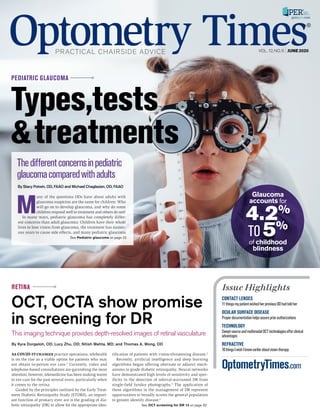
- 1. As COVID-19 changes practice operations, telehealth is on the rise as a viable option for patients who may not obtain in-person eye care.1 Currently, video and telephone-based consultations are garnishing the most attention; however, telemedicine has been making waves in eye care for the past several years, particularly when it comes to the retina. Guided by the principles outlined by the Early Treat- ment Diabetic Retinopathy Study (ETDRS), an import- ant function of primary eyec are is the grading of dia- betic retinopathy (DR) to allow for the appropriate iden- tification of patients with vision-threatening disease.2 Recently, artificial intelligence and deep learning algorithms began offering alternate or adjunct mech- anisms to grade diabetic retinopathy. Neural networks have demonstrated high levels of sensitivity and spec- ificity in the detection of referral-warranted DR from single-field fundus photographs.3 The application of these algorithms in the management of DR represent opportunities to broadly screen the general population to greater identify disease.4 By Kyra Dorgeloh, OD; Lucy Zhu, OD; Nitish Mehta, MD; and Thomas A. Wong, OD RETINA OptometryTimes.com CONTACT LENSES 11thingsmypatientwishedherpreviousODhadtoldher OCULAR SURFACE DISEASE Properdocumentationhelpsassurepriorauthorizations TECHNOLOGY Swept-sourceandmultimodalOCTtechnologiesofferclinical advantages REFRACTIVE 10thingsIwishIknewearlieraboutvisiontherapy Issue Highlights See OCT screening for DR 13 on page 32 OCT, OCTA show promise in screening for DR This imaging technique provides depth-resolved images of retinal vasculature M any of the questions ODs have about adults with glaucoma suspicion are the same for children: Who will go on to develop glaucoma, and why do some children respond well to treatment and others do not? In many ways, pediatric glaucoma has completely differ- ent concerns than adult glaucoma: Children have their whole lives to lose vision from glaucoma, the treatment has numer- ous years to cause side effects, and many pediatric glaucoma By Stacy Potwin, OD, FAAO and Michael Chaglasian, OD, FAAO Thedifferentconcernsinpediatric glaucomacomparedwithadults VOL.12,NO.6 JUNE 2020 PEDIATRIC GLAUCOMA Types,tests &treatments See Pediatric glaucoma on page 22 Glaucoma accounts for 4.2% TO 5% of childhood blindness
- 2. AUTOMATED TRUE COLOR RETINAL IMAGING T H E N E X T G E N E R AT I O N I N RE TINA & GL AUCOMA DIAGNOSTICS Scan, call 888.422.7313, email info@icare-usa.com, or visit www.icarecentervue.com 24-HOUR AT HOME TONOMETRY + Easy to use + Remote diurnal IOP curve + Long term monitoring + Alert notifications AUTOMATED TRUE COLOR RETINAL IMAGING + True color confocal imaging + Fast image acquisition + Ultra-high resolution + No dilation (2.5mm pupil size) NEW!
- 3. As our June issue goes to print, the country is opening up even more, and many more ODs are moving beyond non-emergent care. In that vein, this issue is chock full of solid clinical information. In our cover story this month, Drs. Stacy Potwin and Michael Chaglasian look at pediatric glaucoma and how diagnosis and treatment differs from that of adult patients with the disease. Also featured on our cover this month is a look at how OCT and OCTA show promise in screening for diabetic retinopathy. Drs. Kyra Dorgeloh, Lucy Zhu, Nitish Mehta, and Thomas Wong Dr. Ernie Bowling offers thoughts on better com- munication between patients and ODs to promote better contact lens care compliance. Dr. Crystal Brimer shares 11 things her patient wishes her previ- ous OD had told her. Dr. Brimer asked for only 5! Dr. Jeff Anshel explains the importance of lacto- ferrin in diagnosing dry eye, and Dr. Vin Dang shows how proper documentation helps ODs obtain prior authorizations for dry eye medications. Dr. Kerry Salsberg outlines clinical advantages that swept-source and multimodal OCT technologies offer. In addition, in a story from sister publication Medical Economics, Dr. Sandeep Jain discusses using better tools for telehelath success. Dr. Dori Carlson examined what was important as an activity for her master’s program. She reveals what she would advise her younger self. Dr. Marc Taub outlines 10 things he wishes he knew earlier in his career about vision therapy. And Dr. Leo Semes shares a case in which a resolved cot- ton-wool spot leaves a RNFL defect in its wake. Finally, Chief Optometric Editor Dr. Ben Casaella plans to better address contrast sensitivity with glau- coma patients after a patient comment. Other WHAT A PRACTICE OWNER WOULDWHAT A PRACTICE OWNER WOULD ADVISE HER YOUNGER SELFADVISE HER YOUNGER SELF By Dori M. Carlson, OD An OD looks back 30 years of running a practice and recommends developing CEO skills 26 3 | PRACTICAL CHAIRSIDE ADVICE By Mike Hennessy, Sr. Moving on into the summer Contact Lenses PATIENTS AREN’T HEARINGPATIENTS AREN’T HEARING CONTACT LENS CARE INFORMATIONCONTACT LENS CARE INFORMATION By Ernie Bowling, OD, FAAO Doctors and staff need to better communicate recommendations to contact lens wearers 5 11 THINGS MY PATIENT WISHED HER11 THINGS MY PATIENT WISHED HER PREVIOUS OD HAD TOLD HERPREVIOUS OD HAD TOLD HER By Crystal M. Brimer, OD, FAAO Patients clearly want more, not less, information and data from their eyecare providers 9 OcularSurfaceDisease LACTOFERRIN LEVELS CANLACTOFERRIN LEVELS CAN DIAGNOSE DRY EYE DISEASEDIAGNOSE DRY EYE DISEASE By Jeffrey Anshel, OD, FAAO New test allows distinction between causes of symptoms 11 PROPER DOCUMENTATION HELPSPROPER DOCUMENTATION HELPS ASSURE PRIOR AUTHORIZATIONSASSURE PRIOR AUTHORIZATIONS By Vin T. Dang, OD, FAAO Newer dry eye medications require approval, and charting helps prove the need 12 Technology SWEPT-SOURCE AND MULTIMODALSWEPT-SOURCE AND MULTIMODAL OCT TECHNOLOGIES OFFEROCT TECHNOLOGIES OFFER CLINICAL ADVANTAGESCLINICAL ADVANTAGES By Kerry Salsberg, OD The expenditure of embracing new technologies is worthwhile 15 TELEHEALTH SUCCESS HINGES ONTELEHEALTH SUCCESS HINGES ON BETTER TOOLSBETTER TOOLS By Sandeep Jain, MD, FCCP, FAASM Communication and respect for everyone’s time needed, as well 20 Glaucoma PEDIATRIC GLAUCOMA: TYPES,PEDIATRIC GLAUCOMA: TYPES, TESTS AND TREATMENTSTESTS AND TREATMENTS By Stacy Potwin, OD, and Michael Chaglasian, OD The different concerns in pediatric glaucoma compared with adults 1 CONTRAST SESITIVITY MANIFESTSCONTRAST SESITIVITY MANIFESTS IN GLAUCOMA PATIENT WITH NOIN GLAUCOMA PATIENT WITH NO CATARACTSCATARACTS By Benjamin P. Casella, OD, FAAO A comment from a patient prompts a doctor’s change in testing and discussion 21 Refractive 10 THINGS I WISH I KNEW EARLIER10 THINGS I WISH I KNEW EARLIER ABOUT VISION THERAPYABOUT VISION THERAPY By Marc B. Taub, OD, MS, FAAO, FCOVD, FNAP How to take on the challenge of providing therapy to improve vision 28 Retina OCT, OCTA SHOWS PROMISE INOCT, OCTA SHOWS PROMISE IN SCREENING FOR DRSCREENING FOR DR By Kyra Dorgeloh, OD; Lucy Zhu, OD; Nitish Mehta, MD; and Thomas A. Wong, OD This imaging technique provides depth-resolved images of retinal vasculature 1 RESOLVED COTTON-WOOL SPOTRESOLVED COTTON-WOOL SPOT LEAVES RNFL DEFECT IN ITS WAKELEAVES RNFL DEFECT IN ITS WAKE By Leo Semes, OD, FAAO Imaging reveals diabetic retinopathy, cotton wool spot, and RNFL defect 30 CHAIRMAN’SLETTER Table of contents COMPLIANCEWITHDIRECTIONSFORUSE WAS100%AMONGTHEUSERSOFA ONE-STEP HYDROGEN PEROXIDE LENSCARESYSTEM ASCOMPAREDTO37%AMONG MULTIPURPOSE SYSTEM USERS CONTACT LENS CARE page 5
- 4. 4 Chief Optometric EditorFROM THE Benjamin P. Casella, OD, FAAOChief Optometric Editor Editorial Advisory Board members are optometric thought leaders. They contribute ideas, offer suggestions, advise the editorial staff, and act as industry ambassadors for the journal.ErnestL.Bowling,OD,FAAOEditorEmeritus 2012-2017 JeffreyAnshel,OD,FAAO OcularNutritionSociety Encinitas,CA MelissaBarnett,OD,FAAO,FSLS UCDavisMedicalCenter Sacramento,CA SherryJ.Bass,OD,FAAO SUNYCollegeofOptometry NewYork,NY JustinBazan,OD ParkSlopeEye Brooklyn,NY ErnestL.Bowling,OD,FAAO Gadsden,AL CrystalBrimer,OD,FAAO CrystalVisionServices Wilmington,NC MichaelBrown,OD,MHS-CL,FAAO U.S.Depart.ofVeteransAffairs Huntsville,AL MileBrujic,OD,FAAO PremierVisionGroup BowlingGreen,OH MichaelA.Chaglasian,OD,FAAO IllinoisEyeInstitute Chicago,IL ClarkY.Chang,OD,MSA,MSc,FAAO WillsEyeHospital Philadelphia,PA A.PaulChous,OD,MA,FAAO ChousEyeCareAssociates Tacoma,WA MichaelP.Cooper,OD SolinskyEyeCare WestHartford,CT MelanieDenton,OD,MBA,FAAO SalisburyEyecareandEyewear Salisbury,NC MartaFabrykowski,OD,FAAO ManhattanEye,EarandThroat HospitalOphthalmology NewYork,NY StevenFerucci,OD,FAAO SepulvedaVAAmbulatoryCare Center&NursingHome Sepulveda,CA BarbaraFluder,OD WilliamsEyeInstitute Merrillville,IN LisaFrye,ABOC,FNAO EyeCareAssociates Birmingham,AL BenGaddie,OD,FAAO GaddieEyeCenters Louisville,KY DavidI.Geffen,OD,FAAO GordonWeissSchanzlin VisionInstitute SanDiego,CA JeffryD.Gerson,OD,FAAO WestGlenEyecare Shawnee,KS AlanGlazier,OD,FAAO ShadyGroveEyeandVisionCare Rockville,MD WhitneyHauser,OD SouthernCollegeofOptometry Memphis,TN ScottG.Hauswirth,OD,FAAO UniversityofColorado SchoolofMedicine Aurora,CO JamesHill,OD,FAAO MedicalUniversity ofSouthCarolina Charleston,SC MiltonM.Hom,OD,FAAO Azusa,CA DavidL.Kading,OD,FAAO SpecialtyEyecareGroup Kirkland,WA JenniferLyerly,OD TriangleVisionsOptometry Cary,NC KatherineM.Mastrota,MS,OD,FAAO HotelAssociationofNewYork CityHealthCenter NewYork,NY PamelaJ.Miller,OD,FAAO,JD Highland,CA AndrewS.Morgenstern,OD,FAAO WalterReedNationalMilitary Hosp. Bethesda,MD MohammadRafieetary,OD,FAAO CharlesRetinaInstitute Memphis,TN StuartRicher,OD,PhD,FAAO JamesLovellFederalHealthCare Facility NorthChicago,IL JohnRumpakis,OD,MBA,FAAO PracticeResourceManagement LakeOswego,OR ScottE.Schachter,OD AdvancedEyecare PismoBeach,CA LeoP.Semes,OD,FAAO UniversityofAlabamaatBirming- hamSchoolofOptometry Birmingham,AL DianaL.Shechtman,OD,FAAO NovaSoutheasternUniversity FortLauderdale,FL JosephP.Shovlin,OD,FAAO,DPNAP NortheasternEyeInstitute Scranton,PA DianaCanto-Sims,OD BuenaVistaOptical Chicago JosephSowka,OD,FAAO NovaSoutheasternUniversity CollegeofOptometry FortLauderdale,FL TracyL.SchroederSwartz,OD,FAAO MadisonEyeCare Madison,AL MarcB.Taub,OD,MS,FAAO,FCOVD SouthernCollegeofOptometry Memphis,TN WilliamD.Townsend,OD,FAAO AdvancedEyeCare Canyon,TX WilliamJ.Tullo,OD,FAAO TLCLaserEyeCenters/Prince- tonOptometricPhysicians Princeton,NJ ThomasA.Wong,OD StateUniversityofNewYork CollegeofOptometry NewYork,NY ChrisWroten,OD Bond-WrotenEyeClinic Hammond,LA Editorial Advisory Board JUNE 2020 | Physical and psychological changes of opening post COVID-19 P roceed with caution. This is one of several man- tras I have been attempting to live by lately. As practices begin to “open back up,” the process has been beset with caution. Early on, when the Centers for Disease Control and Prevention (CDC) recommended postponing non-emergent care, ODs knew that dipping our toes into the waters of “rou- tine” clinical life again would be slow. For starters, there are the physical barriers to infection and transmission that need to be in place. We all went on shopping sprees for non-contact forehead thermometers, partitions, filters, wipes, gloves, masks, alcohol, hydrogen peroxide. We updated office protocols: health checks for all who enter, people calling ahead or knocking on the door, questions about COVID-19 specific symp- toms and travel to regions deemed “hot spots,” frequent sanitation of all surfaces, changes to the flow of the building, and on and on. I got used to the physicality pretty quickly. We are used to sanitizing surfaces, anyway. I’m not bothered by the mask and glasses. I will say I’m not smart enough to keep the glasses from fogging. I’m thankful for the slit-lamp partition compli- ments of Zeiss. The air filters aren’t all that noisy. In my head What drives me nuts, however, is the psychology of the whole thing. Did I see any silent carriers today? Were my glasses on tight enough? Does my staff take things as seriously outside the office as I mandate at work? How many gloves are sufficient to store up? How long before I’m comfortable seeing more than a patient every hour? What are my bills going to look like as my Payroll Protection Plan (PPP) loan gets spent down? Will I miss checking a box somewhere on the loan forgive- ness application and have to pay it all back? Will we have that second spike we keep hearing about? I’d be lying if I told you these things weren’t all in my head at the time I penned this editorial. Many of these things are in your heads, too, and I want you to know you are absolutely not alone. As far as being a small business owner during all of this, I have felt alone at times. I was recently on a vir- tual happy hour with 8 friends, and I was the only one who wasn’t working from home. In fact, one friend was quick to say business was better than ever. Instead of countering that I had operated on less than 1 percent of my typical patient load for 2 months, I simply said, “Hear, hear.” My motive is not to be rhetorical and not to solicit answers. It is to be honest with you as my friend in optometry. I firmly believe that brighter days are ahead for us. We are in this together, and it is together that we will overcome. Stay safe, take care, and I sin- cerely hope to see you soon. Dr. Dori Carlson shares what she wishes she knew as a younger OD on page 25. ByBenjaminP.Casella,OD,FAAO Chief Optometric Editor Practices in Augusta, GA, with his father in his grandfather’s practice bpcasella@gmail.com 706-267-2972
- 5. | PRACTICAL CHAIRSIDE ADVICE 5 Contact Lenses By Ernie Bowling, OD, FAAO A n estimated 45 million U.S. residents enjoy the benefit of contact lens wear, but many of them might be at increased risk for complications stem- ming from improper wear and care.1 Unlike daily disposable, single-use contact lenses, those with longer replacement schedules must be main- tained. Contact lens solutions perform the essential functions of disinfecting, cleaning, and preserving the lenses to prevent infection and improve wear- ing comfort.2 Patient-doctor disconnect Far too often, the contact lens care regimen is given too little attention during the annual eye exam. Two surveys conducted to assess contact lens education revealed that one-third (32.9 percent) of contact lens wearers over 18 years of age recalled never hearing any contact lens wear and care recommen- dations, and only 19.8 percent recalled being told to avoid “topping off” their contact lens solutions. Yet there is a disconnect between what the patient hears and what the provider says. The same survey reported that the majority of pro- viders stated they shared care recommendations always or most of the time at initial visits, regu- lar checkups, and at complication-related visits.3 This gap between what providers say and what the patient hears might be a factor in the large pro- portion of contact lens wearers reporting behav- iors that place them at risk for contact lens-re- lated complications.4 Addressing this gap might improve contact lens wear and care practices. Patient recommendations So, how does a busy practice accomplish this? Fortunately, there are resources available. The American Optometric Association makes the fol- lowing recommendations for contact lens wearers:5 – Always wash and dry hands before handling contact lenses – Carefully and regularly clean contact lenses as directed by your eye doctor. Rub the con- tact lenses with fingers and rinse them thoroughly before soaking the lenses overnight in multipurpose solution that com- pletely covers each lens – Store lenses in the proper lens storage case and replace the case every 3 months or sooner. Clean the case after each use, and keep it open and dry between cleanings – Use only products recommended by your eyecare practitioner to clean and disinfect your lenses. Do not use saline solu- tion and rewetting drops to dis- infect lenses—that is not what they are designed to do – Use fresh solution to clean and store contact lenses. Never reuse old solution. Change contact lens solu- tion according to the manufactur- er’s recommen- dations, even if you don’t wear the lenses daily – Always follow the rec- ommended contact lens replacement schedule pre- scribed by your eye doctor – Remove contact lenses before swimming or entering a hot tub – See your eye doctor for regularly sched- uled contact lens and eye examination The American Academy of Ophthalmology also has recommendations for contact lens care,6 and the Centers for Disease Control and Prevention also has poster and patient information sheets available.7 Hydrogen peroxide Finally, a word about an old stand-by: hydro- gen peroxide. Despite its well-established disin- fection and safety benefits, the use of hydrogen peroxide lens care systems remains low in com- parison with multipurpose solution use.8 Hydro- gen peroxide care systems currently account for about 25 percent of lens care recommendations by U.S. practitioners.9 Noncompliant contact lens care behaviors are common among multipurpose solution users, including “topping off” solu- tion, failure to rub and rinse lenses, and infrequent lens case cleaning and replacement.10 Hydro- gen peroxide lens care systems are easy to use and limit the number of steps nec- essary to achieve disinfection. There is evidence of greater compliance with hydrogen perox- ide care systems versus multipurpose care sys- tems. A 2007 survey found compliance with directions for use was 100 percent among the users of a one-step hydrogen peroxide lens care system, in comparison with 37 percent among multipurpose system users.11 Hydrogen peroxide lens care systems provide practitioners with a means to address many of the concerns with lens care noncompliance. It definitely has its place in our arsenal of lens care regimens. REFERENCES 1. Cope JR, Collier SA, Nethercut H, Jones JM, Yates K, Yoder JS. Risk behaviors for contact lens-related eye infections among adults and adolescents—United States, 2016. MMWR Morb Mortal Wkly Rep. 2017 Aug 18;66(32):841-845. Patients aren’t hearing contact lens care information Doctors and staff need to better communicate recommendations to contact lens wearers TAKE-HOME MESSAGE The gap between what providers say about contact lens wear and care practic- es and what the patient hears needs to be addressed in order to prevent infection issues and improve wearing comfort. There is evidence of greater compliance with hydrogen peroxide systems versus multipurpose systems. There is a disconnect between what the patient hears and what the provider says ERNIE BOWLING, OD, FAAO is in practice in Rome, GA, and is past chair of the American Academy of Optometry’s Comprehensive Eye Care Section 32.9% of contact lens wearers over age 18 recalled never hearing any contact lens wear and care recommendations See Contact lens care on page 10
- 6. INDICATIONS AND IMPORTANT SAFETY INFORMATION EYLEA® (aflibercept) Injection 2 mg (0.05 mL) is indicated for the treatment of patients with Neovascular (Wet) Age-related Macular Degeneration (AMD), Macular Edema following Retinal Vein Occlusion (RVO), Diabetic Macular Edema (DME), and Diabetic Retinopathy (DR). CONTRAINDICATIONS • EYLEA is contraindicated in patients with ocular or periocular infections, active intraocular inflammation, or known hypersensitivity to aflibercept or to any of the excipients in EYLEA. WARNINGS AND PRECAUTIONS • Intravitreal injections, including those with EYLEA, have been associated with endophthalmitis and retinal detachments. Proper aseptic injection technique must always be used when administering EYLEA. Patients should be instructed to report any symptoms suggestive of endophthalmitis or retinal detachment without delay and should be managed appropriately. Intraocular inflammation has been reported with the use of EYLEA. EYLEA is a registered trademark of Regeneron Pharmaceuticals, Inc. References: 1. Early Treatment Diabetic Retinopathy Study Research Group. Fundus photographic risk factors for progression of diabetic retinopathy. ETDRS report number 12. Ophthalmology. 1991;98(5 suppl):823-833. 2. Care of the Patient With Diabetes Mellitus: Quick Reference Guide. American Optometric Association website. http://bit.ly/2M22OUJ. Accessed August 7, 2019. 3. Ferrucci S,Yeh B. Diabetic retinopathy by the numbers. Rev Optom. June 15, 2016. http://bit.ly/2KNNJ4E. Accessed August 7, 2019. © 2020, Regeneron Pharmaceuticals, Inc. All rights reserved. 777 Old Saw Mill River Road, Tarrytown, NY 10591 Educate your patients about living with DR and potential treatment options2,3 Refer DR patients for timely intervention • According to the AOA, you should refer patients with2,3 – Severe nonproliferative DR (NPDR) within 2 to 4 weeks – Proliferative DR (PDR) within 1 week Follow up to ensure they have visited a retina specialist Through early detection, monitoring, and timely referral, you can play a pivotal role in managing your DR patients’ vision1-3 HELPDRIVE PATIENT OUTCOMES Help your patients with DIABETIC RETINOPATHY (DR), and IF YOU SEE OR SUSPECT DR:
- 7. Brought to you by REGENERON anti-VEGF = anti–vascular endothelial growth factor; AOA = American Optometric Association. WARNINGS AND PRECAUTIONS (cont’d) • Acute increases in intraocular pressure have been seen within 60 minutes of intravitreal injection, including with EYLEA. Sustained increases in intraocular pressure have also been reported after repeated intravitreal dosing with VEGF inhibitors. Intraocular pressure and the perfusion of the optic nerve head should be monitored and managed appropriately. • There is a potential risk of arterial thromboembolic events (ATEs) following intravitreal use of VEGF inhibitors, including EYLEA. ATEs are defined as nonfatal stroke, nonfatal myocardial infarction, or vascular death (including deaths of unknown cause). The incidence of reported thromboembolic events in wet AMD studies during the first year was 1.8% (32 out of 1824) in the combined group of patients treated with EYLEA compared with 1.5% (9 out of 595) in patients treated with ranibizumab; through 96 weeks, the incidence was 3.3% (60 out of 1824) in the EYLEA group compared with 3.2% (19 out of 595) in the ranibizumab group. The incidence in the DME studies from baseline to week 52 was 3.3% (19 out of 578) in the combined group of patients treated with EYLEA compared with 2.8% (8 out of 287) in the control group; from baseline to week 100, the incidence was 6.4% (37 out of 578) in the combined group of patients treated with EYLEA compared with 4.2% (12 out of 287) in the control group. There were no reported thromboembolic events in the patients treated with EYLEA in the first six months of the RVO studies. ADVERSE REACTIONS • Serious adverse reactions related to the injection procedure have occurred in <0.1% of intravitreal injections with EYLEA including endophthalmitis and retinal detachment. • The most common adverse reactions (≥5%) reported in patients receiving EYLEA were conjunctival hemorrhage, eye pain, cataract, vitreous detachment, vitreous floaters, and intraocular pressure increased. Please see Brief Summary of Prescribing Information on the following pages. 03/2020 EYL.20.02.0082 The more you know about emerging clinical science about anti-VEGF and other potential therapies for DR, the better you can help inform your patients about how treatment may be able to help Refer patients to a retina specialist who can treat DR2,3 Continue to monitor your patients with DR2,3 • The AOA recommends frequent monitoring of patients2 – At least every 6 to 8 months in patients with moderate NPDR and more frequently for patients with greater disease severity2 Visit diabeticretinaldisease.com for additional information and useful patient resources
- 8. 1 INDICATIONS AND USAGE EYLEA is a vascular endothelial growth factor (VEGF) inhibitor indicated for the treatment of: Neovascular (Wet) Age-Related Macular Degeneration (AMD); Macular Edema Following Retinal Vein Occlusion (RVO); Diabetic Macular Edema (DME); Diabetic Retinopathy (DR). 4 CONTRAINDICATIONS 4.1 Ocular or Periocular Infections EYLEA is contraindicated in patients with ocular or periocular infections. 4.2 Active Intraocular Inflammation EYLEA is contraindicated in patients with active intraocular inflammation. 4.3 Hypersensitivity EYLEA is contraindicated in patients with known hypersensitivity to aflibercept or any of the excipients in EYLEA. Hypersensitivity reactions may manifest as rash, pruritus, urticaria, severe anaphylactic/anaphylactoid reactions, or severe intraocular inflammation. 5 WARNINGS AND PRECAUTIONS 5.1 Endophthalmitis and Retinal Detachments. Intravitreal injections, including those with EYLEA, have been associated with endophthalmitis and retinal detachments [see Adverse Reactions (6.1)]. Proper aseptic injection technique must always be used when administering EYLEA. Patients should be instructed to report any symptoms suggestive of endophthalmitis or retinal detachment without delay and should be managed appropriately [see Patient Counseling Information (17)]. 5.2 Increase in Intraocular Pressure. Acute increases in intraocular pressure have been seen within 60 minutes of intravitreal injection, including with EYLEA [see Adverse Reactions (6.1)]. Sustained increases in intraocular pressure have also been reported after repeated intravitreal dosing with vascular endothelial growth factor (VEGF) inhibitors. Intraocular pressure and the perfusion of the optic nerve head should be monitored and managed appropriately. 5.3 Thromboembolic Events. There is a potential risk of arterial thromboembolic events (ATEs) following intravitreal use of VEGF inhibitors, including EYLEA. ATEs are defined as nonfatal stroke, nonfatal myocardial infarction, or vascular death (including deaths of unknown cause). The incidence of reported thromboembolic events in wet AMD studies during the first year was 1.8% (32 out of 1824) in the combined group of patients treated with EYLEA compared with 1.5% (9 out of 595) in patients treated with ranibizumab; through 96 weeks, the incidence was 3.3% (60 out of 1824) in the EYLEA group compared with 3.2% (19 out of 595) in the ranibizumab group. The incidence in the DME studies from baseline to week 52 was 3.3% (19 out of 578) in the combined group of patients treated with EYLEA compared with 2.8% (8 out of 287) in the control group; from baseline to week 100, the incidence was 6.4% (37 out of 578) in the combined group of patients treated with EYLEA compared with 4.2% (12 out of 287) in the control group. There were no reported thromboembolic events in the patients treated with EYLEA in the first six months of the RVO studies. 6 ADVERSE REACTIONS The following potentially serious adverse reactions are described elsewhere in the labeling: • Hypersensitivity [see Contraindications (4.3)] • Endophthalmitis and retinal detachments [see Warnings and Precautions (5.1)] • Increase in intraocular pressure [see Warnings and Precautions (5.2)] • Thromboembolic events [see Warnings and Precautions (5.3)] 6.1 Clinical Trials Experience. Because clinical trials are conducted under widely varying conditions, adverse reaction rates observed in the clinical trials of a drug cannot be directly compared to rates in other clinical trials of the same or another drug and may not reflect the rates observed in practice. A total of 2980 patients treated with EYLEA constituted the safety population in eight phase 3 studies. Among those, 2379 patients were treated with the recommended dose of 2 mg. Serious adverse reactions related to the injection procedure have occurred in <0.1% of intravitreal injections with EYLEA including endophthalmitis and retinal detachment. The most common adverse reactions (≥5%) reported in patients receiving EYLEA were conjunctival hemorrhage, eye pain, cataract, vitreous detachment, vitreous floaters, and intraocular pressure increased. Neovascular (Wet) Age-Related Macular Degeneration (AMD). The data described below reflect exposure to EYLEA in 1824 patients with wet AMD, including 1223 patients treated with the 2-mg dose, in 2 double-masked, controlled clinical studies (VIEW1 and VIEW2) for 24 months (with active control in year 1). Safety data observed in the EYLEA group in a 52-week, double-masked, Phase 2 study were consistent with these results. Table 1: Most Common Adverse Reactions (≥1%) in Wet AMD Studies Baseline to Week 52 Baseline to Week 96 Adverse Reactions EYLEA (N=1824) Active Control (ranibizumab) (N=595) EYLEA (N=1824) Control (ranibizumab) (N=595) Conjunctival hemorrhage 25% 28% 27% 30% Eye pain 9% 9% 10% 10% Cataract 7% 7% 13% 10% Vitreous detachment 6% 6% 8% 8% Vitreous floaters 6% 7% 8% 10% Intraocular pressure increased 5% 7% 7% 11% Ocular hyperemia 4% 8% 5% 10% Corneal epithelium defect 4% 5% 5% 6% Detachment of the retinal pigment epithelium 3% 3% 5% 5% Injection site pain 3% 3% 3% 4% Foreign body sensation in eyes 3% 4% 4% 4% Lacrimation increased 3% 1% 4% 2% Vision blurred 2% 2% 4% 3% Intraocular inflammation 2% 3% 3% 4% Retinal pigment epithelium tear 2% 1% 2% 2% Injection site hemorrhage 1% 2% 2% 2% Eyelid edema 1% 2% 2% 3% Corneal edema 1% 1% 1% 1% Retinal detachment <1% <1% 1% 1% Less common serious adverse reactions reported in <1% of the patients treated with EYLEA were hypersensitivity, retinal tear, and endophthalmitis. Macular Edema Following Retinal Vein Occlusion (RVO). The data described below reflect 6 months exposure to EYLEA with a monthly 2 mg dose in 218 patients following CRVO in 2 clinical studies (COPERNICUS and GALILEO) and 91 patients following BRVO in one clinical study (VIBRANT). Table 2: Most Common Adverse Reactions (≥1%) in RVO Studies CRVO BRVO Adverse Reactions EYLEA (N=218) Control (N=142) EYLEA (N=91) Control (N=92) Eye pain 13% 5% 4% 5% Conjunctival hemorrhage 12% 11% 20% 4% Intraocular pressure increased 8% 6% 2% 0% Corneal epithelium defect 5% 4% 2% 0% Vitreous floaters 5% 1% 1% 0% Ocular hyperemia 5% 3% 2% 2% Foreign body sensation in eyes 3% 5% 3% 0% Vitreous detachment 3% 4% 2% 0% Lacrimation increased 3% 4% 3% 0% Injection site pain 3% 1% 1% 0% Vision blurred 1% <1% 1% 1% Intraocular inflammation 1% 1% 0% 0% Cataract <1% 1% 5% 0% Eyelid edema <1% 1% 1% 0% Less common adverse reactions reported in <1% of the patients treated with EYLEA in the CRVO studies were corneal edema, retinal tear, hypersensitivity, and endophthalmitis. Diabetic Macular Edema (DME) and Diabetic Retinopathy (DR). The data described below reflect exposure to EYLEA in 578 patients with DME treated with the 2-mg dose in 2 double-masked, controlled clinical studies (VIVID and VISTA) from baseline to week 52 and from baseline to week 100. Table 3: Most Common Adverse Reactions (≥1%) in DME Studies Baseline to Week 52 Baseline to Week 100 Adverse Reactions EYLEA (N=578) Control (N=287) EYLEA (N=578) Control (N=287) Conjunctival hemorrhage 28% 17% 31% 21% Eye pain 9% 6% 11% 9% Cataract 8% 9% 19% 17% Vitreous floaters 6% 3% 8% 6% Corneal epithelium defect 5% 3% 7% 5% Intraocular pressure increased 5% 3% 9% 5% Ocular hyperemia 5% 6% 5% 6% Vitreous detachment 3% 3% 8% 6% Foreign body sensation in eyes 3% 3% 3% 3% Lacrimation increased 3% 2% 4% 2% Vision blurred 2% 2% 3% 4% Intraocular inflammation 2% <1% 3% 1% Injection site pain 2% <1% 2% <1% Eyelid edema <1% 1% 2% 1% Less common adverse reactions reported in <1% of the patients treated with EYLEA were hypersensitivity, retinal detachment, retinal tear, corneal edema, and injection site hemorrhage. Safety data observed in 269 patients with nonproliferative diabetic retinopathy (NPDR) through week 52 in the PANORAMA trial were consistent with those seen in the phase 3 VIVID and VISTA trials (see Table 3 above). 6.2 Immunogenicity. As with all therapeutic proteins, there is a potential for an immune response in patients treated with EYLEA. The immunogenicity of EYLEA was evaluated in serum samples. The immunogenicity data reflect the percentage of patients whose test results were considered positive for antibodies to EYLEA in immunoassays. The detection of an immune response is highly dependent on the sensitivity and specificity of the assays used, sample handling, timing of sample collection, concomitant medications, and underlying disease. For these reasons, comparison of the incidence of antibodies to EYLEA with the incidence of antibodies to other products may be misleading. In the wet AMD, RVO, and DME studies, the pre-treatment incidence of immunoreactivity to EYLEA was approximately 1% to 3% across treatment groups. After dosing with EYLEA for 24-100 weeks, antibodies to EYLEA were detected in a similar percentage range of patients. There were no differences in efficacy or safety between patients with or without immunoreactivity. 8 USE IN SPECIFIC POPULATIONS. 8.1 Pregnancy Risk Summary Adequate and well-controlled studies with EYLEA have not been conducted in pregnant women. Aflibercept produced adverse embryofetal effects in rabbits, including external, visceral, and skeletal malformations. A fetal No Observed Adverse Effect Level (NOAEL) was not identified. At the lowest dose shown to produce adverse embryofetal effects, systemic exposures (based on AUC for free aflibercept) were approximately 6 times higher than AUC values observed in humans after a single intravitreal treatment at the recommended clinical dose [see Animal Data]. Animal reproduction studies are not always predictive of human response, and it is not known whether EYLEA can cause fetal harm when administered to a pregnant woman. Based on the anti-VEGF mechanism of action for aflibercept, treatment with EYLEA may pose a risk to human embryofetal development. EYLEA should be used during pregnancy only if the potential benefit justifies the potential risk to the fetus. All pregnancies have a background risk of birth defect, loss, or other adverse outcomes. The background risk of major birth defects and miscarriage for the indicated population is unknown. In the U.S. general population, the estimated background risk of major birth defects and miscarriage in clinically recognized pregnancies is 2-4% and 15-20%, respectively. Data Animal Data In two embryofetal development studies, aflibercept produced adverse embryofetal effects when administered every three days during organogenesis to pregnant rabbits at intravenous doses ≥3 mg per kg, or every six days during organogenesis at subcutaneous doses ≥0.1 mg per kg. Adverse embryofetal effects included increased incidences of postimplantation loss and fetal malformations, including anasarca, umbilical hernia, diaphragmatic hernia, gastroschisis, cleft palate, ectrodactyly, intestinal atresia, spina bifida, encephalomeningocele, heart and major vessel defects, and skeletal malformations (fused vertebrae, sternebrae, and ribs; supernumerary vertebral arches and ribs; and incomplete ossification). The maternal No Observed Adverse Effect Level (NOAEL) in these studies was 3 mg per kg. Aflibercept produced fetal malformations at all doses assessed in rabbits and the fetal NOAEL was not identified. At the lowest dose shown to produce adverse embryofetal effects in rabbits (0.1 mg per kg), systemic exposure (AUC) of free aflibercept was approximately 6 times higher than systemic exposure (AUC) observed in humans after a single intravitreal dose of 2 mg. 8.2 Lactation Risk Summary There is no information regarding the presence of aflibercept in human milk, the effects of the drug on the breastfed infant, or the effects of the drug on milk production/excretion. Because many drugs are excreted in human milk, and because the potential for absorption and harm to infant growth and development exists, EYLEA is not recommended during breastfeeding. The developmental and health benefits of breastfeeding should be considered along with the mother’s clinical need for EYLEA and any potential adverse effects on the breastfed child from EYLEA. 8.3 Females and Males of Reproductive Potential Contraception Females of reproductive potential are advised to use effective contraception prior to the initial dose, during treatment, and for at least 3 months after the last intravitreal injection of EYLEA. Infertility There are no data regarding the effects of EYLEA on human fertility. Aflibercept adversely affected female and male reproductive systems in cynomolgus monkeys when administered by intravenous injection at a dose approximately 1500 times higher than the systemic level observed humans with an intravitreal dose of 2 mg. A No Observed Adverse Effect Level (NOAEL) was not identified. These findings were reversible within 20 weeks after cessation of treatment. 8.4 Pediatric Use. The safety and effectiveness of EYLEA in pediatric patients have not been established. 8.5 Geriatric Use. In the clinical studies, approximately 76% (2049/2701) of patients randomized to treatment with EYLEA were ≥65 years of age and approximately 46% (1250/2701) were ≥75 years of age. No significant differences in efficacy or safety were seen with increasing age in these studies. 17 PATIENT COUNSELING INFORMATION In the days following EYLEA administration, patients are at risk of developing endophthalmitis or retinal detachment. If the eye becomes red, sensitive to light, painful, or develops a change in vision, advise patients to seek immediate care from an ophthalmologist [see Warnings and Precautions (5.1)]. Patients may experience temporary visual disturbances after an intravitreal injection with EYLEA and the associated eye examinations [see Adverse Reactions (6)]. Advise patients not to drive or use machinery until visual function has recovered sufficiently. BRIEF SUMMARY—Please see the EYLEA full Prescribing Information available on HCP.EYLEA.US for additional product information. Manufactured by: Regeneron Pharmaceuticals, Inc. 777 Old Saw Mill River Road Tarrytown, NY 10591 EYLEA is a registered trademark of Regeneron Pharmaceuticals, Inc. © 2019, Regeneron Pharmaceuticals, Inc. All rights reserved. Issue Date: 08/2019 Initial U.S. Approval: 2011 Based on the August 2019 EYLEA® (aflibercept) Injection full Prescribing Information. EYL.19.07.0306
- 9. | PRACTICAL CHAIRSIDE ADVICE 9 Contact Lenses By Crystal M. Brimer, OD, FAAO O Ds often talk about the state of the contact lens indus- try and patient behavior in an attempt to shape healthy habits. Positive clinical outcomes occur when ODs identify noncompliance hurdles before they appear, and steer patients clear of them. Therefore, it is important that optom- etrists occasionally spend time con- necting with patients to determine where gaps in knowledge lie. Doing so lets us discover our strengths and weaknesses and helps us become better educators. I recently asked a contact lens wearer to tell me 5 things she wished her eye doctor had told her during previous visits. She replied not with 5—but with 11! The most significant takeaway from our dis- cussion was this: The topics that interested her most were the topics ODs address the most. I asked her to rank them in order of importance. EDUCATE ME ON THE IMPORTANCE OF SUNGLASSES FROM INFANCY TO ELDERLY I love that she added “from infancy to elderly.” Not unlike many of our patients, she cares for two children and elderly parents, and she wants to be a responsible caregiver. Additionally, remem- ber that the mother is the CEO of the house. One piece of advice: Never forget her nurturing and logistical obligations because they make up what and who she represents to your practice. ODs often incentivize patients to add sunwear to their contact lens purchases. It is a target in many offices, but it still gets overlooked among the hustle and bustle. I recall another patient conversation in which, despite our yearly persuasion attempts, it was the patient’s buddy’s cool sunglasses that moti- vated him to finally get a pair of glasses at all, enabling him to reduce his contact lens wear time and dependence. Perhaps we should keep in mind that though glasses, contact lenses, dilation, pressure checks, and refractions may be fun for us (or not), they are defi- nitely not enjoyed by patients. So, let’s start talking up the impor- tance of sun protection! It is an area that is seen as both functional and fashionable. Many patients also see sunglasses as something extra they are doing to be good to themselves ver- sus the functional necessity of con- tact lenses and back-up glasses. Sun- glasses are a breath of fresh air. EDUCATE ME ABOUT THE IMPORTANCE OF NUTRITION IN CARING FOR MY EYES She also wants to know what foods to incorporate into her diet, how much, how often, and why? Again, I am so excited that a patient wants to hear this! But I fear that in many offices, patients want to know more than the doctor is equipped to answer when it comes to nutrition. Nutrition is very trendy among younger gen- erations. It is common for young patients to be into the effects of nutrition on all aspects of the body. Older generations also have an interest in nutrition, mainly for the preservation and recov- ery of the body. This puts pressure on ODs to continually learn more and be able to provide information to patients. We have long been accustomed to recommending Age-Related Eye Disease Study (AREDS) supplements and more recently, omega supplements, to specific patients. But I challenge optometrists to create more holistic value into their care by spending a cou- ple of hours researching foods that are benefi- cial to patients. EDUCATE ME ON PREVENTATIVE THINGS I CAN DO TO KEEP MY EYES HEALTHY, LIKE TEA TREE EYEWASH Disclosure: This came closely following a breakout of lice at her child’s school. Many times, ODs focus energy educating the “enti- tled” contact lens wearer who believes there are no consequences for abusing lenses. ODs can become labored by repetitive discussions with such a patient. If nothing more, perhaps this patient’s com- ment can serve as a ray of hope and encourage- ment to not become cynical and to keep learning and digging for more ways to educate patients because they generally do want to be healthy and proactive. WHEN SHOULD I CALL MY EYE DOCTOR IF I THINK I HAVE SCRATCHED MY EYE OR HAVE REDNESS? The patient’s pre-existing belief was that she just needed to rinse and rest. But still, she wondered how to judge that (particularly on a weekend when she can’t get in touch with her doctor). Another great question! It motivates me to cre- ate a postcard or rack card to educate contact lens patients on the importance of good care solu- tions, proper care, what to do before calling, and when to call. It is also reminder to have a triage card at the front desk to guide staff when a caller needs to be seen emergently, urgently, or at the next avail- able opening. This is especially important when the doctor is not in the office because once the patient calls, the practice is liable for what is said. EDUCATE ME ON THE IMPACT OF LONG- TERM CONTACT LENS WEAR She wanted to know if her eyes need a break, more than overnight. The fact that this ques- tion’s answer is not common knowledge at this point is disappointing, and it compels me to dis- close that she is not a patient of mine. (I had to throw that in). I hope most ODs are encouraging their contact lens wearers to take their lenses out as soon as they get home and have awake hours with their glasses on. I assume her doctor did too, but maybe it needed to be said by the technician as well, and perhaps even the front desk when she checked out and again when she picked her lenses up. This is another great chance to restate import- ant messages in writing. More than I asked Without previous discussion I had asked her for 5 things she wished her doctor had told her, and she spit out 11 concerns faster than you can imagine. Here are the other 6: – Are there exercises I can do for my eyes? 11 things my patient wished her previous OD had told her Patients clearly want more, not less, information and data from their eyecare providers TAKE-HOME MESSAGE Communication and education are highly correlated with better patient adherence and when ODs spend time connecting with contact lens wearers one-on-one, the degree to which patients follow contact lens recommendations increas- es. Establishing rapport, asking questions and sharing knowledge with contact lens patients is a great way to guarantee clinical success. BY CRYSTAL M. BRIMER, OD, FAAO In private practice in Wilmington, NC We need a way to communicate with our patients that does not automatically put them on the defense See Previous OD on page 10
- 10. 10 Contact Lenses disinfection-49856. Accessed 5/26/20. 10. Guthrie S, Dumbleton K, Jones L. Is there a relationship between care system and compliance? Contact Lens Spectr. 2016 Apr;31(4):40-43. 11. Dumbleton K, Richter D, Bergenske P, Jones LW. Compliance with lens replacement and the interval between eye examinations. Optom Vis Sci. 2013 Apr;90(4):351-8. 2. Bloise L. Contact lens care and maintenance. J Fr Ophtalmol. 2017 Apr;40(4):329-337. 3. Konne NM, Collier SA, Spangler J, Cope JR. Healthy contact lens behaviors communicated by eye care providers and recalled by patients – United States, 2018. MMWR Morb Mortal Wkly Rep. 2019 Aug 16;68(32):693-697. 4. Cope JR, Collier SA, Rao MM, Chalmers R, Mitchell GL, Richdale K, Wagner H, Kinoshita BT, Lam DY, Sorbara L, Zimmerman A, Yoder JS, Beach MJ. Contact lens wearer demographics and risk behaviors for contact lens-related eye infections—United States, 2014. MMWR Morb Mortal Wkly Rep. 2015 Aug 21;64(32):865-870. 5. American Optometric Association. What You Need to Know About Contact Lens Hygiene & Compliance. Available at: https://www.aoa.org/patients-and-public/ caring-for-your-vision/contact-lenses/what-you-need- to-know-about-contact-lens-hygiene-and-compliance. Accessed 5/26/20. 6. Boyd K. How to Take Care of Contact Lenses. Am Acad Ophthalmology. Available at. https://www.aao. org/eye-health/glasses-contacts/contact-lens-care. Accessed 5/26/20. 7. Centers for Disease Control and Prevention. CDC-INFO On Demand—Publications. Available at: https://wwwn.cdc.gov/pubs/cdcinfoondemand. aspx?ProgramID=192. Accessed 5/26/20. 8. Nichols JJ. Contact lenses 2016. CL Spectrum. 2017 Jan;32(1):22-25,27,29,55. 9. Chalmers RL. A fresh look at one-step hydrogen peroxide disinfection. Rev Optom. 2014 Aug. Available at: https://www.reviewofoptometry.com/article/a- fresh-look-at-one-step-hydrogen-peroxide-lens- JUNE 2020 | – Is there a health difference between the var- ious lenses I can choose from? – Is there a difference between name brand and generic contact lens products? Answer honestly, and not because you get free samples. – What eye drops should I use for moisturiz- ing, allergies, red eyes when I’m wearing contact lenses versus when I’m not? Should I limit how often and for how long I wear them? – Is there any risk with wearing cosmetic con- tact lenses? What?? These are things I want to tell patients about but sometimes encounter resistance when I try. She wants this information? So where is the disconnect? Perhaps ODs need a better way to deliver infor- mation in which the recommendation is not always directly linked to a sale. Also, ODs need a way to communicate with patients that does not automatically put them on the defense, some- thing nonintimidating. We could start with video education in the waiting room and online, but I think one of the best ways is to invest a few hours into writing key messages and printing them to accompany every contact lens exam. If nothing else, it will give the impression that you care about your patients’ wellbeing and wearing success. Updated care This past year, I have made a lot of changes to the physical appearance and functionality of my practice. Physical changes have transformed the tone of the practice, and this in turn affects patient demeanor. When patients walk through my prac- tice’s door, they experience an undeniable sen- sation that I care. I provide bound booklets with information to help them, not only through products and treat- ments that we offer, but with lifestyle changes that will improve stress levels, quality of sleep, exercise, and diet. I chose a a three-tiered, good-better-best for- mat for these booklets, which allows patients the option to choose a lesser form of over-the-counter (OTC) treatment or go directly for the best care my office can offer. This helps keep patient expectations in line with the treatments they have chosen and ensures that patients are constantly aware of the advanced options I offer without me “selling” a specific contact lens or treatment. I am trying to continually improve, listen to patients, and deliver what they need in a way that will encourage them to listen, absorb, and implement. Perhaps this will have to be a life-long quest, but I would like to think I can get there sooner than that! Contact lens care Continued from page 5 Previous OD Continued from page 9 crystalbrimerod@gmail.com Dr.Bowling receivedhisDoctorofOptometryandMasterofScienceinPhysiologi- calOpticsfromtheUABSchoolofOptometry. erniebowling@icloud.com Have a triage card at the front desk to guide your staff when a caller needs to be seen emergently, urgently, or at the next available opening recalled being told to avoid “topping off” their contact lens solutions 25PERCENTof lens care recommendations by U.S. practitioners are hydrogen peroxide care systems 19.8PERCENT (Photocredit:LIGHTFIELDSTUDIOS@AdobeStock)
- 11. | PRACTICAL CHAIRSIDE ADVICE 11 Ocular Surface Disease By Jeffrey Anshel, OD, FAAO I t should come as no surprise to eye- care practitioners who address dry eye in their practices that a nutri- tional approach to this disorder is effective. Unfortunately, most practi- tioners do not adequately test for the source of dry eye and instead attempt to offer a “blanket” approach that might or might not work. However, with proper testing, a com- prehensive nutritional approach can be valuable for addressing both aqueous deficient dry eye (ADDE) and evaporative dry eye (EDE). This process would be more expedi- ent if we could distinguish between the two forms quickly and accurately, as well as confirm the diagnosis of dry eye disease (DED) versus aller- gic conjunctivitis. Clinical tests Because there are many causes of DED, it is a chal- lenge to efficiently test for the cause of the disor- der. Most practitioners rely on one of the many questionnaires that are available, but these take some time to complete and depend on patient recall of their symptoms. Dry eyes and ocular allergies can have many overlapping complaints, making it more challenging to determine the specific dis- order needing therapy. Many clinical tests are available to determine the source of DED. These include tear film breakup time (TBUT), which determines tear film integrity; Schirmer’s test and Menicon Zone-Quick (phenol red thread) for tear volume; TearLab Osmolarity System (TearLab) for osmolarity; vital dyes rose bengal and lissamine green for cell integrity; lid wiper epitheliopathy to observe an increase the friction between the lid margin conjunctiva and the ocular surface; and Advanced Tear Diagnos- tics TearScan 300 microassay to measure lactofer- rin protein and IgE levels in the tear film. Evaluating the tear layer involves using more than just any one of these tests. Because of the variety of causes and several factors involved in tear film instability, practitioners should incor- porate these tests into a pre-examination routine. Any patient who complains of excessive or defi- cient tearing, redness, irritation, discharge, or any other typical anterior ocular complaint should be screened prior to seeing the doctor. Getting to the “root” While much of the media surrounding DED focuses on lipid layer enhancement due to mei- bomian gland dysfunction, just adding “fish oil” to a tear layer is not adequate to resolve the underlying source of the disease process. One analogy to consider is a visit to the dentist with a cavity in one tooth. The dentist would not think of just “cap- ping” the tooth without treating the underlying root to address the source of the degeneration. Likewise, simply enhancing the lipid layer of the tears without addressing the “root” of the tear layer (the mucin layer) will not manifest a complete solution to the problem. Lactoferrin a DED indicator Lactoferrin is an antiviral, antibacterial iron-bind- ing glycoprotein that is vital to tear production. It is also a mucus-specific anti-inflammatory mole- cule. Serum lactoferrin is produced by acinar cells in the lacrimal gland and possibly also from tear neutrophils during infection and inflammation. By binding iron, lactoferrin prevents the patho- gen from obtaining sufficient iron, which it relies upon for growth.1-3 The name comes from two root words: “lacto,” referring to milk (and specifically “first milk” from lactation), and “ferrin,” referring to its iron-binding nature. Due to its action in mucosal tissue, lacto- ferrin in tear fluid has been shown to be decreased in DEDs such as Sjögren’s syndrome.4,5 Lactofer- rin was found to be negatively correlated to rose bengal staining, indicating that reduced lactofer- rin was a marker of ocular surface damage. How- ever, in EDE in the absence of epithelial defects, tear lactoferrin was also found to be reduced.6 A rapid, portable test utilizing microfluidic tech- nology has been developed (TearScan 300 MicroAs- say System) to enable measurement of lactoferrin levels in human tear fluid at the point of care, with the aim of improving diagnosis of Sjögren’s syn- drome and other forms of DED.7 Lactoferrin’s primary role is to bind to free iron and, in doing so, remove the substrate required for bacterial growth.1 The antibacterial action of lactoferrin is also explained by the presence of specific receptors on the cell surface of microor- ganisms. Lactoferrin binds to bacterial walls, and the oxidized iron part of the lactoferrin oxidizes bacteria. This affects membrane permeability and results in cell lysis. Lactoferrin has other antibacterial mechanisms, such as stimulation of phagocytosis.7 It not only disrupts the membrane but can also penetrate into the cell. Its binding to the bacterial wall is associ- ated with the specific peptide lactoferricin, and it is produced by splitting lactoferrin with another protein, trypsin.2,3 Lactoferrin also has antiviral actions. One com- mon mechanism of antiviral activity of lactofer- rin is its separation of viruses from their target cells. Many viruses tend to bind to the lipopro- teins of cell membranes and then penetrate into the cell.8 Lactoferrin binds to the same lipopro- teins, thereby repelling the virus particles. Besides interacting with the cell membrane, lactoferrin directly binds to viral particles.8 Lactoferrin also suppresses virus replication after the virus has penetrated into a cell.3,9 Such an indirect antivi- ral effect is achieved by affecting natural killer cells, which play a crucial role in the early stages of viral infections. Lactoferrin and lactoferricin, a similar but dif- ferent protein, also act as antifungal agents, inhib- iting the growth of fungi.10 Lactoferrin also acts against Candida albicans, a form of yeast that causes oral and genital infections.11 Lactoferrin seems to bind the plasma membrane of C. albi- cans, inducing apoptosis.12 Allergic conjunctivitis is also a common clin- ical presentation, especially during the spring. While the hallmark of itching can be useful in proper diagnosis, the condition features overlap- ping symptoms with dry eye. The immune sys- tem produces IgE antibodies in allergic reactions. These antibodies travel to cells that release chem- icals which cause itching and other reactions. An increased total IgE level indicates that it is likely that a patient has one or more allergies. A test with high specificity can help with diagnosis and treatment protocol. Summary Differentiation of severe dry eye versus allergic conjunctivitis is important in a clinical setting. New technology is available to facilitate this pro- Lactoferrin levels can diagnose dry eye disease New test allows distinction between causes of symptoms TAKE-HOME MESSAGE Effective diagnosis of dry eyes versus ocular allergies enables the correct treatment to be administered. A rapid test to measure lactoferrin levels in tear fluid, with results available while the patient is still in the chair, allows a diagnosis of dry eye disease to be made with confidence. JEFFREY ANSHEL, OD, FAAO, is in private practice in Encinitas, CA See X on page 17
- 12. 2020 | 12 Ocular Surface Disease JUNE 2020 | By Vin T. Dang, OD, FAAO P rior authorizations, or PAs, are currently every medical optometrist’s nightmare, but there is hope if you know to plan ahead. A PA according to the Center for Medicare and Medicaid Services (CMS) is “an approval...before you get care or fill a prescription.” The doctor must contact the patient’s insurance plan to show medical necessary reason for a particular drug or treatment for it to be covered. Simply put, when ODs decide the medicine our patients need, the insurance is asking, “Do they really need it?” Addressing PAs can become a tiresome and frustrating pro- cess for the doctor, the staff, and the patient. However, with proper chart- ing techniques and knowing the right language, ODs may be able to obtain approvals faster, with more consis- tentcy, and with fewer appeals. Medical need A successful prior authorization pro- cess begins long before hitting the e-pre- scribe button. To prove a clear, medi- cal need for a medication, detailed charting must back up the doctor’s recommendation. A PA can frequently be denied simply for a lack of support- ing evidence. The purpose of the PA is to prove all other viable options have been exhausted, and there is a medical need for the medication. With new medications, therapies, and treatments continually coming to market, it is more import- ant than ever to document any and all prior treat- ments. From 2016 to 2019, 181 new medications were approved by the FDA,1,2 most notably for dry eye Xiidra (lifitegrast 5%, Novartis) and Cequa (cyclosporine 0.09%, Sun Pharma). The recent increase in PA requests can be linked to increases in medication choices and more Rxs being written, leading to increased costs to med- ical insurances. Because of this, new medications are held to higher scrutiny during the PA process. By adding additional steps to approve Rxs and requiring generics or lower-cost mediations to be used first, insurers reduce upfront costs as well as make it harder to prescribe new medications. Medications A well-documented chart ready for PA approval shows that all medications, including over-the- counter (OTC) treatments, have been tried and listed clearly in the chart. Avoid non-specific doc- umentation such as “continue present manage- ment.” Note under medications and in chart notes what medication the patient is using. The adage, “If you don’t note it in the chart, it didn’t happen,” applies perfectly here. Dry eye PAs will often get denied for simple things like not trying over-the-counter tears at qid dosage. The doctor will counter saying the patient has been using them for years, but noth- ing is noted in the chart—which means there is no documentation to prove that statement. Make sure doctors, technicians, or other staff write all medications by name and dose—and be specific. Do not list “artificial tears PRN,” or you are guaranteed a denial. Most insurances require documentation of 2 different OTC artificial tears used at least 2 weeks each with a dosage of 4 times a day. Without this documented in the chart, the PA will be denied, the patient will need to return to document failure of previous artificial tears, and the care plan goes backward. It is important to list in the chart note adverse reactions to medications the patient may have. If the patient cannot handle the stinging from ben- zalkonium chloride (BAK) or has a preservative sensitivity, note it. This is especially helpful when the insurance compnay has a preferred formulary that does not work for the patient. A separate point about medication charting is to describe why the doctor is switching medica- tion. It can be as simple as “Formulary medica- tion ineffective” or “Still dry with tears alone.” Proper documentation helps assure prior authorizations Newer dry eye medications require approval, and charting helps prove the need VIN T. DANG, OD, FAAO, practices in a multispecialty clinic in Bakersfield, CA See Prior authorizations on page 14 Figure 1. Be sure to evaluate and document corneal or conjunctival staining with fluorescein or lissamine green and tear break-up time.
- 13. Indication Xiidra® (lifitegrast ophthalmic solution) 5% is indicated for the treatment of signs and symptoms of dry eye disease (DED). Important Safety Information • Xiidra is contraindicated in patients with known hypersensitivity to lifitegrast or to any of the other ingredients. • In clinical trials, the most common adverse reactions reported in 5-25% of patients were instillation site irritation, dysgeusia and reduced visual acuity. Other adverse reactions reported in 1% to 5% of the patients were blurred vision, conjunctival hyperemia, eye irritation, headache, increased lacrimation, eye discharge, eye discomfort, eye pruritus and sinusitis. • To avoid the potential for eye injury or contamination of the solution, patients should not touch the tip of the single-use container to their eye or to any surface. • Contact lenses should be removed prior to the administration of Xiidra and may be reinserted 15 minutes following administration. • Safety and efficacy in pediatric patients below the age of 17 years have not been established. Please see Brief Summary of Prescribing Information on adjacent page. Novartis Pharmaceuticals Corporation One Health Plaza East Hanover, New Jersey 07936-1080 © 2020 Novartis 5/20 XIA-1388813 SHE MAY NEED MORE THAN ARTIFICIAL TEARS TO DISRUPT INFLAMMATION IN DRY EYE DISEASE1,2 Her eyes deserve a change. References: 1. U.S. Food and Drug Administration. Code of Federal Regulations, Title 21, Volume 5 (21CFR349). https://www.accessdata.fda.gov/scripts/cdrh/cfdocs/ cfcfr/CFRSearch.cfm?CFRPart=349&showFR=1. Accessed April 17, 2020. 2. Jones L, Downie LE, Korb D, et al. TFOS DEWS II Management and Therapy Report. Ocul Surf. 2017;15(3):575-628. 3. Xiidra [prescribing information]. East Hanover, NJ: Novartis Pharmaceuticals Corp; November 2019. *In some patients with continued daily use. One drop in each eye, twice daily (approximately 12 hours apart). † Xiidra is an LFA-1 antagonist for the treatment of dry eye disease. Pivotal trial data: The safety and efficacy of Xiidra were assessed in four 12-week, randomized, multicenter, double-masked, vehicle-controlled studies (N=2133). Patients were dosed twice daily. Use of artificial tears was not allowed during the studies. The study endpoints included assessment of signs (based on Inferior fluorescein Corneal Staining Score ICSS on a scale of 0 to 4) and symptoms (based on patient-reported Eye Dryness Score [EDS] on a visual analogue scale of 0 to 100).3 A larger reduction in EDS favoring Xiidra was observed in all studies at day 42 and day 84. Xiidra reduced symptoms of eye dryness at 2 weeks (based on EDS) compared to vehicle in 2 out of 4 clinical trials. Effects on signs of dry eye disease ICSS (on a scale from 0-4; 0=no staining; 4=coalescent) was recorded at each study visit. At day 84, a larger reduction in inferior corneal staining favoring Xiidra was observed in 3 of the 4 studies.3 Choose twice-daily Xiidra for lasting relief that can start as early as 2 weeks.3 *†
- 14. 2020 | Callout.2019 lorem sus volorporio demperf erspereres qui deliquos simet perepel t nulla pariatucepteur sint 14 Ocular Surface Disease JUNE 2020 | When completing the PA, these words can help make the case for the patient. Testing Testing is an integral part of charting for dry eye PA approval. In my experience, the more testing to docu- ment the condition, the better. At the very least, make sure to evaluate corneal or conjunctival staining with flu- orescein or lissamine green and tear break-up time (see Figure 1). Chart notes must show how the patient has responded before and after treatment. This can demon- strate why the patient needs a change in therapy. List treatments or procedures the patient has used. Prior authorizations Continued from page 12 Dr.Dang focusesonocularsurfacediseaseanddry eye.HeearnedhisDoctorateofOptometrydegreefrom SouthernCaliforniaCollegeofOptometry.In2016,he receivedhisFellowshipwiththeAmericanAcademyof Optometry.Dr.DangisfluentinEnglish,French,andCan- toneseandenjoyscaringforpatientsofallagesand backgrounds.Whenheisnotpursuinghispassionswithin eyecare,hetravelstheworldwithhiswifeandtwo youngsons.Inhisfreetime,Dr.Dangusedtoplaycom- petitivetabletennisbuthasnowmovedontopickleball, thefastestgrowingsportinAmerica.Dr.Dangspeakson behalfofJohnson&JohnsonVisionCare,Novartis,and ScienceBasedHealth. vdang@empireeyeandlaser.com The more documentation of failed treatment, the more likely the patient will receive a quick approval. Some- thing as simple as trying and failing warm compresses or punctal plugs can make a difference. While waiting Fortunately, most pharmaceutical companies will offer coupons and product samples to help patients through the PA process. Some com- panies, such as Novartis, have set up an extensive program (Xiidra iinsider within Ask iiris) to help with cost lowering and PA approval. This com- pany, as well as many others, also offers a patient assistance program to deliver the medication to patients who cannot afford it. Summing up With thorough documentation and notes, the PA process can be faster and less frustrating. However, some insurance companies will fight PAs. Clearly document what the patient has tried and why the doctor thinks those treatments have failed. When the insurance company is review- ing charts for keywords to base its approval or denial, it is important to use the terminology it is looking for. Such terminology varies based on the medication, but the insurance com- pany is looking for proven therapeu- tic failure or intolerance of accepted formulary options. In the case of exceptionally diffi- cult PAs, the doctor may appeal the decision, and a well-worded letter of appeal along with exemplary chart- ing will get the job done. REFERENCES 1. U.S. Food & Drug Administration. Novel Drug Approvals for 2019. Available at: https://www.fda.gov/drugs/new-drugs-fda- cders-new-molecular-entities-and-new- therapeutic-biological-products/novel-drug- approvals-2019. Accessed 5/27/20. 2. Dalton M. Understanding prevalence, demographics of dry eye disease. Ophthalmology Times. Available at: https:// www.ophthalmologytimes.com/article/ understanding-prevalence-demographics- dry-eye-disease. Accessed 5/27/20. XIIDRA® (lifitegrast ophthalmic solution), for topical ophthalmic use Initial U.S. Approval: 2016 BRIEF SUMMARY: Please see package insert for full prescribing information. 1 INDICATIONS AND USAGE Xiidra® (lifitegrast ophthalmic solution) 5% is indicated for the treat- ment of the signs and symptoms of dry eye disease (DED). 4 CONTRAINDICATIONS Xiidra is contraindicated in patients with known hypersensitivity to lifitegrast or to any of the other ingredients in the formulation [see Adverse Reactions (6.2)]. 6 ADVERSE REACTIONS The following serious adverse reactions are described elsewhere in the labeling: • Hypersensitivity [see Contraindications (4)] 6.1 Clinical Trials Experience Because clinical trials are conducted under widely varying conditions, adverse reaction rates observed in clinical trials of a drug cannot be directly compared to rates in the clinical trials of another drug and may not reflect the rates observed in practice. In five clinical studies of DED conducted with lifitegrast ophthalmic solution, 1401 patients received at least one dose of lifitegrast (1287 of which received lifitegrast 5%). The majority of patients (84%) had ≤ 3 months of treatment exposure. One hundred-seventy patients were exposed to lifitegrast for approximately 12 months. The majority of the treated patients were female (77%). The most common adverse reactions reported in 5%-25% of patients were instillation- site irritation, dysgeusia, and reduced visual acuity. Other adverse reactions reported in 1%-5% of the patients were blurred vision, conjunctival hyperemia, eye irritation, headache, increased lacrimation, eye discharge, eye discomfort, eye pruritus, and sinusitis. 6.2 Postmarketing Experience The following adverse reactions have been identified during post- approval use of Xiidra. Because these reactions are reported volun- tarily from a population of uncertain size, it is not always possible to reliably estimate their frequency or establish a causal relationship to drug exposure. Rare cases of hypersensitivity, including anaphylactic reaction, bron- chospasm, respiratory distress, pharyngeal edema, swollen tongue, and urticaria have been reported. Eye swelling and rash have been reported [see Contraindications (4)]. 8 USE IN SPECIFIC POPULATIONS 8.1 Pregnancy Risk Summary There are no available data on Xiidra use in pregnant women to inform any drug-associated risks. Intravenous (IV) administration of lifitegrast to pregnant rats, from pre-mating through gestation Day 17, did not produce teratogenicity at clinically relevant systemic exposures. Intravenous administration of lifitegrast to pregnant rabbits during organogenesis produced an increased incidence of omphalocele at the lowest dose tested, 3 mg/kg/day (400-fold the human plasma exposure at the recommended human ophthalmic dose [RHOD], based on the area under the curve [AUC] level). Since human sys- temic exposure to lifitegrast following ocular administration of Xiidra at the RHOD is low, the applicability of animal findings to the risk of Xiidra use in humans during pregnancy is unclear [see Clinical Phar- macology (12.3) in the full prescribing information]. Data Animal Data Lifitegrast administered daily by IV injection to rats, from pre-mating through gestation Day 17, caused an increase in mean pre-implantation loss and an increased incidence of several minor skeletal anomalies at 30 mg/kg/day, representing five, 400-fold the human plasma expo- sure at the RHOD of Xiidra, based on AUC. No teratogenicity was observed in the rat at 10 mg/kg/day (460-fold the human plasma exposure at the RHOD, based on AUC). In the rabbit, an increased incidence of omphalocele was observed at the lowest dose tested, 3 mg/kg/day (400-fold the human plasma exposure at the RHOD, based on AUC), when administered by IV injection daily from gesta- tion Days 7 through 19. A fetal no observed adverse effect level (NOAEL) was not identified in the rabbit. 8.2 Lactation Risk Summary There are no data on the presence of lifitegrast in human milk, the effects on the breastfed infant, or the effects on milk production. However, systemic exposure to lifitegrast from ocular administration is low [see Clinical Pharmacology (12.3) in the full prescribing infor- mation]. The developmental and health benefits of breastfeeding should be considered, along with the mother’s clinical need for Xiidra and any potential adverse effects on the breastfed child from Xiidra. 8.4 Pediatric Use Safety and efficacy in pediatric patients below the age of 17 years have not been established. 8.5 Geriatric Use No overall differences in safety or effectiveness have been observed between elderly and younger adult patients. Manufactured for: Novartis Pharmaceuticals Corporation One Health Plaza East Hanover, NJ 07936 T2019-110
- 15. | PRACTICAL CHAIRSIDE ADVICE 15 Technology By Kerry Salsberg, OD C ontemporary optical coherence tomography (OCT) devices are providing clinicians with incredible visual data, reporting tools, and operational ben- efits. Although “trading up” comes with a consid- erable (and often intimidating) price tag, in my experi- ence the clinical and business value inherent to these instruments justifies the expenditure. The modern OCT devices available today—whether those devices offer swept-source (SS) or spectral-domain (SD) imaging technology—are multi- modal, meaning clinicians can cap- ture an incredible amount of infor- mation both quickly and easily. In one scan, for instance, ODs can observe a patient’s macular thick- ness, ocular nerve health, retinal nerve fiber layer (RNFL) thickness, cup-to-disc ratio, and more. Beyond image acquisition, these systems often come equipped with highly sophisticated reporting functions. Software collates key images and data streams in one place, compares results to normative databases (where possible), and, ultimately, allows ODs to make a diagnos- tic decision with deep and wide data readily available. The net sum of this functionality is an enhanced ability to quickly and confidently diagnose and monitor poste- rior segment and glaucomatous disease, as well as other applications in the anterior segment. Here, I’ll review some of the ways we have put modern OCT to work in our practice. SS imaging benefits posterior segment Many physicians use OCT to image, diagnose, and mon- itor posterior segment disease, such as diabetic macu- lar edema (DME), retinal vein occlusion (RVO), and dia- betic retinopathy. In this domain, SS-OCT imaging tech- nologies, which scan at higher speeds and use a longer wavelength light source than SD-OCT devices, provide compelling clinical advantages. The primary advantage of SS-OCT, as evidenced by recent literature and my own clinical experience, is supe- rior visualization of deep anatomical topography, includ- ing the choroid.1,2 The longer wavelength compared to Swept-source and multimodal OCT technologies offer clinical advantages The expenditure of embracing new technologies is worthwhile TAKE-HOME MESSAGE Investing the time, money and resources in new technologies is vitally important, allowing earlier diagnosis, increasing patient confidence and ensuring that decisions are made based on the best data available. KERRY SALSBERG, OD, is in group practice in Toronto. Figure 1. Small, full-thickness macular hole prompts immediate referral for pars plana vitrectomy. Figure 2. 12.x9 mm widefield scan collated with B-scan, RNFL, and GCL +/++ thickness scans OS. 1 2 See OCT on page 16 SD-OCT, combined with the increased sig- nal-to-noise ratio inherent in SS-OCT, bet- ter penetrates ocular tissue and challenging media, such as blood and cataracts, to pro- duce high-resolution images of key posterior segment structures. Although contemporary SD-OCT devices remain capable of captur-
- 16. 16 Technology ing quality, high-fidelity B-scans, SS-OCT offers consistency across a broader range of anatomical structures. As an added benefit, Topcon’s DRI OCT Triton swept- source device is capable of capturing 12×9 mm widefield scans, helping us assess both disc and macula in one scan. OCT and glaucoma diagnosis Another exciting area in OCT application lies in the realm of glaucoma, in which increasingly sophisticated imaging and reporting tools may facilitate confident, early diagno- ses. Multimodal platforms available on the market today can produce a wide range of crucial images and data points, pro- viding information we can use to find abnormalities asso- ciated with early glaucomatous disease. More specifically, practitioners experienced at diagnos- ing glaucoma can use these systems to (among other appli- cations) quantify circumpapillary RFNL thickness, iden- tify defects of the retinal ganglion cell complex (RGCC), and assess optic nerve health. Clinical research has demon- strated that pathological changes to the RNFL and ganglion cell layer (GCL) in the macula can occur before noticeable changes to the optic nerve head and before identifiable visual field (VF) changes, making early, routine diagnostics work- ups valuable.3,4 The reporting capabilities offered by the most recent gen- eration of OCT devices are remarkable in both scope and pre- sentation, and these capabilities are especially pertinent in the area of glaucoma, where clinicians must lean on many data points—many of which, as we are learning, are not directly correlative—to make a confident, early diagnosis. The dense data sets enabled by the scanning speed of SS-OCT devices further strengthen this capability. Modern report- ing tools collate a large amount of data and compare data to established normative databases in real time, increasing analytical confidence at the point of care. Case studies Macular analysis leads to immediate surgical referral A female patient, age 46, presented with “blurred vision.” Visual acuity OS registered at 20/40-2 . Using our swept-source system’s macula report function, we identified a small, full-thickness macular hole (see Figure 1). We immediately referred the patient for a pars plana vitrectomy. Postopera- tively, the patient now sees 20/25+2 OS. JUNE 2020 | OCT Continued from page 15 3 4 Figure 3. Widefield scan with RNFL circular thickness scan, retina thickness map, and other reports in OD. Figure 4. Disc report and topography scan captured by swept-source imaging platform. WEST PALM BEACH, FL— LVI Intermediate and 17 of its af- filiates including the Laser Vision Institute and TLC Laser Eye Center brands, doing business as Vision Group Holdings, filed for voluntary protec- tion from creditors in Delaware under Chapter 11 of the U.S. Bankruptcy Code. The filing will facilitate the sale of the compa- ny’s business as a going concern. The company’s plan is to quickly transition to a new ownership group through an expected 90-day process and use the time to restructure and strengthen its balance sheet and debt profile, while continuing to oper- ate normally. The company’s investment banking advisor reports strong interest from multiple po- tential acquirors, including the company’s current financial backers. The company expects to close a sale sometime in September. “The action we are taking is largely the result of the negative impact that the COVID-19 pandemic has had on the economy,” says Lisa Melamed, in- terim CEO for Vision Group Holdings. Based in West Palm Beach, Vision Group Hold- ings oversees and manages 2 of the leading LASIK surgery providers in the nation: The LASIK Vision Institute and TLC Laser Eye Centers. The company has performed more than 3.2 million LASIK eye procedures and serves multiple markets—63 cit- ies—in the U.S. and Canada. The planned sale will position the company for future growth, provide access to capital, and cement the company’s position, according to the company. Melamed confirmed that stay-in-place orders and the mandatory closure of non-essential businesses including elective medical procedures forced the company to close all locations and temporarily lay off most of its team members. She advised how- ever, “while our centers were closed in response to the pandemic, we are excited to report that we have started to reopen and are currently treating new patients.” “We expect to emerge from the proceedings stronger a than ever,” she added. “Already we are seeing strong demand for our services at the cen- ters we’ve reopened.” TLC parent company Vision Group Holdings files for bankruptcy IN BRIEF
- 17. | PRACTICAL CHAIRSIDE ADVICE 17 Technology cess, which produces results within the span of a single patient visit.13 Thus, a practitioner can be confident of the diagnosis and proper treatment protocol on the first visit and while the patient is still in the chair. The reimbursable tests can be performed by a technician and can help to estab- lish the practice as a leader in dry eye resolution. REFERENCES 1. Farnaud S, Evans RW. Lactoferrin – a multifunctional protein with antimicrobial properties. Mol Immunol. 2003 Nov;40(7):395-405. 2. Xanthou M. Immune protection of human milk. Biol Neonate. 1998;74(2):121-133. 3. Kuwata H, Yip TT, Yip CL, et al. Bactericidal domain of lactoferrin: detection, quantitation, and characterization of lactoferricin in serum by SELDI affinity mass spectrometry. Biochem Biophys Res Comm. 1998 Apr 28;245(3):764-773. 4. Levay PF, Viljoen M. Lactoferrin: a general review. Haematologica. 1995 May-Jun;80(3):252-267. 5. Ohashi Y, Ishida R, Kojima T, et al. Abnormal protein profiles in tears with dry eye syndrome. Am J Ophthalmol. 2003 Aug;136(2):291-299. 6. Versura P, Nanni P, et al. Tear proteomics in evaporative dry eye disease. Eye (Lond). 2010 Aug;24(8):1396-1402. 7. Karns K, Herr AE. Human tear protein analysis enabled by an alkaline microfluidic homogeneous immunoassay. Anal Chem. 2011 Nov 1;83(21):8115-8122. 8. Sojar HT, Hamada N, Genco RJ. Structures involved in the interaction of Porphyromonas gingivalis fimbriae and human lactoferrin. FEBS Lett. 1998 Jan 30;422(2):205-208. 9. Nozaki A, Ikeda M, Naganuma A, et al. Identification of a lactoferrin-derived peptide possessing binding activity to hepatitis C virus E2 envelope protein. J Biol Chem. 2003 Mar 21;278(12):10162-10173. 10. Puddu P, Borghi P, Gessani S, et al. Antiviral effect of bovine lactoferrin saturated with metal ions on early steps of human immunodeficiency virus type 1 infection. Int J Biochem Cell Biol. 1998 Sep;30(9):1055-1062. 11. Wakabayashi H, Uchida K, Yamauchi K, et al. Lactoferrin given in food facilitates dermatophytosis cure in guinea pig models. J Antimicrob Chemother. 2000 Oct;46(4):595-602. 12. Lupetti A, Paulusma-Annema A, Welling MM, et al. Synergistic activity of the N-terminal peptide of human lactoferrin and fluconazole against Candida species. Antimicrob Agents Chemother. 2003 Jan;47(1):262-267. 13. Chao C, Tong L. Tear lactoferrin and features of ocular allergy in different severities of meibomian gland dysfunction. Optom Vis Sci. 2018 Oct;95(10):930-936. a new OCT system, the value of new iterations cannot be overstated. Even the most experi- enced clinicians benefit from having deep and wide topographical data at their disposal. Ultimately, contemporary OCT offers a com- pelling argument for why clinicians should continue to invest in new and innovative sys- tems as a whole, and imaging devices in par- ticular. REFERENCES 1. Miller AR, Roisman L, Zhang Q, Zheng F, Rafael de Oliveira Dias J, Yehoshua Z, Schaal KB, Feuer W, Gregori G, Chu Z, Chen CL, Kubach S, An L, Stetson PF, Durbin MK, Wang RK, Rosenfeld PJ. Comparison between spectral-domain and swept-source optical coherence tomography angiographic imaging of choroidal neovascularization. Invest Ophthalmol Vis Sci. 2017 Mar;58(3):1499-1505. 2. Qiao Y, Tan C, Zhang M, Sun X, Chen J. Comparison of spectral domain and swept source optical coherence tomography for angle assessment of Chinese elderly subjects. BMC Ophthalmol. 2019;19:42. 142. 3. Liu T, Tatham AJ, Gracitelli CP, Zangwill LM. Weinreb RN, Medeiros FA. Rates of retinal nerve fiber layer loss in contralateral eyes of glaucoma patients with unilateral progression by conventional methods. Ophthalmology 2015 Nov;122(11):2243-2251. 4. Medeiros FA, Zangwill LM, Bowd C, Mansouri K, Weinreb RN. The structure and function relationship in glaucoma: implications for detection of progression and measurement of rates of change. Invest Ophthalmol Vis Sci. 2012 Oct 5;53(11):6939-6946. Dr.Salsberg hasbeeninpracticefor21years,andhisinterestsinclude pediatrics,dryeyedisease,andocularnutrition.HeearnedhisDoctor- ateofOptometryfromtheUniversityOfWaterlooandisamemberofthe OntarioAssociationofOptometristsandregionaladministratorofVision- SourceCanada.Dr.SalsbergisalsotheCEOof3conX,ahealthcaresoft- warecompanyspecializingindigitalsignageandpatientcommunication tools.Inhisfreetime,helikestotravelandexperiencedifferentcountries, cultures,andfoods. eyeker@gmail.com Suspected glaucoma revealed as nutritional deficiency Another 46-year-old female patient presented to our prac- tice without complaint. A fundus exam revealed sub- tle temporal optic nerve head (ONH) pallor OU. OCTs revealed bilateral temporal RNFL thinning as well as bilateral diffuse macular GCL thinning (characterized by reduced average thickness). The patient’s best-cor- rected VA was 20/25+2 OD/OS. The patient also exhib- ited difficulty with color vision testing. No other neuro- logical symptoms were apparent (Figures 2-5). The patient is a vegetarian and adhering to a low-cal- orie diet. The patient was sent for blood tests and results indicated severely low vitamin B12 levels, most likely resulting in this optic neuropathy. The patient was imme- diately placed on weekly intramuscular injections of cya- nocobalamin for 5 weeks, then oral B12 and folic acid. Important patient and practice benefits Beyond raw imaging power and usability, modern OCT devices provide several advantages in the patient expe- rience and practice development. Chiefly, the fast image acquisition speed inherent to newer devices, especially swept-source devices, reduces patient chair time. In addi- tion to expediency, this is typically more comfortable for patients and also helps reduce the number of “retakes” required in my experience. Tangentially, we have found that implementing state- of-the-art technology increases patient confidence. The visualization inherent to our SS-OCT device makes patient education easy and engaging, and we find that patients often respond well to our perceived technological sav- viness. As a practice that emphasizes patient–physician dialogue and constructive consultation, our device has been a boon in this regard. From a business standpoint, our device’s rapid speed and ease of use allows us to provide comprehensive, high-quality care to more patients in a shorter amount of time. We are able to accelerate a key part of our work- flow, meet with more patients, and positively impact our bottom line. New horizons for imaging technology As an eyecare professional who is passionate about deliv- ering my patients the best care possible, I view the assess- ment and integration of new technology as imperative. Although it inevitably costs time and resources to onboard 5 Figure 5. Color fundus photograph, red-free photography, and disc topography metrics. Jump Head Continued from page 11 Dr.Anshel sitsontheadvisoryboardsofAdvancedTearDiagnosticsandInno- vativeMedicalSupplies. jeffanshel@gmail.com
- 20. 20 Technology JUNE 2020 | By Sandeep Jain, MD, FCCP, FAASM W hile the practice of telemedicine is hardly a new concept, there is no question that the current COVID-19 health care crisis has jump-started this new approach to patient care. Due to the virus, it is clear that telemedicine, once viewed as simply a convenience, is now an essential method of treat- ing patients. At its best, tele- medicine is an efficient way for doctors to manage time, make appointments, “meet” with patients and next of kin, and provide treatment such as medications, referrals, and further investigations. However, there is a risk of overwhelming doc- tors with demands on their time at all hours of the day. The key is in the development of a com- munications platform that is available to the doc- tor at all times and yet limits distractions. New way of communicating For telemedicine to succeed, a totally new way of communicating is necessary that is direct and respects the patient’s needs as well as the doctor’s time. The communication may be synchronous, as in a video call, or asynchronous, with messaging and sharing information that balances patient con- venience with minimal distraction for the doctor. It is most efficiently implemented through phone apps because everyone—doctors, patients, next of kin, and hospital personnel—are familiar with these devices and use them all the time. The following are some of the criteria that must be met if the practice of telemedicine is to succeed during the current crisis and beyond: – All parties should be connected on one plat- form. Today, doctors use their own electronic health record (EHR) system to conduct tele- medicine or traditional medicine, then fax or electronically send their consultations to other doctors. These voluminous notes have to be signed off, even if not urgent, and cre- ates more work. At times, the essential point is lost in the long notes written mainly to jus- tify the billing. – The platform should allow any doctor to send a message about any office or hospital patient to any other doctor on that patient’s care team, no matter what location, organization, or elec- tronic health record. The messages should be prioritized and sent in an optimal way to avoid unnecessarily disturbing that doctor. The receiving doctor should get this message in a delayed but reliable manner to reduce distraction but yet be able to request this message sooner if the need arises. A feature such as this can encourage doctors to opt in to connect with their colleagues and provide timely attention to their patient’s needs with- out getting overwhelmed. – The platform should allow direct commu- nication among doctors in addition to data sharing to reduce data overload and avoid errors. Though EHR systems are in the pro- cess of becoming interoperable, simply allow- ing any doctor to access to data from other doctors will cause data overload unless the doctors communicate. – Patients must be an integral of this platform and be able to add all of their health care pro- viders and give consents to encourage com- munication among their doctors. The patients should share their appointments and treat- ments with all of their doctors on a single app rather than sign into different portals to connect with different doctors. They can thus encourage their doctors to talk to each other. – The platform should be usable anytime from anywhere and yet respect the doctor’s time and privacy. Communication with patients should not be limited only to the time a doc- tor is in the office. The doctors should be able to communicate with patients at all times as necessary and still maintain a record of that communication to add to the EHR later. – The platform must allow doctors to secure consent from the patient and create a note for the EHR to assure payment from the insur- ance company, Medicare, or Medicaid. – Doctors should have access to a platform that allows simultaneous secure video communi- cation with patients inside isolation rooms as well as the next of kin at home during the viral epidemic and beyond. This same platform could be used for hospital rounds to communicate with other doctors and for charge capture. – The platform should be private, secure, and Health Insurance Portability and Account- ability Act (HIPAA)-compliant to allow video telemedicine consultations without concerns about loss of privacy. This is not the case today because parties are using WhatsApp or Facetime on their phones or other com- puters without proper security protections. Extend the visit This type of practice management and communi- cations app is much more than technology. It gives doctors the ability to “visit” with clients not only during scheduled video appointments but also at other times when a patient needs help but without creating undue distraction to the doctor. Today, due to the extreme levels of contagion, doctors are likely to make in-office visits short. They are wearing protective equipment that hides the face and limits the ability to “connect” with a patient. Doctors can use a video chat after the in-office visit to discuss care with patients and family at the same time via app. While this doesn’t replace the actual face-to-face meetings, it does relieve stress, isolation, and pos- sible depression that COVID-19 patients experience. The current situation has identified the urgent need for universal distraction-free communica- tion technology that literally allows physicians a connection with their patients and their doctors at their fingertips. Telehealth success hinges on better tools Communication and respect for everyone’s time needed as well TAKE-HOME MESSAGE While modern day health- care is in the early phase of digitization, it is also on the cusp of a significant conversion. The key to telehealth’s success lies in the tools available for practitioners to support it. The most important tool needed for tele- health to succeed is a platform that connects patients to medical providers as well as their electronic health records, ideally in the form of a mobile phone application or messaging service. The platform should allow direct communication among doctors in addition to data sharing to reduce data overload and avoid errors SANDEEP JAIN, MD, FCCP, FAASM, is the developer of ListenMD, a HIPAA- compliant messaging app Dr.Jain isaDiplomateoftheAmericanBoardofInternalMedicine,Pulmonary, SleepandCriticalCareMedicine.HehasbeenpracticinginBrowardCountyfor over20years.ListenMDgiveshealthcareprofessionalstheabilitytosecurely connectwithpatientsandtheirdoctorsatalltimesontheirphones.Thepat- entedappisanintelligentpatient-centereduniversalhealthcarecommunica- tionplatformdesignedfordoctors,officestaff,andpatients. FROM THE PAGES OF
- 21. | PRACTICAL CHAIRSIDE ADVICE 21 Glaucoma By: Benjamin P. Casella, OD, FAAO I have spoken to glaucoma patients and suspected glaucoma patients about the risk of visual field loss many times for several reasons. They need to know, they have a right to know, it is part of my fiduciary obligation, and it may improve compliance. I usually end the conversation by saying “if glau- coma goes untreated for long enough, it can affect your central vision, as well”. A recent conversation with a glau- coma patient of mine led me to the unfortunate conclusion that I really need to rephrase my concluding remarks in this conversation (which I have often). Initial presentation A Caucasian male who is now 51 years old first presented to me in 2015 for a comprehensive eye examination. He was a low myope and an early presbyope looking to update his current glasses. Best-corrected visual acuity (BCVA) was 20/20 OU. His family history was noncontributory. Of note, his intraocular pressures (IOP) were 20 mm Hg OD and 21 mm Hg OS in the early afternoon. Upon posterior segment examination through dilated pupils, his right optic nerve head appeared suspicious for glaucoma with a slightly larger cup than his left optic nerve head. I thought his retinal nerve fiber layer, as examined with the use of a pre-corneal lens and a red-free fil- ter, appeared asymmetric as well. There was no frank history of trauma. I took photos of his optic nerves and invited him back in a week or two for baseline glaucoma testing. Follow up He eventually returned in October 2019. He apologized and stated he remem- bered our conversation about the need for glaucoma testing well but that he had just gotten busy and put it off until the present time. At that visit, his BCVA was 20/20 in each eye. IOPs were mea- sured at 32 mm Hg OD and 16 mm Hg OS in the mid-morning. Unfortunately, his right optic nerve appeared to be obviously glaucomatous with a vertical cup-to-disc ratio of 0.8 and a clearly evi- dent notch superiorly at 11 a.m. His left optic nerve looked unchanged from his previous exam in 2015. Gonioscopy showed relatively symmetric angles open to the ciliary body OU with mid pigment. There was no evidence of angle recession. Cen- tral corneal thickness values were 533 µm OD and 537 µm OS. Spectral-domain optical coherence tomogra- phy (OCT) studies OU showed a thin ganglion cell complex and diffuse retinal nerve fiber layer thinning OD. The OS study was essentially clear. Visual field studies were conducted, and the OD showed dense arcuate defects superiorly and infe- riorly. OS was unremarkable. I told the patient that he had unilateral open-an- gle glaucoma OD and that he needed to be treated. He consented to treatment, and I started him on a prostaglandin analog at bedtime OD with a tar- get of 50 percent pressure reduction in that eye. When he returned in a month IOPs were 14 mm Hg OD and 17 mm Hg OS at the same time of day. The dense arcuate defects in the OD visual field were repeatable, and the OS field remained unremarkable. I congratulated him on his right eye’s remark- able response to glaucoma monotherapy and invited him to return again in 3 months for an IOP check. At that visit in February 2020, his IOPs were unchanged. It was at that visit that he intrigued me with a comment and a piece of paper he brought with him. Contrast sensitivity This man is a highly intelligent and astute observer. A comment from a patient prompts a doctor’s change in testing and discussion TAKE-HOME MESSAGE Contrast sensitivity eval- uates how well a person can distinguish an object from its background and it is an often overlooked symptom of glaucoma. Contrast sensitivity is an often-overlooked aspect of one’s visual quality of life BY BENJAMIN P. CASELLA,OD,FAAO Practices in Augsta, GA with his father in his grandfather’s practice Figure 1. The print which the patient reported having difficulty seeing with his right eye due to reduced contrast sensitivity. See Contrast sensitivity on page 24 Contrast sensitivity manifests in glaucoma patient with no cataracts