

Recommended
Recommended
More Related Content
What's hot
What's hot (20)
Similar to SRS SBRT WORKFLOW.pptx
Similar to SRS SBRT WORKFLOW.pptx (20)
More from Kanhu Charan
More from Kanhu Charan (20)
Recently uploaded
Recently uploaded (20)
SRS SBRT WORKFLOW.pptx
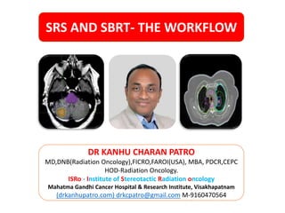
- 1. MANAGEMENT OF DIFFUSE GLIOMAS 11/8/2023 1 DR KANHU CHARAN PATRO MD,DNB(Radiation Oncology),FICRO,FAROI(USA), MBA, PDCR,CEPC HOD-Radiation Oncology. ISRo - Institute of Stereotactic Radiation oncology Mahatma Gandhi Cancer Hospital & Research Institute, Visakhapatnam (drkanhupatro.com) drkcpatro@gmail.com M-9160470564 SRS AND SBRT- THE WORKFLOW
- 4. MANAGEMENT OF DIFFUSE GLIOMAS 11/8/2023 4 Errors
- 9. SBRT WORKFLOW Simulation Planning TX Deliver y Motion Verification Localization Motion Management Delivery
- 10. Steps
- 11. Steps SBRT
- 12. Steps
- 13. MANAGEMENT OF DIFFUSE GLIOMAS 11/8/2023 13 SRS – Immobilization –frame to frameless
- 14. MANAGEMENT OF DIFFUSE GLIOMAS 11/8/2023 14 SBRT – Immobilization
- 15. MANAGEMENT OF DIFFUSE GLIOMAS • Brain • Head and neck • Lymph node • Prostate • Spine • Extremity bone 11/8/2023 15 Motion management not required
- 16. MANAGEMENT OF DIFFUSE GLIOMAS • Lung • RIB • Adrenal • Liver • PVTT • Pancreas • CBD 11/8/2023 16 Motion management required Hepatopancreatic biliary
- 17. Everybody is a king when everything is inside the ring [PTV]
- 18. BECAUSE ORGANS DO NOT FOLLOW STATUE GAME
- 21. 11/8/2023 Various Motion management systems
- 24. Elekta ABC
- 25. Elekta symmetry for ITV generation
- 33. 11/8/2023
- 34. WHICH MOTION MANAGEMENT SYSTEM IS BETTER?
- 35. GOSSIP- WHOSE SPOUSE IS BETTER?
- 36. ANSWER- WHAT MANAGEMENT ACQUIRES
- 39. MANAGEMENT OF DIFFUSE GLIOMAS 11/8/2023 39 GTV-ITV-PTV
- 40. MANAGEMENT OF DIFFUSE GLIOMAS 11/8/2023 40 TRAGET DELINEATION 1. WHAT YOU SEE THAT IS GTV 2. NO CTV 3. DETERMINE ITV IF NO MOTION MANAGEMNET IN EXTRACRANIAL SBRT 4. GIVE 1-2MM PTV TO GTV
- 41. MANAGEMENT OF DIFFUSE GLIOMAS 11/8/2023 41 OAR
- 42. 11/8/2023 42 Review your contour
- 43. MANAGEMENT OF DIFFUSE GLIOMAS 11/8/2023 43 Notes to physics
- 45. MANAGEMENT OF DIFFUSE GLIOMAS 11/8/2023 45 Isocentric vs Nonisocentric
- 46. MANAGEMENT OF DIFFUSE GLIOMAS 11/8/2023 46 FFF vs no FFF
- 47. 11/8/2023 47 Junction volume Accept under dosage in one of the Subvolumes
- 48. RVR 1. For plan optimization, additional dose may be dumped in RVR. 2. High absorbed dose in RVR
- 52. GOAL
- 53. 53
- 54. PUSHING BACKWARD AND FORWARD AT A TIME DIFFICULT BUT NOT IMPOSSIBLE OAR TARGET 54
- 55. MANAGEMENT OF DIFFUSE GLIOMAS 11/8/2023 55 Michael Goitein
- 56. MANAGEMENT OF DIFFUSE GLIOMAS 11/8/2023 56 MLC and CONE
- 57. 11/8/2023 57 Dose displaying 1. Isodose Contours: Set of closed contours linking voxels of equal dose 2. Color Wash: The coding of CT and Dose in the same voxel through the modulation of both intensity (CT) and color (Dose) 3. Isodose Surfaces: The Shaded surface (pseudo 3D) representation of the dose level and selected VOI
- 58. MANAGEMENT OF DIFFUSE GLIOMAS 11/8/2023 58 Basics – DVH
- 59. PLAN CONFLICTS
- 61. 11/8/2023 61 DVH pitfalls 1. Insensitive to hot spot and cold spot 2. Shape of DVH alone can be misleading 3. DVH is the most direct and informative representation of a treatment plan available 4. 3D dose distribution are large and cumbersome to analyze quantitatively 5. User interactivity is essential to extract the most information from dose distribution. 6. Clinical studies have shown that DVH metrics correlate with patient toxicity outcomes. 7. A drawback of the DVH methodology is that it offers no spatial information; i.e., a DVH does not show where within a structure a dose is received.
- 63. MANAGEMENT OF DIFFUSE GLIOMAS 11/8/2023 63 Plan evaluation
- 64. MANAGEMENT OF DIFFUSE GLIOMAS 11/8/2023 64 MLC and CONE
- 65. 11/8/2023 65 Dose displaying 1. Isodose Contours: Set of closed contours linking voxels of equal dose 2. Color Wash: The coding of CT and Dose in the same voxel through the modulation of both intensity (CT) and color (Dose) 3. Isodose Surfaces: The Shaded surface (pseudo 3D) representation of the dose level and selected VOI
- 66. MANAGEMENT OF DIFFUSE GLIOMAS 11/8/2023 66 CBCHOP Mary Dean/Applied Radiation Oncology/2017
- 67. COSID INDEX
- 69. 11/8/2023 69 COSID INDEX Patro K C/Journal of Current Oncology/2022 C COVERAGE INDEX O OAR INDEX S SPILLAGE INDEX I IMAGING INDEX D DELIVERY INDEX
- 70. 11/8/2023 70 Coverage Index Patro K C/Journal of Current Oncology/2022 PTV/CTV/GTV D2/D98 95-107 Dmax
- 71. 11/8/2023 71 OAR INDEX Patro K C/Journal of Current Oncology/2022 Max dose in series organ Mean dose in parallel organ Volumetric analysis
- 72. Basics of plan evaluation – Serial vs Parallel
- 73. 11/8/2023 73 Basics of plan evaluation – Spillage Index Patro K C/Journal of Current Oncology/2022 Conformity index Homogeneity index Gradient index
- 74. 11/8/2023 74 Basics of plan evaluation – Imaging Index Patro K C/Journal of Current Oncology/2022 Axial view Coronal view Sagittal View
- 75. 11/8/2023 75 Basics of plan evaluation – Delivery index Patro K C/Journal of Current Oncology/2022 Complexity of plan MU Complexity of Delivery
- 76. Example
- 77. SL NO PARAMETER VALUE 1 D MAX 36.43Gy 2 D95% 31.01Gy 3 D100% 28.23Gy 4 V95% 99.99% 5 V30 Gy[V100%] 99.56% 6 V110% 44.45% 7 V120% 0.03% 8 V130% 0% 1. Prescription Isodose level is usually not 100% PD covering 100% PTV 2. Often 95% PD covering 95% PTV or higher 3. Or 100% PD covering 95% PTV or higher. Michael Torrens,/J Neurosurg (Suppl 2)/2014 PTV coverage index
- 78. Conformity
- 79. • Is your desired defined dose is confined to PTV ? • FORMULA • VOLUME OF PRESCRIPTION ISODOSE/PTV VOLUME • 43.798/37.491=1.17 • DESIRABLE=1 [Sonja Petkovska Proceedings of the Second Conference on Medical Physics and Biomedical Engineering] RTOG conformity index
- 80. • FORMULA (VOLUME OF PRESCRIPTION ISODOSE IN AREA OF INTEREST)2 PTV VOLUME X VOLUME OF PRESCRIPTION ISODOSE • =39.764 x 39.764 /37.494 x43.798 =0.96 • IDEAL= > 0.85. AND <1 Michael Torrens,/J Neurosurg (Suppl 2)/2014 Paddick conformity index
- 81. Homogenous vs heterogenous NON STEROTAXY HOMOGENOUS PLAN STEROTAXY HETEROGENOUS PLAN FOR EXAMPLE MARGINAL DOSE IS 20 Gy AT 80% MEANS YOU CAN ACCEPT HOT SPOT INSIDE 125% i.e. 25Gy 80% = 18Gy 100%= 18/80 X 100 = 25Gy
- 82. • How homogeneous your dose inside the PTV? • FORMULA • MAXIMUM DOSE/PRESCRIPTION DOSE • 36.43Gy/30Gy=1.21 • DESIRABLE = 1.1-1.3 HOMOGENITY index
- 83. • Dose fall off observation is very much needed in this evaluation under headings • Gradient index • Difference between various isodose lines • e.g between 80% and 60%- ideal- <2mm • Between 80% and 40%- ideal- < 8mm • For that reason, we must calculate equivalent radius Dose fall off- Gradient index
- 84. • To evaluate dose gradient, we must find out difference between radius of various isodose line • But none is iso spherical • We must find out equivalent radius from formula • First find out the specified isodose volume • Then calculate the radius • V=4/3 πr3 • r= (3V/4π)1/3 Equivalent radius
- 85. SL NO PARAMETER VOLUME RADIUS 1 100% ISODOSE 43.79CC 2.19mm 2 80% ISODOSE 64.45CC 2.49mm 3 60% ISODOSE 101.19CC 2.89mm 4 50% ISODOSE 130.84CC 3.15mm 5 40% ISODOSE 177.96CC 3.49mm r= (3V/4π)1/3 Equivalent radius
- 86. • FORMULA • Difference of equivalent radius of prescription isodose and equivalent radius of 50% isodose • 2.19mm-3.15mm=0.96mm • It should be between 0.3 to 0.9 Gradient index
- 87. • BETWEEN 80% AND 60%- IDEAL-<2mm • HERE- 0. 4mm • BETWEEN 80% AND 40%- IDEAL- <8mm • HERE- 1mm EORTC-22952-26001 Distance between various isodose lines
- 88. Isodose line COLOUR ISODOSE LINE Dark green 100% Light green 80% Sky green 60% Pink 50% Blue 40% ISODOSE LINES
- 89. SL NO ORGAN DESIRABLE ACHIEVED 1 RT. EYE MAX <22.5Gy 1.97Gy 2 LT. EYE MAX <22.5Gy 4.4Gy 3 RT. OPTIC NERVE MAX <22.5Gy 2.3Gy 4 LT. OPTIC NERVE MAX <22.5Gy 5.5Gy 5 OPTIC CHIASM MAX <22.5Gy 7.5Gy 8 BRAIN STEM MAX 23-31Gy 10.01Gy 9 RT. COCHLEA MEAN <25Gy <1Gy 10 LT. COCHLEA MEAN <25Gy <1Gy GG HANNA/CLINICAL ONCOLOGY/2016 OAR coverage
- 90. 11/8/2023 90 Low dose bath
- 92. 11/8/2023 92 BEAM entry exit point
- 93. 11/8/2023 93 3D vs beamlet
- 94. MANAGEMENT OF DIFFUSE GLIOMAS 11/8/2023 94 BEV vs REV
- 95. 11/8/2023 95 Basics of plan evaluation – Check list
- 96. Basics of plan evaluation – Check list TARGET COVERAGE D2 D98 AXIAL SAGGITAL CORONAL GTVp GTVn CTV PTV OAR LIMIT PHASE 1 PHASE 2 TOTAL VARIATION OC-[0.03cc] RON [0.03cc] LON [0.03cc] EYE_R MEAN EYE_L MEAN PAROTID_R MEAN PAROTID_L MEAN SPINAL CORD [0.03cc] BRAIN STEM [0.03cc]
- 97. Patient name N VIJAYA LAKSHMI TOLERANCES UMR UMR56950 Sex and Age 50 YEARS & FEMALE Technique VMAT Dose per fraction (Gy) 8.5 No. of fractions 3 Total dose (Gy) 25.5 Volume of PTV (cc) 25.143 Volume of prescription (100%) Isodose (cc) 122.3 Target volume covered by prescription isodose (cc) INTERSECTION VOLUME 24.665 Volume of 80% isodose (cc) 37.54 Volume of 60% isodose (cc) 54.33 Volume of 50% isodose (cc) 482 Volume of 40% isodose (cc) 90.22 Eqv.radius of 100% isodose (cm) 3.08 Eqv.radius of 80% isodose (cm) 2.08 Eqv.radius of 60% isodose (cm) 2.35 Eqv.radius of 50% isodose (cm) 4.86 Eqv.radius of 40% isodose (cm) 2.78 Volume received by 100% isodose (%) 97.99 Maximum dose (Gy) 32.14 Conformity index as per RTOG[ VOLUME OF PRESCRIPTION ISODOSE/VOLUME OF PTV] 4.86 IDEALLY 1 Conformity index as per Paddic 0.20 Homogeneity index [ MAX DOSE/PRESCRIPTION DOSE] 1.26 BETWEEN 1-1 TO 1.3 Gradient index [ EQUIVALENT RADIUS OF50%-EQUIVALENT RADIUS OF 100%] 1.78 BETWEEN 0.3 TO 0.9 Distance b/w 80% iso and 60% iso (cm) 0.27 LESS THAN 2MM Distance b/w 80% iso and 40% iso (cm) 0.71 LESS THAN 8MM OAR DOSES BRAIM-PTV [V27Gy] OPTIC CHIASMA RT OPTIC NERVE LT OPTIC NERVE BRAIN STEM Patient specific QA <10% deviation Patient 4 . Excel shhet
- 98. QA IS MANDATORY
- 99. For setup Is localizer box is mandatory?
- 100. MANAGEMENT OF DIFFUSE GLIOMAS 11/8/2023 10 0 2D verification vs 3D verification
- 101. MANAGEMENT OF DIFFUSE GLIOMAS 11/8/2023 10 1 Hexapod couch ROLL YAW PITCH
- 102. TRAIN YOUR BRAIN TO DECREASE THE DOSES TO OARS STRACTURES BUT NOT AT THE COST OF PTV 11/8/2023 102
- 103. Take care of OAR otherwise rare will not be rare
- 104. RESTRAIN YOURSELF FROM GIVING STRICT CONSTRAIN OTHERWISE TUMOR WILL SUSTAIN. 11/8/2023 104
- 105. Stereotaxy class
- 107. Radiology class
- 108. Neuro-Oncology Class
- 109. SRS/SBRT
- 111. Kanhu 11/8/2023 111