Git for lessons gmf
•Download as PPT, PDF•
0 likes•340 views

Gastrointestinal tract for the lessons

Recommended
More Related Content
What's hot
What's hot (20)
Viewers also liked
Viewers also liked (14)
Similar to Git for lessons gmf
Similar to Git for lessons gmf (20)
Recently uploaded
Recently uploaded (20)
Git for lessons gmf
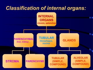
- 1. Classification of internal organs: INTERNAL ORGANS viscera, splanchna PARENCHYMAL liver, kidney… TUBULAR Oesophagus, trachea GLANDS TUBULAR (SIMPLE, COMPOUND) ALVEOLAR (SIMPLE, COMPOUND) STROMA PARENCHYME
- 2. General plan of the structure ofGeneral plan of the structure of cavitas (tubular) organscavitas (tubular) organs:: • Mucose membrane (inner); • Muscular layer (middle); • Serous or adventitial (outer).
- 3. Mucous, muscular & serous layersMucous, muscular & serous layers of the stomachof the stomach
- 4. Mucose membrane of the stomach.
- 5. The structure of the wall of different departments of the alimentary tube
- 6. The serous layer of the abdominalThe serous layer of the abdominal cavity is the peritoneumcavity is the peritoneum • It has two layers: visceral, covering internal organs & parietal, lying walls of the abdominal cavity.
- 7. The relation of organs with theThe relation of organs with the peritoneumperitoneum By the relation with the peritoneum all organs of the abdominal cavity are grouped as: intraperitoneal, mesoperitoneal & extraperitoneal (or retroperitoneal)
- 8. DEPARTMENTS OF THE ALIMENTARYDEPARTMENTS OF THE ALIMENTARY CANALCANAL • Oral cavity, • pharynx, • esophagus, • stomach, • Small & • large intestine.
- 9. FUNCTIONS OF THE ALYMENTARY SFUNCTIONS OF THE ALYMENTARY SYYSTEMSTEM • it provides storage,processing of ingest, and elimination of unabsorbed component (faeces) • it secretes enzymes and lubricants which facilitate the passage of the ingest • it absorbs nutrients and other materials and passes them into the body • it propels material within it by muscular action • it has defensive, immunology properties • it houses microorganisms • its epithelial lining forms barrier between its lumen and the true interior of the body.
- 10. Motor function:
- 11. л
- 14. Internal organs develop fromInternal organs develop from endoderm & mesodermendoderm & mesoderm
- 15. DEVELOPMENT OF THEDEVELOPMENT OF THE ALIMENTARY SYSTEMALIMENTARY SYSTEM • Alimentary system is developed on 3-4th weeks of the development from the primitive gut, which is closed on the cranial and caudal ends
- 16. FORMATION OF THE ORAL ANDFORMATION OF THE ORAL AND ANAL PITSANAL PITS • They appear on the cranial and anal ends on the 1st month of embryonic development • They form two layered membranes from ectoderm and endoderm: pharyngeal and anal • They are broken till end of the 5th week of the development Pharyndeal membrane Anal membrane entoderm ectoderm Viteline duct
- 17. s u p e r io r , p r o x im a l h a lf o f d e s c e n d in g p a r t o f d u o d e n u m , p a n c r e a s a n d d u c ts b u c c a l c a v ity , p h a r y n x , o e s o p h a g u s , s to m a c h , r e s p ir a to ry s y s te m , liv e r, g a llb la d d e r , F O R E G U T r e m a in in g d u o d e n u m , je ju n u m , ile u m c a e c u m , a p p e n d ix , a s c e n d in g c o lo n M ID G U T d e s c e n d in g c o lo n , s ig m o id c o lo n , r e c tu m a n d a n a l c a n a l to th e a n a l v a lv e s H IN D G U T P R I M I T I V E G U T foregut midgut hindgut Oral cavity anus
- 18. BUCCAL CAVITY DERIVES FROM BOTHBUCCAL CAVITY DERIVES FROM BOTH ECTODERMAL AND ENDODERMALECTODERMAL AND ENDODERMAL REGIONSREGIONS • The pharyngeal arches form the mandibular and the maxillary prominences • The opening of the stomodeum bounded cranially by the frontonasal prominence, caudally by the mandibular prominences and laterally by the maxillary prominences
- 19. ROTATION OF THE STOMACH AND FETAL GUTROTATION OF THE STOMACH AND FETAL GUT • The stomach has two rotations. The first 90° clockwise, viewed from the cranial end, the second 90° clockwise about an anteroposterior axis, so its right surface becomes dorsal and its left ventral • The proximal limb of the primary intestinal loop moves to the right and the distal limb to the left having already rotated through an angle of 90°
- 21. • Caecum may remains below the liver • Rotation of organs on the left side is called situs viscerus inversus