Calcium and phosphorous metabolism
•
12 likes•7,725 views

Indian Dental Academy: will be one of the most relevant and exciting training center with best faculty and flexible training programs for dental professionals who wish to advance in their dental practice,Offers certified courses in Dental implants,Orthodontics,Endodontics,Cosmetic Dentistry, Prosthetic Dentistry, Periodontics and General Dentistry.

Recommended
More Related Content
What's hot
What's hot (20)
Viewers also liked
Similar to Calcium and phosphorous metabolism
Similar to Calcium and phosphorous metabolism (20)
More from Indian dental academy
More from Indian dental academy (20)
Recently uploaded
Recently uploaded (20)
Calcium and phosphorous metabolism
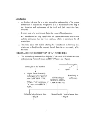
- 1. Introduction - As dentists, it is vital for us to have a complete understanding of the general metabolism of calcium and phosphorous as it is these minerals that help in the formation and maintenance of the teeth and their supporting bony structure. - 2 points need to be kept in mind during the course of this discussion. 1. Ca++ metabolism is a very complicated and controversial topic on which no definite conclusion has yet been reached, which is acceptable by all researchers. 2. This topic deals with factors affecting Ca++ metabolism in the body as a whole and it should not be assumed that all these factors necessarily affect the teeth. IMPORTANCE AND DISTRIBUTION OF Ca++ IN THE BODY - The human body contains about 1kg of Ca++ of which 99% is in the skeleton and remaining 1% in soft tissues and ECF (990gms) and (10gms). of 990 gms in the skeleton - 10 gms forms the readily exchangeable Ca++ pool of bone (MISCIBLE POOL) - 980 gm slow exchange of Ca++ takes place (STABLE POOL) 10.0-10.5mg/dl NORMAL SERUM CALCIUMLEVEL Remaining in soft tissues Diffusible/ ultrafilterable form 5.5mg/dl Non-diffusible / protein bound form 4.5mg/dl 1 10gms
- 2. Ionized Ca 5.0mg/dl Bound to PO4, HCO3, Citrate 0.5mg/dl Bound to albumin 4.0mg/dl Bound to globulin 0.5mg/dl FUNCTIONS OF CALCIUM 1. Contributes to hardness of bone and is a major component of teeth. 2. Stabilises the cell membrane and their permeability. 3. Maintenance of excitability of nerve and muscles. 4. Normal skeletal and cardiac muscle contraction. 5. Helps in the neurotransmitter release. 6. Secretion of granular material from exocrine and endocrine glands. 7. Hormone release and activity – discharge of non-adrenaline in the nerve terminal cell. 8. Synthesis of nucleic acid and protein. 9. Blood coagulation – Ca++ is required for the conversion of many inactive enzymes in the coagulation process. DAILY REQUIREMENTS OF CALCIUM (WHO estimate) 6 months – 2 year = 0.5-0.6 g/day 1200 mg/day 16 years to adults = 0.5-0.6g/day Pregnancy and lactation = 1.0-2gm/day Children = 800mg/day SOURCES OF CALCIUM AND PHOSPHOROUS - Milk + milk products. - Egg. 2
- 3. - Meat - Fish - Leafy vegetables - Hard water PHOSPHOROUS DISTRIBUTION OF PHOSPHOROUS IN THE BODY - The human body contains 500-600 gms of PO4 -- out of which 85% (425g) is in the bone. - Remaining phosphorous is present in the liver, pancreas and brain. - Phosphorous is essential for the formation of teeth. Normal serum PO4 -- level is 2.5 – 4.5 mg/dl. Serum inorganic form Organic phosphorous 3-4 mg/dl in adults remaining in the form of phospholipids & glycerophosphates 5-6mg/dl in children FUNCTIONS OF PHOSPHOROUS Formation of bone and teeth. It is the essential constituent of all cells. Important constituent of high energy phosphate compounds like ATP, creatine phosphate, cyclic AMP, hexose phosphate, 2-3DPG, phospholipids, nucleotides. Helps in the regulation of glycolysis. Phosphorylation of lipids and sugar i.e. absorption, transportation and metabolism. Urinary buffer, which regulates urinary pH. DAILY REQUIREMENTS OF PHOSPHOROUS – 1gm/day CONCEPT OF CALCIUM BALANCE This term is used to describe the amount of Ca++ either stored or lost by the body over a specific period of time. This can be calculated by deducting the amount of Ca in the urine from the Ca taken in the diet. 3
- 4. Ca in diet - Ca in fasces Ca absorbed – Ca in urine Ca lost / gained e.g., If 1.0gm Ca is in the diet – 0.7 gm in fasces 0.3g absorbed. 0.1g net gain 0.2g in urine The Ca balance values are said to change with age. In a growing child, there is a Net gain for growing and mineralizing skeleton. In an aging adult, there is a Net loss as Ca from bone is lost too due to conditions like osteoporosis. Hence, amount of Ca lost is greater than Ca in intake. ABSORPTION OF CALCIUM AND PHOSPHOROUS It is seen that almost all the food taken in three the diet is almost completely absorbed in the gut whreas the amount of minerals absorbed is very negligible. This could be due to the various factors affecting the absorption of Ca++ and PO4 -- . The factors can be studied under: Factors affecting mucosal cell Factors influencing Ca absorption in the gut FACTORS INFLUENCING MUCOSAL CELLS 1. Vitamin D and Ca absorption It is seen and proven that the active form of vitamin D or 1,25 DH CC (di- hydroxy cholecalciferol) increases the absorption of Ca in the gut, kidney and bone. This increase in absorption of Ca is directly related to the increased concentration of Ca binding protein produced by increasing synthesis of mRNA. 1,25 DHCC ↓ Increases synthesis of mRNA ↓ Increases level of Ca binding protein CBP ↓ Increases Ca plasma level 4
- 5. 2. Effect of dietary Ca intake and Ca need It is seen by means of experiments that the amount of Ca STORED in the body is a factor which influences Ca absorption. This factor can be better understood with the help of an example. e.g., Rottenson in 1938 used 2 groups of rats for his experiment He fed 1st group 2nd group 0.15% Ca for 4 weeks 0.8% Ca for 4 weeks 4 weeks later he observed that Stored - 190mg Ca was stored (↓) (↑↑) 570mg Ca was stored Thus suggesting that greater the dietary intake of Ca, greater amount of Ca is stored. In the 2nd stage of experiment, both groups received 0.4% Ca for 5 weeks. 0.4% Ca 0.4% CA 5 weeks 5 weeks 5 weeks later ABSORBED AMOUNT 900 mg (↑↑) 600 mg (↓) This suggested that the group with a low Ca store absorbed more Ca than the group with the high Ca store. Thus, suggesting that the amount of Ca absorbed is directly dependent on the amount of Ca present in the store. This kind of an adaptation mechanism is directly under the control of formation of 1,25 DHCC. 3. Effect of pregnancy and growth - During pregnancy, the amount of dietary Ca absorbed increases especially in the later months of pregnancy. Half of the Ca absorbed during pregnancy goes to the developing foetus and the remaining half is stored in the mother’s skeleton as a reserve for lactation. - 2 hormones are active during pregnancy 5
- 6. PLACENTAL LACTOGEN OESTROGEN Increases Ca absorption Acts by increasing release of PTH which increases Ca absorption indirectly. Hence, in later stages of pregnancy increased PTH levels are reported. Growth Period - During growth of children, the growth hormone levels are high. This acts by increasing Ca absorption and reducing amount of endogenous Ca excretion. Parathyroid hormone - Is one of the main hormones controlling Ca absorption. It mainly acts by controlling the formation of 1,25 DHCC which it active form of vit. D which is responsible for increased Ca absorption. CALCIUM : PHOSPHOROUS RATIO An increase in the plasma Ca level causes a corresponding ↓ in absorption of phosphorous and vice-versa. The product of Ca and inorganic phosphate of the blood is always constant (ratio is 2 Ca : 1 PO4). II FACTORS INFLUENCING AVAILABILITY OF CALCIUM IN THE GUT 1. pH of the intestine • It is seen that the acids of the gastric juices dissolve most of the Ca salts and inorganic phosphate in the diet hence allowing greater amount of Ca available for absorption. • In the lower part of the small intestine, the pH is said to be more alkaline, thus causing Ca salts to undergo precipitation. Hence Ca absorption occurs only in the upper part of the small intestine where the pH is acidic.In patients wth achlorhydria/gastrectomy, less Ca absorption takes place. 2. Amount of dietary Ca and PO4 As discussed before, as the levels of dietary Ca and PO4 is increased, the amount of Ca and PO4 absorbed also increases but only upto certain limit beyond which no more absorption takes place 6
- 7. This is because the active transport system in the mucosal cells can deal with only a certain load of Ca. Once this load is exceeded, the remaining Ca is either excreted or passively diffuses in the lower part of the gut.This a homoestatic mechanism for preventing excessive concentration of Ca in the blood or tissue which could have a hazardous effect. 3. Phytic acid and phytates Are found in foods like – Oat meal Whole wheat Cereals They are considered to be anti- calcifying factors as they combine with the Ca of the diet thereby forming insoluble salts of Ca which are not absorbable. Hence, if these phytates are taken in a diet, where the Ca intake is low or vit. D intake is low, it is seen that severity of the deficiency disease worsens.The effect of phytates is said to be quantitative and can be neutralized by adding sufficient extra Ca to the diet. 4. Effect of Oxalates Oxalates is another substance present in certain foods like spinach and rhubarb leaves. These oxalates precipitate significant amounts of Ca from the diet or from the digestive juices thereby decreasing the amount of Ca available for absorption.Hence, it may be noticed that after eating spinach, a certain roughness can be experienced on the surface of the teeth which is due to the precipitation and deposition of Ca oxalate crystals (Ca from the saliva) on the teeth. 5. Effect of fat Fat combines with Ca forming insoluble Ca soaps which decreases Ca absorption but increases the availability of phosphorous for absorption.Fat combines with Ca and liberates PO4 for absorption. Hence, excess fats hinder Ca absorption but favour phosphorous absorption. 6. Effects of proteins and Aminoacids It is seen that a considerable increase in the Ca absorption occurs with a high protein diet. Proteins and Amino acids effect Ca in 2 ways: Proteins form soluble complexes thus allowing Ca to remain in a form which is Protein metabolism leads to formation of certain acids that encourages the removal 7
- 8. easily absorbable of Ca from the skeleton. Hence, urinary Ca level increase with a richer protein diet Hence a high protein diet may be considered as a possible factor for osteoporosis. But under such conditions, the increased absorption is of no value as this absorbed phosphorous cannot be retained without Ca. Hence the excess phosphorous is quickly excreted. 7. Effect of carbohydrates It is seen that certain carbohydrates like lactose create an acidic pH in the gut by favouring growth of acid producing organisms. This acidic environment of gut favours Ca absorption. Contradictory studies show that an increased level of Ca is seen in the body not by increase in Ca absorbed but by decrease in Ca excreted in urine. 8. Bile salts favours Ca and phosphorous absorption as it dissolves fats thereby allowing greater amounts of Ca+ phosphorous to be available for absorption. HORMONAL CONTROL OF CALCIUM METABOLISM A complex set of interlocking mechanisms takes place in order to allow man to survive major dietary Ca intake fluctuations. These mechanisms are mainly controlled by the endocrine systems. Three main hormones acting at 3 different sites are responsible for Ca metabolism. 1. Vit. D3 Bone. 2. Parathormone Kidney 3. Calcitonin Intestine 1) Vit. D3 is a steroid derivative 8
- 9. Vit. D3 – cutaneous synthesis – important in Ca metabolism. Vit. D2 – Ergocalciferol Dietary source is fish, milk and eggs. • Vit. D is also known as cholecalciferol. • Besides dietary intake, cutaneous synthesis of vit.D. is another important source of Vit. D in the body. • Present within the inner layers of the epidermis, lies an enzyme known as 7-dehydrocholesterol. UV Rays PRE VITAMIN D3 Bound Binding protein (DBP) to Vit. D and taken to liver IN LIVER Taken to kidney Pre Vit. D3 IN LIVER 25, OH cholecalciferol (main circulating subs, but it is relatively inactive as it is bound to globulin. 25-OH-CC Undergoes Hydroxylation by 1--hydroxylase enzyme whose formation is under the control of PTH 1,25, Dihydroxycholecalciferol or calcitriol (active form of Vit. D3 which is considered to be a horomone) - Daily requirements of vit. D 400 I.U. Action of Vit. D. Vit. D. acts on the intestine, kidney and bone thereby bringing about an increase in the Ca absorbed. The active form of vit. D which is produced in the kidney has its action on many tissues away from the site of its production. Hence 1,25DHCC is often known as a hormone.Acts mainly by increasing the synthesis of mRNA which directly increases the concentration of (CBP) Ca binding protein mainly in the tissues of the intestinal mucosa. The CBP thereby binds with increased amount of Ca and allows increased Ca to be absorbed. 1. Action on the intestinal 9
- 10. 1,25 DHCC increases the synthesis of CBP in the intestinal epithelium thereby increasing the transportation of Ca from the intestinal lumen to the epithelium. Hence, the amount of Ca absorbed is directly proportional to the amount of CBP present in the mucosal cells. 2. Action the kidney 1,25 DHCC increases the amount of Ca absorbed from the renal tubules. 3. Action on the bone Before studying the action of vit. D on the bone we must understand the term Osteocytic Membrane System. As we all know, the cells of the bone are the: Osteocytes Osteoblasts Osteoclasts The osteocytes / resting bone cells are present within the bone.The osteoblasts / bone forming cells are found in the endosteum lining the bone. The osteocytes and the osteoclasts communicate with each other via the long cytoplasmic processes. Hence, the osteoblasts present outside the bone and osteocytes present within the bone form a system of interconnected cells that spread all over the bone suface.This extensive system is known as osteocytic membrane system (OCM). This system separates the bone from the E.C.F. Bone fluid is present between bone and OCM). Action Vit. D. / 1,25 DHCC is said to mediate the pump that actively transports Ca from within the OCM to the ECF thereby raising plasma Ca levels. Deficiency of vit. D in the body either due to a deficient diet or due to insufficient solar exposure results in disorders known as Rickets in younger children or Osteomalacia in adults. Rickets - Delayed eruption 10
- 11. - Malallignment of teeth REGULATION OR CONTROL OF SYNTHESIS OF 1,25 DHCC OR FEED BACK MECHANISM The synthesis of active vit. D3 or 1,25DHCC is said to be tightly and directly regulated by the parathyroid hormone. Hence, an increase in the dietary Ca results in increased levels of 25 (OH) CC viz., the inactive form. Any fall in the plasma Ca levels cause release of PTH. This PTH activates enzyme 1- - hydroxylase which then converts vit. D3 to active 1,25 DHCC.Increases synthesis of CBP hence, there is an increase in Ca absorbed. During pregnancy, hormones like placental lactogen and oestrogen also have an effect on 1- -hydroxylase 2) PARATHYROID HORMONE (PTH) PTH is secreted by the parathyroid glands which acts located on the posterior surface of the thyroid gland. They are 4 in number. PTH acts mainly on 2 sites i.e. a) bone b) kidney It acts by a cAMP mechanism to increase Ca levels and decrease in organic phosphate levels in the plasma. a) Action on the bone Fast phase Slow phase i) Fast phase Within few minutes of injection of a large dose of PTH, the plasma Ca levels is said to increase. Mechanism PTH mainly acts by increasing the permeability of osteocytic membrane system to Ca so that Ca from the bone fluid diffuses into the osteocytes and osteoblasts from where vit. D mediated active transport transfers the Ca from within the OMS to the ECF thereby increasing the plasma Ca levels.Any decrease in the concentration of Ca within the bone fluid leads to dissolution of Ca salts from the nearby bones without affecting the bone matrix (indirectly activates osteoclastic activity or bone resorption). ii) Slow phase 11
- 12. Several days of increased PTH activity is required to demonstrate the slow phase. PTH acts by Activating the bone resorbing cells / osteoclasts Forming new osteoclasts from the osteoprogenitor cells Osteoclasts are multinucleated giant cells which produces resorption of bone surfaces in contact with them. The osteoclasts send out long finger-like-projections towards the bone. These finger like projections release a. PROTEOLYTIC ENZYMES which digests the organic matrix. b. ACIDS like citric / lactic acid which dissolves the bone minerals. The Ca and PO4 released are poured into the ECF. b) Action on kidney (↑↑Ca++ ) - Increases the resorption of Ca from the distal tubules (↓↓ PO4 – – ). - Decreases the resorption of PO4 from PCT thereby causing phosphaturia and hypophosphatameia. - Increases the synthesis of 1--hydroxylase which is responsible for the active form of Vit. D which increases Ca absorption. REGULATION OF PTH SECRETION The plasma Ca levels are said to have a direct influence on the PTH secretion.Any fall in the Ca levels causes a release of the PTH which brings back the level to normal. Similarly increase in plasma Ca level inhibits the secretion of PTH. 12
- 13. 3.CALCITONIN Is secreted by the parafollicular cells by the thyroid glands. Mainly acts to maintain the integrity of the bone. Actions It mainly acts by decreasing the permeability of OMS to Ca thereby increasing the amount of Ca deposited in the readily EXCHANGEABLE CALCIUM POOL OF BONE.It prevents the formation of new osteoclasts from osteoprogenitor cells thereby preventing bony resorption. REGULATION OF SECRETION OF CALCITONIN The release of calcitonin is regulated by the plasma calcium levels. A rise in the plasma Ca level above 9mg % stimulates the release of calcitonin.Hence, plasma Ca levels controls the direction of PTH and calcitonin.In adults, role of calcitonin is very minor and normal Ca level is maintained chiefly by PTH.Calcitonin plays a major role in pregnancy. It protects the bones of the mother from excessive loss of Ca as during pregnancy PTH and 1,25DHCC levels are high. EXCRETION OF CALCIUM AND PHOSPHOROUS Ca is said to be excreted both in the faeces and in the urine. About 90% of the total amount of Ca is excreted in the faeces.Ca of the urine is excreted as calcium chloride (CaCl2) and (CaPO4) 10% remaining.The approximate daily turnover rates of Ca in an adult are as follows: Intake 1000mg Intestinal absorption 350mg Secretion in GIT juice 250mg Net absorption over secretion 100mg Loss in the faeces 100mg Excretion in the urine 100mg BUFFER ACTION OF PHOSPHOROUS IN BLOOD 2 varieties of phosphates are present in the blood Alkaline Acidic H2PO4 HPO4 13
- 14. Concentration is about Concentration is about 1.05m mol/lit 0.26 m mol/lit In the plasma they are in a ratio of alkaline 4 : 1 acidic. Where pH of extra-cellular fluid becomes more acidic there is a relative increase in H2PO4 – and HPO4 – – and the opposite happen when fluid is alkaline. The average total quantity of inorganic phosphorous ranges between normal limits of 3- 4mg/dl in adults and 4-5mg/dl in children. IMPAIREMENTS IN BLOOD CALCIUM I] Hypercalcemia Increased level of Ca in the blood. Symptoms - Tiredeness - Loss of appetite. - Nausea, vomitting. - Constipation. - Polyuria. - Dehydration. - Loss of muscle tone. - Decreased excitability of muscles and nerves. Conditions in which it occurs - Hyperparathyroidism. - Acute osteoporosis. - Vit. D intoxication. - Thyrotoxicosis. II] Pathologic calcifications of soft tissues 3 theories have been forwarded for the mechanism of calcification a) Nucleation theory 14
- 15. Ca is deposited both inside and outside the cells (in mitochondria) which later coalesce together to form calcified tissues. This proposes that organic matrix components facilitate precipitation by acting as heterogenous nucleating agents like collagen, phospholipids which act on specific phosphorylating sites. b) Booster theory This theory proposes the existence of a mechanism to boost the local Ca and PO4 ion product to a point where precipitation occurs. c) Matrix vesicles Small round vesicular extracellular matrix vesicles about 100nm in diameter are involved in the initiation of calcification. They are probably arrived from cell processes originating from the plasma membrane. i) Dystrophic calcification - Precipitation of Ca in degenerating and dead tissues occur. - Common sites are - Blood vessels - Kidneys ii) Metastatic calcification - Due to excess Ca in the circulating blood, they are deposited in previously undamaged tissue - Usually occurs in – Hyper vitaminosis D Hyperparathyroidism Destructive lesions of the bones. - Common sites are - Blood vessels - Kidney iii) Calcinosis - Is calcification occurring in / under skin and can be associated with scleroderma. 15
- 16. III] Hypocalcemia - Decreased levels of Ca in the blood (below 4mg is lethal). Symptoms - Tetany (Carpopedal spasm). a) Chvotek’s sign - Contraction of ipsilateral facial muscles elicited by tapping over the facial nerve at the angle of the jaw. b) Trosseau sign - Spasms of the muscles of the upper extremity that causes flexion of wrists and thumb and extension of fingers. - This is elicited by occluding the circulation of fingers for a few minutes. c) Erbs sign - Is hyperexcitability of muscles to electrical stimulation. - This occurs in cases of – - Insufficient Ca in the diet. - Hypoparathyroidism. - Insufficient vit. D in the diet. - Increase in calcitonin levels. 16
- 17. IV] VIT D. DEFICIENCY ↓↓ Ca++ level Rickets Osteomalacia - In children. - Mainly affected the long bones in the body, ribs. - Occurs due to failure in mineralization, due to lack of Ca level, the cartilaginous form of bone is said to persist. - In adults. - Mainly affects the flat bones in the body. - Especially seen in post and menopausal women who have a decreased dietary Ca intake and decreased exposure to the sun resulting in increased removal of Ca from the bone causing softening of the skeleton and its distortion. - The cartilage continuous to grow and proliferate thus resulting in localized areas of cartilage proliferation (rickety rosary, beaded bone formation) - The weight of the body on the uncalcified long bones result in the bowing of legs – knock kness. - 17
- 18. Dental - Development of abnormality in enamel and dentin. - Delayed eruption of teeth. - Mal-alignment of teeth. - Higher caries index. - Wider predentine zone. - Increased amount of inter- globular dentin. Dental Findings - Severe periodontitis Treatment: Treatment: Supply adequate amount of Ca and phosphate in diet and administration of large amounts of Vit. D. V] PARATHYROID HORMONE a) Hyperparathyroidism - Increases levels of PTH due to an adenoma of the parathyroid glands. C/F: - Pathologic fracture of bones due to increased mobilization of Ca from bone resulting in osteitis fibrosa cystica. - Large punched out cystic area of bone. o Joint stiffness. o Urinary tract stones. o Muscle weakness. o Thirst. o Polyuria o Anorexia. 18
- 19. o Weight loss. Dental findings - Malocclusion seen due to definite drifting of teeth causing spacing (this occurs as a result of increased loss of Ca from the bone, there is an absolute attempt to new bone formation and repair resulting in spacing). Radiographically - Areas of radiolucency seen in bone. - Loss of lamina dura present. Treatment - Removal of cause. b) Hypoparathyroidism - Seen to develop due to excision of the parathyroid glands. - This is characterized by a fall in the serum Ca levels and an increase in the serum inorganic phosphate levels. C/F: - Similar to hypocalcemia. - Tingling and numbness in the fingers, face and toes. - Extreme cases – cramps in the hands and feel which is very painful. - Carpopedal spasm. - Spasm of muscles of respiration resulting in the dread of suffocation. - Signs of tetany present. - In infants, symptoms of tetany may be confused for epilepsy, though there is no loss of consciousness. Dental Findings - Aplasia / hypoplasia of teeth seen when hypoparathyroidism develops before teeth formation. c) Parathyroid poisoining 19
- 20. - In rare cases, the PO4 level of body fluids rise rapidly when the kidneys cannot excrete rapidly. Ca and PO4 get supersaturated and lead to metastatic deposition in lungs, alveoli, kidneys, thyroid gland, gastric mucosa etc. - Elevation of blood Ca levels before 17 mg/dl along with concurrent elevation of PO4 – – death can occur in few days. Osteoporosis - In adults (old age) due to decreased bone matrix rather than poor bone calcification. Summary Ca and PO4 are important minerals that are required in minimal amounts for basic activities in the body like the normal ionic Ca levels in the plasma is required for proper bone mineralization and maintenance of cell membrane integrity. The plasma Ca levels need to be regulated within a very narrow range because of its marked effect on neuromuscular and cardiac excitability. Three hormones which are – PTH - Calcitonin - Vit. D Play important role in maintaining Ca and PO4 levels to a normal. CALCIUM AND PHOSPHOROUS METABOLISM CONTENTS Calcium Phosphorous Introduction Distribution of Ca in the body Functions of Ca Daily requirements Sources Absorption of Ca+ PO4 Concept of Ca balance Distribution of PO4 in the body Function of PO4 Daily requirements Sources 20
- 21. Factors controlling the absorption of Ca+ PO4 Hormonal control of Ca metabolism Vit. D3 PTH Calcitonin Excretion of Ca+ PO4 Summary 21