Acute Appendicitis
•
4 likes•1,592 views

This document discusses the anatomy, clinical presentation, diagnosis, and treatment of acute appendicitis. It notes that the appendix is commonly located retrocecally and is supplied by the appendicular artery. The classic symptoms of acute appendicitis are known as Murphy's triad of periumbilical pain shifting to the right lower quadrant, anorexia, and fever. Diagnosis is made through physical exam finding tenderness at McBurney's point and confirmed through blood tests, urine analysis, and imaging like ultrasound or CT scan. Treatment is through open or laparoscopic appendectomy, while appendicular masses may be initially treated conservatively with antibiotics using the Ochsner-Sherren regimen before interval appendectomy

Recommended
More Related Content
What's hot
What's hot (20)
Similar to Acute Appendicitis
Similar to Acute Appendicitis (20)
More from J.J.M.Medical College,Davangere
More from J.J.M.Medical College,Davangere (20)
Recently uploaded
Recently uploaded (20)
Acute Appendicitis
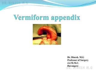
- 1. Dr. Dinesh. M.G Professor of Surgery J.J.M.M.C. Davangere
- 2. Introduction Considered by most to be a vestigial organ Its importance in surgery Acute appendicitis Carcinoid tumour
- 3. Anatomy Blind muscular tube Mucosa, submucosa, muscularis propria and serosa Positions Retrocaecal 74% Pelvic 21% Paracaecal 2% Pre ileal 1% Post ileal 0.5% Subcaecal 1.5%
- 4. Anatomy Appendicular artery runs in mesoappendix It is an ‘end-artery’ & a branch of ileocolic artery Microscopic anatomy Lined by columnar cell of colonic type Crypts are present in the mucosa in which lie the argentaffin (Kultschitzsky) cells
- 5. Acute appendicitis Reginald Fitz first published paper on appendicular perforation in 1886 Charles McBurney described clinical features including the point of maximum tenderness in Rt. iliac fossa McBurney’s point
- 6. Aetiology No definite single aetiology Obstruction of lumen by Faecolith Pin worms Carcinoma caecum etc.
- 7. Clinical features Murphy’s triad Periumbilical pain shifting to Rt.iliac fossa Anorexia, nausea and vomiting Fever Signs Tachycardia, pyrexia Tenderness or rebound tenderness in Rt. iliac fossa Guarding or rigidity Rovsing’s sign Positive ‘Psoas sign’ Positive ‘Obturator test’
- 8. Psoas test
- 10. Differential diagnosis Children Acute gastroenteritis Mesenteric lymphadenitis Meckel’s diverticulitis Intussusception Lobar pneumonia and pleurisy Adults Right ureteric colic Rt. Acute pyelonephritis Perforated peptic ulcer Testicular torsion Acute pancreatitis
- 11. Differential diagnosis Adult females Salpingitis Mittelschmerz Torsion /heamorrhage of an ovarian cyst Ectopic pregnancy Elderly Carcinoma of caecum
- 12. Investigations CBC leucocytosis Urine analysis Serum creatinine and electrolytes X ray erect abdomen in diffuse peritonitis Usg abdomen CT abdomen Urine pregnancy test
- 14. CT scan
- 15. Treatment Appendicectomy Open Laparoscopic
- 16. Appendicular mass May form by 3rd day of acute appendicitis Consists of inflamed appendix, greater omentum, oedematous caecal wall & oedematous coils of small intestine It may form an abscess or resolve with treatment Differential diagnosis of appendicular mass Carcinoma caecum Ileocaecal tuberculosis
- 17. Management of appendicular mass Ochsner-Sherren regimen Conservative treatment with Nil by mouth Ryle’s tube aspiration Antibiotic therapy Cephalosporins Aminoglycoside Metronidazole Recording of size of mass daily Recording of TPR chart 4hourly Input & output chart
- 18. Management of appendicular mass Interval appendicectomy if mass resolves after 6-8 weeks Early laparotomy if appendicular abscess develops Management of appendicular abscess Controversial Early laparotomy and drainage of abscess with appendicectomy in one sitting Percutaneous US or CT guided catheter drainage followed by elective appendicectomy 8-12 weeks later
- 19. Complications of appendicitis Perforation Postoperative wound infection Intra abdominal and pelvic abscess Pyelephlebitis Enterocutaneous fistula Small bowel obstruction
- 20. Points to remember Diff. diagnosis of acute appendicitis in adult males include Right ureteric colic Rt. acute pyelonephritis Perforated peptic ulcer Testicular torsion Acute pancreatitis Diff diagnosis of acute appendicitis in adult females include Salpingitis ovarian torsion ectopic pregnancy
- 21. Points to remember Complications of acute appendicitis include Perforation Postoperative wound infection Intra abdominal and pelvic abscesses Pyelephlebitis Enterocutaneous fistula Small bowel obstruction Treatment of choice in acute appendicitis is Open or laparoscopic appendicectomy Regimen for conservative treatment of appendicular mass is Ochsner- Sherren regimen
- 22. Thank you