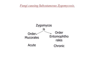
1. Fungi causing Subcutaneous Zygomycosis.
Zygomycos
is
Order
Mucorales
Order
Entomophtho
rales
Acute Chronic
2. 1. Zygomycosis (Mucorales).
Mucorales infections Definition:
•Angiotropic (blood vessel-invading)
•The most common genera causing disease are:
Rhizopus
Rhizomucor
Mucor
Absidia
•Fast growing non-septate molds
3. Geographical distribution & normal habitat
•World-wide
•Soil and decomposing organic matter
•Found in outdoor and indoor air
•Food and dust
Clinical forms
The infection typically involves the:
•Rhino-facial-cranial area
•Lungs, gastrointestinal tract or skin
•Other parts can also be affected
4. Risk factors
•The disease is associated with:
Diabetic ketoacidosis
Malnourished children
Severely burned patients
•It is also seen in patients with
Leukemia
Lymphoma
AIDS
In patients using corticosteroids
5. Laboratory diagnosis
•Specimens:
Aspirated material from sinuses
Sputum in pulmonary disease
Biopsy material
Management of mucormycosis
•The prognosis is bad
•Most cases of gastric and pelvic disease are diagnosed at
autopsy
•Cases occurred in patients with pulmonary disease, leukemia, or
with lymphomas, are usually fatal
•Control of the diabetes, aggressive surgical debridement of
involved tissue, and high doses of Amphotericin B are
recommended
7. •Not like Mucorales:
No vascular invasion or infarction
Chronic inflammatory response
Basidiobolus infection
•Chronic inflammatory or granulomatous disease
•Subcutaneous tissue of the limbs, chest, back or buttocks
•Mostly in children (predominance in males)
8. Conidiobolus infection
•Chronic inflammatory or granulomatous disease
•Nasal submucosa
•Characterized by polyps or palpable subcutaneous masses
•Occur mainly in adult (80% of cases)
Laboratory diagnosis
•Specimens:
1.Aspirated material from sinuses
2.Biopsy material
9. The following steps are used for the both types of
Zygomycosis:
1.10 or 20% KOH:
1.Typically contain thick-walled aseptate hyphae
2.Swollen cells (up to 50 um) and distorted hyphae
may be present
2. Culture:
1.SDA without cycloheximide at 30°C
2. Rapid growth
3- A diagnosis can also be made by examining a biopsy
for granuloma formation and the presence of aseptate
hyphae, eosinophils, neutrophils, and fibroblasts. Tissue
for culture should be send in a dry sterile container, and
biopsies should be fixed and send in formal saline.