Ecosystem Interactions Class Discussion Presentation in Blue Green Lined Styl...
Renaissance
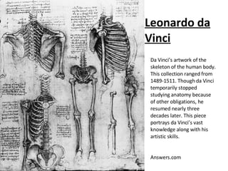
1. Leonardo da Vinci Da Vinci’s artwork of the skeleton of the human body. This collection ranged from 1489-1511. Though da Vinci temporarily stopped studying anatomy because of other obligations, he resumed nearly three decades later. This piece portrays da Vinci’s vast knowledge along with his artistic skills. Answers.com
2. Andreas Vesalius The two most outer layers of muscle. Found in Vesalius’ Epitome. Basel, 1543. Vesalius drew his bodies in very realistic poses. Each hand shows a different muscle layer, comparing the two in a single picture. Universityofglasgow.com
3. Henry Gray This descriptive picture of the inner body was one of Gray’s 363 drawings of his 750 page book, Gray’s Anatomy . He published this in 1858, he was only 31 years old. Gray took on a great challenge in detailing each drawing, showing his dedication to the art and science. Factropolis.com
4. Bartolomeo Eustachi This is a page from Eustachi’s publication, Tubulae Anatomicae. This book was published in 1714. Eustachi showed the body in unique poses to best show the muscles he wished to explain. Aucklandcity.com