
Recommended
Recommended
More Related Content
What's hot
What's hot (20)
Similar to Bone Scanning Predicts Nasal Bone Graft Viability Early
Similar to Bone Scanning Predicts Nasal Bone Graft Viability Early (20)
Recently uploaded
Recently uploaded (20)
Bone Scanning Predicts Nasal Bone Graft Viability Early
- 1. and hence viability at a relatively early stage for both cortical (12,15) and cancellous (9,10,16—18) grafts. It therefore provides a way of predicting graft failure before X-ray or clinical changes are apparent. This allows intervention before nonviable bone tissue be comes necrotic, infected, or the region fibroses. Bone scanning has been studied in grafts to other sites in the facial skeleton in humans (17-19), but it has not pre viously been systematically assessed in rhinoplasty. Rhi noplasty may well represent a different situation as bone grafts are being placed in a highly vascular region that is not contiguous with other bony structures. The current study had a number of purposes. First, it looked at the timing of revascularization of free autologous cancellous nasal bone grafts as measured by radionucide bone scanning. Second, it examined the usefulness of bone scanning in the early assessment of the ultimate success or failure of these grafts. Third, it compared the results ofgrafts from calvarial donor sites withthosefromiliacsitesto determinewhethereither site produced better graft survival. MATERIALS AND METHODS Patients Twenty patients (9 males and 11 females aged 17 to 43) who presented consecutivelyfor augmentation rhinoplasty using bone grafts were studied. Only one patient had had previous surgery,and twice he had undergone insertion of silasticprostheseswith an unsatisfactoryresulton each occa sion. Eleven subjects had partial thickness bone grafts har vested from the calvaria, while the other nine had similar grafts harvested from the iliac crest. Bone Scanning Each patient had three-phase bone scintigraphy ofthe facial skeleton performed both preoperatively and postoperatively. Thefirst 10patientsalsohada delayedsingle-photonemission computed tomography (SPECI') study ofthe head performed. For the radionucideangiography,the patientwas posi tionedwiththe nosecenteredin the fieldofview ofa GE 400 AC analogcamerainterfacedwith a PDP I1/34 computer. All studies were acquired using a LEAP collimator. A bolus of 1 GBq of technetium-99m-MDP (methylene diphospho nate) was injected via an antecubital vein and a 2-sec/frame dynamic image was collected for 30 sec. Immediately follow This study examinedthe role of radiOnuClidebone scanning intheearlyassessmentoffreeautologouscancellousbone grafts in augmentation rhinoplasty and compared the fail ure rates of grafts taken from the calvariaand ilium.Twenty patients had three-phase bone scanning of the facial re glons performed between 2 and 15 wk after rhinoplasty, and a comparison was made with the results of clinical assessmentandX-rayfindings3 moaftersurgery.Eleven patients had grafts taken from the calvana and nine had iliac grafts. On lateral views, bone graft uptake of isotope, whichwas lessthanor equalto the adjacentsoft tissue, wasfoundin2 outof 20 patientsandthisfindingpredicted subsequent graft failure as measured by clinical assess ment and X-ray evidence of bone resorption. Both failures had grafts taken from the calvana while none from the ilium failed. These failure rates, however, were not signifi candy different. J NucI Med 1991; 32:33—36 ugmentation rhinoplasty is employed after trauma, after resection of malignancy, for repair of congenital defects, and in cosmesis. The use of autolo gous cancellous bone grafts in this procedure has been shown to have significant advantages over the use of either cartilaginous grafts or the use of nonorganic materials such as silastic (1—5).These grafts are usually harvested from the iliac crest (2,6) or calvaria (1,3, 7,8), with the calvaria being considered superior by some (1,3,8). The results of such bone grafts have usually been assessedby the degreeof patient and surgeonsatisfac tion with the final result and by radiologic assessment ofthe extent ofbone graft resorption (1—3,5-7).How ever, these methods cannot provide an early assessment of revascularization of the bone fragments: a factor which ultimately determines graftoutcome (9—14). Radionucide bone scanning has been shown to be a useful method of determining bone graft vascularity ReceivedJan. 26, 1990; revIsionaccepted Jun. 11, 1990. Forrepilntscontact:Dr.StuartRamsey,DepartmentofNuclearMedl@ cine,St.Vincent'sHospital,Darllnghurst,NSW,Australia. 33Three-PhaseBone Scanning and Bone Graft Viability •Ramsey et al Bone Scanning in the Early Assessment of Nasal Bone Graft Viability StuartC.Ramsey,Michael0. Yeates,andLawrenceC.Y.Ho Department ofNuclear Medicine, St. Vincent'sHospital, Sydney, Australia and Department of Surgery, ConcordHospital, Sydney, Australia by on September 21, 2017. For personal use only.jnm.snmjournals.orgDownloaded from
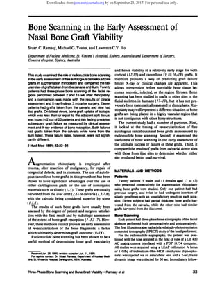
- 2. TABLE1Number of Patients from Each Donor Site GroupwithEach Bone ScanGradeDonor site Bone scan grade Calvaria* Iliaccrest* ing this, 400,000 count blood-pool images ofthe anterior face positionedidenticallyto the blood flowimagesand 300,000 count images of both laterals of the face were performed. Three hundred thousand count views of the anterior face and similar images ofright and left laterals ofthe skull were taken 3 hr later. The initial patients had normal size delayed images, however, it was found that in patients with grafts harvested fromthe calvariathe harvestsiteappearedin the fieldof view interfering with acquisition. Images ofthe face only, magnified times 2, were therefore acquired to exclude the donor site. The SPECT images were acquired over 64 angles with each angle collected for 30 sec. These scans were reconstructed using an in-house developed Metz prefilter (20) and Philips Gamma- 11 software and were then displayed as standard transaxial, sagittal, and coronal slices. Toallowassessmentofthe timecourseofrevascularization, the timing of the bone scan was varied after surgery. Five patients had their bone scans performed 2—3wk postopera tively, 10had scans 4—8wk postoperatively, and the remaining 5 werescanned10—15wk aftersurgery.Two patientshadtwo postoperativescansperformed,one becauseof failureof the initial scan to visualize the grafted bone. An assessmentof the successof revascularizationand the volume of revascularized bone was obtained by dividing the bone scans into four categories. This grading system was based on the uptake by the graft fragments compared with that of the frontal bones on lateral views (Fig. 1). FIGURE 1 Lateralfacialviewsshowingbonescangradingsystem.(A) Grade 0: Graft uptake equal to or less than adjacent soft tissue. (B) Grade 1: Graft activity greater than soft tissue but less than frontal bones. (C) Grade 2: Graft activity equal to frontalbones.(0) Grade3: Graftactivitygreaterthanfrontal bones. X-rays Thirteen patients had radiographs of the nasal bones per formed 3 mo after surgery as part oftheir routine postoperative follow-up. The graft was classified as either resorbed or intact, depending on its appearance on lateral views. Clinical Progress Patients were routinely reviewed by the operating surgeon, who assessed their progress clinically 3 mo after surgery. At this time, the grafts were classified according to their success or failure. Statistics The failure rates ofgrafts taken from the calvaria and ilium were compared using Fisher's exact probability test. The bone scan grades for the two donor sites were compared using the Wilcoxon two-sample test. RESULTS The bone scan results according to donor site are shown in Table 1. Their correlation with clinical results and X-ray assessment of the degree of bone graft re sorption 3 mo after surgery is shown in Table 2. Only two patients were considered to have failure of their grafts when assessed by the surgeon 3 mo after surgery (Table 3). In both ofthese cases, bone scanning showed uptake by the grafts that was less than or equal to the adjacent soft tissue (grade 0). These two patients were also the only two who showed resorption of the B bone grafts on X-ray. All other patients were regarded by the surgeon as having successful grafts clinically; all had isotope uptake by the grafts of greater than the adjacent soft tissues (grades 1to 3), and the 11who had X-raysshowed no evidence ofbony resorption.In none of the 10 SPECT studies was graft uptake discordant with that seen on the planar views. Of the 11 patients whose graftswere harvested from the calvaria, 2 had grafts which were regarded as failures clinically and both had grade 0 uptake and X-ray evi dence of bone graft resorption. One was scanned at D both 4—8wk and 10—15wk after surgery, while the other was scanned at 10—15wk only. Of those grafts taken from the ilium, all were clinically successful and none had grade 0 uptake. The failure rates of the grafts A •1 RT LATERAL nos• ,nign x 2.@ 3B@k / 197s.cs RLATERFIL MAGX2 300K,' l4Osec C R.LAT SKULL R •LATERAL 300K,6e ‘@ MA02 3eeK/29 I― 020144221334 *Nosignificantdifferencebetweendonorsites(p>0.10). The Journal of Nuclear Medicine •Vol. 32 •No. 1 •January 199134 by on September 21, 2017. For personal use only.jnm.snmjournals.orgDownloaded from
- 3. 00202180702302037020 TABLE3Success Ratesof BoneGrafts According to DonorSiteDonor siteSuccessFailure TABLE 2 Comparison of Bone Scan Grade with Clinical Progress and X-Ray Assessment of Bone Graft Resorption Clinical X-ray Bonescan grade Success Failure Normal Resorbed This was true for scans performed at 4—8wk and 10- 15 wk after surgery. Scans performed 2—4wk after surgery were able to predict graft success. However, no failures occurred in this patient group so the efficacy of bone scanning at 2—4wk after surgery in the prediction ofgraft failure is inferred rather than proven. Delayed SPECT studies of the head did not contrib ute any extra information in the prediction of graft failure in the first 10 patients, including one patient with a failed graft. This differs from some other pub lished work (14,21—23),which suggests that the skull and facial region is best assessed by SPECT scanning. The fact that nasal bone grafts are positioned away from the complicated bony anatomy of the skull, and can be projected off the skull using lateral views, probably explains why SPECT was not helpful. Radionuclide angiography also proved unhelpful. However, no postoperative infections were encountered and dynamic studies would be expected to be most helpful in differentiating osteomyelitis from cellulitis or normal postoperative change. Reports using animal models show that grafts taken from the calvaria maintain their volume better than those taken from the ilium, especially when grafted to skull or facial sites (10, 16,24), and it has been suggested this is also true for humans (1,3). In the current study, only two grafts were considered to be inadequate and both were taken from the calvaria. One ofthese patients had previously undergone insertion ofsilastic prostheses twice and this may have resulted in scarring, reduced vascularity, and, hence, inadequate invasion ofthe bone grafts by new blood vessels. The other patient could not be distinguished preoperatively from patients who had successful grafts. However, no statistically significant difference was found between the failure rates of grafts taken from the two sites nor the volume of revascular ized bone as assessed by bone scan grades. In conclusion, planar bone scanning is a useful method in the early detection ofnasal bone graft failure. A larger study is required to answer the question of whether calvarial or iliac donor sites produce lower failure rates in humans. REFERENCES 1. Jackson IT, Smith J, Mixter RC. Nasal bone grafting using split skullgrafts.Ann P/as.'Surg I983:11:533—540. 2. Goodman WS, Gilbert RW. Augmentation in rhinoplasty: a personalview.J Oto/aryngo1985;14:107—112. 3. Lejour M, Duchateau J, Potznik A. Routine reinsertion of the hump in rhinoplasty. Scand J P/ast Reconstr Surg l986;20:55—59. 4. Goga D, Robier A, Mateau J, et al. Surgical correction of saddle nose. Apropos of 23 cases. Ann Oto/aryngo/ Chir Cervicofacl988;l05:l23—l25. 5. Meunker R. The bilateral conchal cartilage graft: a new technique in augmentation rhinoplasty. AestheticP/ast Surg 1984;8:37—42. 6. Keller EE, Tiplett WW. Iliac bone grafting: review of 160 taken from the two different sites, however, were found not to be significantly different (p>O.25). As a measure of the volume of revascularized bone, and hence of the success of the graft, the bone scan grades of the grafts taken from the calvaria and ilium were compared. Again no significant difference was found between grafts taken from each site (p>'O.10). DISCUSSION Radionuclide bone scanning is an accepted method for the early assessment of bone graft viability in many sites (9, 12, 13, 15, 17—19).Bone grafts to various sites in the facial skeleton have been assessed by bone scanning, and it has been suggested that SPECT adds extra infor mation in this region (14,21). The nose, however, offers a different environment. First, it is highly vascular and revascularization of grafts might therefore be expected to occur earlier in this region than in other sites in the face. Second, most of the graft is not contiguous with adjacent bone, hence, interpretation of bone scans should be less difficult. The current study shows that bone scan evidence of revascularization of free cancellous bone grafts in the nasal region is present at least as early as 2—3wk after surgery compared with the 6 wk (18) and 3 wk (13) reported for iliac grafts to the mandible in the dog model or the 4 wk reported for split rib grafts to the mandible in the human (17,19). It also shows that radionuclide bone scanning can be used as an early predictor of graft outcome in the nasal region. When assessed on delayed lateral facial views, uptake of iso tope by the bone grafts which was equal to or less than the adjacent soft-tissue uptake was predictive of subse quent graft failure assessed 3 mo after surgery by clinical means and radiographic evidence of bony resorption. Calvana92*Iliac crest90* *Nosignificantdifferencebetweenfailurerates(p>0.25) 35Three-PhaseBoneScanningandBoneGraftViability•Ramsayet al by on September 21, 2017. For personal use only.jnm.snmjournals.orgDownloaded from
- 4. consecutive cases. J Oral Maxilofac Surg l987;45:l 1—14. 7. Jackson IT, Adham M, Bite U, et al. Update on cranial bone grafts in craniofacial surgery. Ann Plast Surg l987;l8:37—40. 8. Jackson IT, Helden 0, Marx R. Skull bone grafts in maxil lofacial and craniofacial surgery. J Oral Maxiiofac Surg l986;44:949—955. 9. BerbbrenA, WeilandAJ, Ostrup LT, et al. Bone scintigraphy in evaluatingthe viabilityof compositebonegraftsrevascu larizedby microvascularanastomoses,conventionalautoge nousbone grafts,and freenon-revascularizedperiostealgrafts. I BoneJoint Surg l982;64:799—809. 10. KUSiakJF, Zins JE, Whitaker LA.The earlyrevascularization ofmembranousbone.P/as:ReconsirSurg1985;76:510—514. 11. Heiple KG, Goldberg VM, Powel AE, et al. Biologyof can cellousbonegrafts.OrthopClinNorthAm l987;l8:l79—185. 12. Dee P. Lambruschi P0, Heibert JM. The use of Tc-99m- MDP bone scanningin the study of vascularizedbone im plants: concise communication. J Nuci Med 198122:522- 525. 13. Kelly JF, Cagle JD, Stevenson JS, et al. Technetium-99m radionucideboneimagingforevaluatingmandibularosseous allografts.JOralSurg1975;33:l1—17. 14. MoskowitzOW, Lukash F. Evaluation ofbone graftviability. SeminNuciMed1988;28:246—254. 15. DzebloNN, Dick HM, Feldman F, et al. Serialbone scanning in patients with extremity bone grafts [Abstract). J NucI Med l982;23:P50. 16. Zins JE, WhitakerLA. Membranousversusendochondral bone: implications for craniofacial reconstruction. Plast Re constr Surg l983;72:778—784. 17. Bergstedt HF, Korlof B, Lind MG, et al. Scintigraphy of humanautologousribtransplantstoa partiallyresectedman dible.Scandf PlastReconstrSurg l978;l2:15l—156. 18. Triplett RG, Kelly JF, Mandenhall KG, et al. Quantitative radionucide imaging for early determination of fate of man dibular bone grafts.J NudMed 1979;20:297—302. 19. FrameJW, EdmondsonHD, O'KaneMM. A radioisotope studyof the healingmandibularbonegraftin patients.BrJ Oral Surg l983;2 1:277—289. 20. McGeeK, Eberl5, WalkerP, et al. A comparisonof two dimensional preffitering techniques for single-photon corn puted tomography (SPED') studies [Abstract). Aust NZ J Med 1988;18:500. 21. Brown ML, KeyesJW, Leonard PF, et al. Facial bone scan flingby emissiontomography.J NuclMed l977;l8:l184— 1188. 22. Collier BD, Carrera GF, Messer EJ, et al. Internal derange rnent of the temporornandibular joint: detection by single photon emission computed tomography. Radiology 1983; 149:557—561. 23. Mitnick RI, PostleyJE, EsserPD, et al. Comparison of planar bone scintigraphy and single-photon computed tomography (SPECT)in evaluationofpatientswithparanasalsinusdisease [Abstract].J NuclMed l983;24:P58. 24. Smith JD, Abramson M. Membranous versus endochondral bone autografts.Arch Otolaryngol l974;99:203—205. 36 The Journal of Nuclear Medicine •Vol. 32 •No. 1 •January1991 by on September 21, 2017. For personal use only.jnm.snmjournals.orgDownloaded from
- 5. 1991;32:33-36.J Nucl Med. Stuart C. Ramsay, Michael G. Yeates and Lawrence C.Y. Ho Bone Scanning in the Early Assessment of Nasal Bone Graft Viability http://jnm.snmjournals.org/content/32/1/33 This article and updated information are available at: http://jnm.snmjournals.org/site/subscriptions/online.xhtml Information about subscriptions to JNM can be found at: http://jnm.snmjournals.org/site/misc/permission.xhtml Information about reproducing figures, tables, or other portions of this article can be found online at: (Print ISSN: 0161-5505, Online ISSN: 2159-662X) 1850 Samuel Morse Drive, Reston, VA 20190. SNMMI | Society of Nuclear Medicine and Molecular Imaging is published monthly.The Journal of Nuclear Medicine © Copyright 1991 SNMMI; all rights reserved. by on September 21, 2017. For personal use only.jnm.snmjournals.orgDownloaded from