Upper gastrointestinal bleeding
•Download as PPTX, PDF•
0 likes•101 views

Es aquel sangrado que ocurre por encima del ángulo de Treitz

Recommended
More Related Content
What's hot
What's hot (20)
Similar to Upper gastrointestinal bleeding
Similar to Upper gastrointestinal bleeding (20)
More from Fredy Ivan SUCARI CALLOHUANCA
More from Fredy Ivan SUCARI CALLOHUANCA (15)
Recently uploaded
Recently uploaded (20)
Upper gastrointestinal bleeding
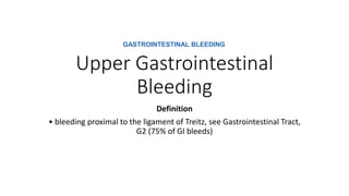
- 1. Upper Gastrointestinal Bleeding Definition • bleeding proximal to the ligament of Treitz, see Gastrointestinal Tract, G2 (75% of GI bleeds) GASTROINTESTINAL BLEEDING
- 2. Etiology • • above the GE junction • ■ epistaxis • ■ esophageal varices (10-30%) • ■ esophagitis • ■ esophageal cancer • ■ Mallory-Weiss tear (10%) • • stomach • ■ gastric ulcer (20%) (see Peptic Ulcer Disease, G11) • ■ erosive gastritis (e.g. from EtOH or post-surgery) (20%) • ■ gastric cancer • ■ gastric antral vascular ectasia (rare, associated with cirrhosis and CTD) • ■ Dieulafoy’s lesion (very rare) • • duodenum • ■ ulcer in bulb (25%) • ■ aortoenteric fistula: usually only if previous aortic graft (see sidebar) • • coagulopathy (drugs, renal disease, liver disease) • • vascular malformation (Dieulafoy’s lesion, AVM) If there is a history of abdominal aortic aneurysm repair in the past 6 months to 1 year, consider aortoenteric fistula.
- 3. Clinical Features • in order of decreasing severity of the bleed: hematochezia (brisk upper GI bleed) > hematemesis > coffee ground emesis > melena > occult blood in stool Clinical Presentation. Typically, upper GI bleed presents with black stool or melena, while lower GI bleed presents with red blood in the stool. In upper GI bleed, • occult blood–positive brown stool can occur with as little as 5–10 mL of blood loss. • Melena develops when at least 100 mL of blood has been lost. Diagnosis. Endoscopy is the most accurate test to determine the etiology of both upper and lower GI bleed. Barium study is always less accurate. Should biopsy be needed, an endoscopy must be performed.
- 4. Treatment • stabilize patient (1-2 large bore IVs, IV fluids, monitor) • send blood for CBC, cross and type, platelets, PT, PTT, electrolytes, BUN, Cr, LFTs • keep NPO • consider NG tube to determine upper vs. lower GI bleeding in some cases • IV PPI: decrease risk of rebleed if endoscopic predictors of rebleeding seen (see prognosis section) ■ given to stabilize clot, not to accelerate ulcer healing ■ if given before endoscopy, decreases need for endoscopic therapeutic intervention • for variceal bleeds, octreotide 50 μg loading dose followed by constant infusion of 50 μg/h • consider IV erythromycin (or metoclopramide) to accelerate gastric emptying prior to gastroscopy to remove clots from stomach • endoscopy (OGD): establish bleeding site + treat lesion ■ if bleeding peptic ulcer: most commonly used method of controlling bleeding is injection of epinephrine around bleeding point + thermal hemostasis (bipolar electrocoagulation or heater probe); less often thermal hemostasis may be used alone, but injection alone not recommended ■ endoclips ■ hemospray
- 5. Prognosis • 80% stop spontaneously • peptic ulcer bleeding: • low mortality (2%) unless rebleeding occurs (25% of patients, 10% mortality) • endoscopic predictors of rebleeding (Forrest classification): spurt or ooze, visible vessel, fibrin clot • can send home if clinically stable, bleed is minor, no comorbidities, endoscopy shows clean ulcer with no high risk predictors of rebleeding • H2-antagonists should not be used since they impact minimally on rebleeding rates and need for surgery • esophageal varices have a high rebleeding rate (55%) and mortality (29%)