SlideShare a Scribd company logo
1 of 54
ARTICLE IN PRESS
http://www.elsevier.de/protis
Published online date 7 August 2006
1
Correspondin
fax +81 29 853
e-mail iinouye
2Current addr
Parkville, Victo
& 2006 Elsev
doi:10.1016/j
157, 401—419, August 2006
Protist, Vol.
ORIGINAL PAPER
Hatena arenicola gen. et sp. nov., a Katablepharid
Undergoing Probable Plastid Acquisition
Noriko Okamoto2, and Isao Inouye1
Graduate School of Life and Environmental Sciences,
University of Tsukuba, 1-1-1, Tennodai, Tsukuba,
Ibaraki 305-8572, Japan
Submitted February 27, 2006; Accepted May 27, 2006
Monitoring Editor: Robert A. Andersen
Hatena arenicola gen. et sp. nov., an enigmatic flagellate of the
katablepharids, is described. It shows
ultrastructural affinities to the katablepharids, including large
and small ejectisomes, cell covering,
and a feeding apparatus. Although molecular phylogenies of the
18S ribosomal DNA support its
classification into the katablepharids, the cell is characterized
by a dorsiventrally compressed cell
shape and a crawling motion, both of which are unusual within
this group. The most distinctive feature
of Hatena arenicola is that it harbors a Nephroselmis symbiont.
This symbiosis is distinct from
previously reported cases of ongoing symbiosis in that the
symbiont plastid is selectively enlarged,
while other structures such as the mitochondria, Golgi body,
cytoskeleton, and endomembrane
system are degraded; the host and symbiont have developed a
morphological association, i.e., the
eyespot of the symbiont is always at the cell apex of Hatena
arenicola; and only one daughter cell
inherits the symbiont during cell division, resulting in a
symbiont-bearing green cell and a symbiont-
lacking colorless cell. Interestingly, the colorless cells have a
feeding apparatus that corresponds to
the location of the eyespot in symbiont-bearing cells, and they
are able to feed on prey cells. This
indicates that the morphology of the host depends on the
presence or absence of the symbiont. These
observations suggest that Hatena arenicola has a unique ‘‘half-
plant, half-predator’’ life cycle; one cell
divides into an autotrophic cell possessing a symbiotic
Nephroselmis species, and a symbiont-lacking
colorless cell, which later develops a feeding apparatus de novo.
The evolutionary implications of
Hatena arenicola as an intermediate step in plastid acquisition
are discussed in the context of other
examples of ongoing endosymbioses in dinoflagellates.
& 2006 Elsevier GmbH. All rights reserved.
Key words: Hatena arenicola;
Katablepharidophyta/Kathablepharida; Nephroselmis symbiont;
plant
evolution; plastid acquisition via secondary endosymbiosis;
ultrastructure.
Abbreviations: EM ¼ electron microscopy; ER ¼
endoplasmic reticulum; ICBN ¼ International Code
of Botanical Nomenclature; ICZN ¼ International
Code of Zoological Nomenclature; LM ¼ light mi-
croscopy; SEM ¼ scanning electron microscopy;
SSU rDNA ¼ small subunit ribosomal DNA; TEM ¼
transmission electron microscopy.
g author;
4533
@sakura.cc.tsukuba.ac.jp (I. Inouye).
ess: School of Botany, University of Melbourne,
ria, Australia.
ier GmbH. All rights reserved.
.protis.2006.05.011
http://www.elsevier.de/protis
mailto:[email protected]
dx.doi.org/10.1016/j.protis.2006.05.011
ARTICLE IN PRESS
402 N. Okamoto and I. Inouye
Introduction
Eukaryotes are currently classified into five or six
supergroups (Baldauf et al. 2000; Baldauf 2003;
Bapteste et al. 2002; Nozaki et al. 2003; Simpson
and Roger 2002), and eukaryotic autotrophs (e.g.,
plants and algae) randomly scatter across those
supergroups. Eukaryotic autotrophs comprise
nine distinct divisions in cell architecture, and this
enormous diversity is explained by several en-
dosymbiotic events (Bhattacharya et al. 2004;
Falkowski et al. 2004; McFadden 2001). It is
widely accepted that a primary endosymbiosis
between a eukaryote and a cyanobacterial sym-
biont gave rise to the three extant primary
eukaryotic autotrophs, Glaucophyta, Rhodophyta,
and Viridiplantae ( ¼ land plants plus green algae)
(see Marin et al. (2005) for an alternative primary
endosymbiosis). Subsequently, secondary endo-
symbioses occurred between green or red algae
and heterotrophic eukaryotic hosts. Two algal
divisions (Euglenophyta and Chlorarachniophyta)
acquired the plastids of green algae, while four
algal divisions (Heterokontophyta, Haptophyta,
Cryptophyta, and Dinophyta) and one parasitic
phylum (Apicomplexa) acquired those of red algae
(although some Dinophyta lost their original
plastid and remained colorless or re-acquired
different plastids as discussed below). An esti-
mated two-thirds of today’s algal diversity resulted
from secondary endosymbioses (Falkowski et al.
2004; Graham and Wilcox 2000), and thus this
process is important in understanding the evolu-
tionary process of plant and algal diversification.
The transition of a symbiont to a plastid involves
a series of changes in both the host and the
symbiont (Cavalier-Smith 2003; Hashimoto 2005;
van der Giezen et al. 2003), which include the
establishment of a specific partner alga, lateral
gene transfer from the symbiont to the host’s
nucleus (Katz 2002), the development of protein-
transport machinery to carry proteins from the
host cytoplasm to the symbiont (van Dooren et al.
2001), and synchronization of cell cycles so that
the symbiont can be passed to host daughter cells
during host cell division.
Evidence about plastid integration is accumu-
lating (Andersson and Roger 2002; Archibald et al.
2003; Hackett et al. 2004b; Huang et al. 2003;
Martin and Herrmann 1998; Martin et al. 2002;
Martin 2003a, b; Nozaki et al. 2004; Stegemann
et al. 2003), however, the intermediate steps in this
process remain largely unknown. Some organisms
appear to be in an intermediate stage of plastid
acquisition, the best-known examples of which
are the Cryptophyta and Chlorarachniophyta,
whose plastids contain a vestige of the symbiont
nucleus termed a nucleomorph (e.g. Douglas et al.
2001; Gilson et al., 2006). They are thought to
represent a late stage of integration. Early
stages of plastid acquisition can be found in
the dinoflagellates (for reviews, Hackett et al.
2004a; Morden and Sherwood 2002; Schnepf and
Elbrächter 1999), where the most dramatic
changes are ongoing. The original plastids of
dinoflagellates have been of red algal origin,
though some dinoflagellates subsequently lost
their original red-algal plastids, which were re-
placed by new ones via extra secondary or tertiary
endosymbioses. These examples probably reflect
stepwise changes in symbiotic conditions during
integration (e.g. Hackett et al. 2004a), and are
useful to understand the plastid acquisition
process.
We discovered an undescribed flagellate, Hate-
na arenicola gen. et sp. nov., in October 2000, in
an intertidal sandy beach in Japan. The organism
appears to be in the process of plastid acquisition.
Most cells of H. arenicola in the natural population
have a green plastid-like structure with a red
eyespot at the cell apex, though it is inherited by
only one of the daughter cells during cytokinesis
(Okamoto and Inouye 2005a). Molecular phyloge-
netic analysis of small subunit ribosomal DNA
(SSU rDNA) and ultrastructural observations of the
plastid-like structure reveal that it is not a plastid
but an autotrophic endosymbiont belonging to the
genus Nephroselmis Stein (Prasinophyceae, Vir-
idiplantae). We previously reported the symbiotic
nature of this association (Okamoto and Inouye
2005a). This paper describes the organism as a
new genus and species of katablepharid, a group
of flagellates recently designated the phylum
Kathablepharida, division Katablepharidophyta
(Okamoto and Inouye 2005b). We compare the
symbiosis of H. arenicola with other examples of
secondary symbioses in dinoflagellates to help
elucidate the intermediate steps in the plastid
acquisition process.
Results
Description
Hatena arenicola Okamoto et Inouye gen. et sp.
nov.
ARTICLE IN PRESS
403Hatena arenicola: Halfway to a Plant?
Latin Diagnosis
Cellulae oblongae secus axem dorsiventrem valde
appresae, sine chromatophoro nec vacuola con-
tractili; 30-40mm longae; 15-20mm latae; ventraliter
subapicali cum sulco vadoso longitudinali 3-4mm
longa et ca. 2mm lata; flagellis crassis binis
inaequalibus in sulco insertis; flagello anteriore
longiore, altero posteriore breviore; ejectisomis
conspicuis distichis prope flagellas longitudinaliter
positis; plerumque cum 1-4 endosymbiontis viridis;
uni stigma endosymbionti situm ad apicem cellulae.
Holotype: Figure 1A
Type locality: Isonoura, Wakayama, Japan (Fig.
1 B-C)
Etymology:
Hatena ¼ ‘enigmatic’ in Japanese
arenicola ¼ ‘inhabiting sand’ in Latin
Light Microscopy
General Morphology: The cell is flattened along
the dorsiventral axis. In the ventral view, it is ovoid,
30-40mm long and 15-20mm wide (Fig. 1 A, D-F).
Figure 1. Hatena arenicola gen. et sp. nov. A. Ventral v
and an eyespot of the symbiont (arrowhead). B,C. Sam
showing two rows of conspicuous Type I ejectisomes
‘‘immature’’ symbiont. G-L. Cell division in Hatena arenic
symbiont. Each panel shows a different individual at a di
The scale bar is 10mm in A, D-L.
The cell has a furrow in the subapical region, 3-
4mm long and ca. 2 mm wide (Fig. 1 D). The long
anterior flagellum and shorter posterior flagellum
emerge from this furrow, and two rows of
ejectisomes are easily visible near the posterior
end of the furrow (Fig. 1 D). One large nucleus is
located in the middle posterior region of the cell,
and the rest of the cytoplasm is mostly occupied
by the plastid of the green symbiont. Cells only
rarely lacked the symbiont (Fig. 1 E), though some
symbionts were not fully developed (Fig. 1 F; see
Discussion).
Cell Division: During cell division, one daughter
cell inherits the Nephroselmis symbiont while
the other does not and becomes colorless. Figure
1 G-L shows cell division in H. arenicola (ventral
view). First, the host nucleus moves to the apex
between the flagellar insertion and the eyespot
(Fig. 1 G). The symbiont contracts to the left side of
the host cell (on the viewer’s right in figures) so that
the left half of the cell remains green, while the right
half becomes colorless (Fig. 1 G). Two new flagella
are formed, and one set moves from the right side of
the nucleus to the left (flagellar transformation;
iew of a symbiont-bearing cell showing two flagella
pling site. D. The same cell in a different focal plane,
. E. A cell lacking the symbiont. F. A cell with an
ola, where the arrowhead indicates an eyespot of the
fferent stage in cell division. N: nucleus. S: Symbiont.
ARTICLE IN PRESS
Figure 2. Hatena arenicola and its symbiont. DIC
images are shown in upper column (A, C, E, G) and
the fluorescent images of the same cells are shown
in lower column (B, D, F, H respectively). Arrow-
heads indicate the eyespot. Blue: DAPI-stained
nuclei of Hatena arenicola (large fluorescence in
the center of the cell) and of the symbiont (smaller
dots). Red: Autofluorescence of the symbiont
plastid. The scale bar is 10mm.
0
0.2
0.6
1
ab
so
rp
tio
n
450 500 550 600 650 700
wavelength
Symbiont of
Hatena arenicola
Chl. b
Chl. b
Chl. a
Chl. a
Figure 3. Microspectrophotometry of the symbiont
plastid. Spectrogram shows absorbance similar to
chlorophyll a/b-containing plastids.
404 N. Okamoto and I. Inouye
Fig. 1 H). The chromosomes separate (Fig. 1 I).
Following nuclear division (Fig. 1 J), cytokinesis
results in one green cell with the symbiont and
one colorless cell without it (Fig. 1 K-L).
Fluorescence Microscopy: Figure 2 A-H shows
DIC and fluorescence images of the same cells.
DAPI label (blue) indicates a large host nucleus in
the cell center and one to four smaller symbiont
nuclei (Fig. 2 B,D,F,H). Each symbiont nucleus is
independent of all others and is surrounded by
plastid(s), shown in red (due to autofluorescence).
Interestingly, the cell with multiple symbiont nuclei
has only a single eyespot at the apex of the host
cell (Fig. 2 E-G; arrowheads).
Microphotometry: Microphotometry of seven
cells shows an absorption pattern characteristic
of plastids with chlorophyll a/b. Average absorp-
tion is shown in Figure 3. The prominent peaks at
435 and 678 nm represent chlorophyll a absorp-
tion, while the smaller peaks at 470 and 650 nm
correspond to chlorophyll b absorption.
Uptake of Prey Cells and Symbiont Specifici-
ty: Molecular phylogenetic analysis of 16S rDNA
indicates that the symbiont is a member of
Nephroselmis (Okamoto and Inouye 2005a). In
feeding experiments using a Nephroselmis strain
(NIES1417; different from that of the symbiont in
16S rDNA sequence; data not shown), colorless
cells of H. arenicola phagocytotically engulfed the
alga (Fig. 4 A-I) and tentatively maintained it.
However, none developed fully, likely because
the strain used in the experiments was not the
exact symbiont of H. arenicola. This suggests that
symbiont specificity is at the species or strain
level. As the H. arenicola cells which engulfed
Nephroselmis NIES1417 died, it is unclear whether
the Nephroselmis cells were digested.
Crawling Motion: Hatena arenicola displays a
conspicuous crawling motion that makes it easy
to recognize this organism in a crude sample.
Figure 5 A-H illustrates cell motion and flagellar
movement. The anterior flagellum produces most
of the propulsion, while the posterior flagellum is
used to change direction. To move forward, a cell
casts the anterior flagellum, which adheres to the
substratum by its tip (Fig. 5 A-C), and pulls itself
forward (Fig. 5 D, E). It then anchors itself with the
posterior flagellum (Fig. 5 F-H), and repeats the
process. The tip of the flagellum is sharply bent
while it is attached to the substratum, as shown in
the supplemental material and in Figure 5 I. The
cell bends the posterior flagellum to change
direction (not shown).
Electron Microscopy
Electron microscopy (EM) revealed that the green
plastid-like structure in the cell is a eukaryotic
ARTICLE IN PRESS
Figure 4. Uptake of Nephroselmis (NIES1417) by Hatena
arenicola. A-H were taken at 6-s intervals. I:
corresponds to Hatena arenicola and the symbiont of each
frame.
Figure 5. Crawling motion of Hatena arenicola.
High-speed video images recorded at 0 ms (A),
60 ms (B), 100 ms (C), 130 ms (D), 220 ms (E),
280 ms (F), 300 ms (G), 340 ms (H), respectively. I.
SEM image showing the anterior flagellum sharply
bent at the distal end (arrowhead). The scale bar is
10 mm in I.
405Hatena arenicola: Halfway to a Plant?
endosymbiont. A single nucleus, usually dorsiven-
trally flattened, is located in the middle posterior
region of the cell, and its chromatin is always
condensed and electron dense (Fig. 6 A,B). Multi-
ple mitochondrial profiles are present throughout
the cytoplasm, though these could be different
sections of a single, large reticulate mitochon-
drion. Mitochondrial cristae are tubular (Fig. 6 C),
and there is a single large Golgi body near the
groove of flagellar insertion between the nucleus
and the flagellar apparatus (Fig. 6 D).
Ejectisomes: There are two types of ejecti-
somes beneath the membrane: large, Type I
ejectisomes sensu Vørs 1992b (Fig. 6 D-E) that
are arrayed in two rows near the flagellar insertion
(Figs 1 D, 6 F-G); and smaller Type II ejectisomes
sensu Vørs 1992b (Fig. 6 H-I) that are arrayed in
numerous rows all over the cell (Fig. 6 F-G). Each
ejectisome consists of a coiled ribbon with a
gradual depression (Fig. 6 D, H), and bound by a
single membrane. Discharged ribbons are
slightly curved (Fig. 6 J; type I ejectisome). The
ribbon does not have any kink, unlike those of
Cryptophyta.
Type II ejectisomes are situated beneath the
plasma membrane of the cell (Fig. 7 A,B) except
for a small smooth area in the apical region, below
which the eyespot is situated (asterisk in Fig. 6 G).
The ejectisomes are spaced regularly between
longitudinally oriented cytoskeletal microtubular
bundles (arrowheads in Fig. 7 A).
Hatena arenicola lacks Type III ejectisomes that
are characteristic of the genus Leucocryptos (Vørs
1992b).
Cellular and Flagellar Covering: The plasma
membrane of each cell has a characteristic
cellular covering composed of a thick inner basal
layer (asterisk in Fig. 7 B) and an outer layer of
electron-opaque material (arrowhead in Fig. 7 B).
This structure extends to the flagellar surface. The
outer layer comprises a regularly arrayed and
spiraling envelope around the whole flagellum
(Fig. 7 C).
Endoplasmic Reticulum: The endoplasmic re-
ticulum (ER) does not form a conspicuous stack
but is loosely distributed throughout the cell. The
rough ER extends beneath the surface of the cell
(double arrow in Fig. 7 B).
Flagella and Basal Bodies: The long anterior
and shorter posterior flagella emerge from a
ARTICLE IN PRESS
Figure 6. Ultrastructure of Hatena arenicola. A. A longitudinal
section of a Hatena arenicola cell showing a
nucleus (N). Most of the nuclear content is condensed. The rest
of the cytoplasm is occupied by the symbiont
(Sym). The symbiont has a large plastid region with multiple
pyrenoids. B. A transverse section of Hatena
arenicola showing host nucleus (N) and the symbiont (Sym)
with the vestigial cytoplasm (asterisk). C.
Mitochondrial profiles showing tubular cristae. D. A Golgi body
near the flagellar basal bodies (arrows) and
Type I ejectisomes sensu Vørs 1992 (nearly longitudinal view).
E. A transverse section of a Type I ejectisome
(sensu Vørs 1992). F. SEM image of Hatena arenicola showing
Type I ejectisomes near the flagellar insertion
as well as smaller ejectisomes (Type II ejectisomes sensu Vørs
1992) regularly arrayed over the cell surface.
G. Magnified SEM image of the same cell. Note that the apical
region of the cell (asterisk) lacks ejectisomes.
H. A longitudinal section of Type II ejectisome. I. A transverse
section of Type II ejectisome. J. A whole mount
TEM image of a discharged type I ejectisome. The scale bar is
10 mm in A, E; 2 mm in B, G; 50 nm in C; 500 nm
in D, F; 1mm in I; 200 nm in H, I.
406 N. Okamoto and I. Inouye
ARTICLE IN PRESS
Figure 7. Surface structure and flagellar transition
region of Hatena arenicola. A. A tangential section of
the surface of Hatena arenicola. Cytoskeletal micro-
tubular bundles (arrows) are arrayed longitudinally
below the cell surface; Type II ejectisomes are
situated between them. Each pore corresponds to
the position of an ejectisome, where the surface
sheath is thin. B. A transverse section of the cell
periphery showing a bilayered surface sheath. An
arrowhead indicates the outer layer. An asterisk
indicates the thick basal layer. An arrow indicates
the plasma membrane. A double arrowhead indi-
cates a profile of rough ER. C. A tangential section of
the flagellum shows the outer layer of the surface
sheath enveloping the flagellum in a spiraling
fashion. D-J. Flagellar transition region of Hatena
arenicola. D. Diagram of flagellar transition region
reconstructed based on serial ultrathin sections.
Each letter in D indicates which part corresponds to
one of the transverse sections (E-J). The scale bar is
500 nm in A, C; 200 nm in B, E-J.
407Hatena arenicola: Halfway to a Plant?
shallow subapical furrow. The flagella are coated
by a ‘‘surface sheath’’. The diagram of the basal
body and the flagellar transition zone (Fig. 7 D) is
based on serial sections of the flagellar transition
zone (Fig. 7 E-J). At the proximal end of the basal
body, there is a cartwheel structure with a central
tube. The triplet microtubules of the basal body
terminate below the plasma membrane (Fig. 7 E),
whereas the doublet microtubules attach to the
plasma membrane by a connecting fiber (Fig. 7 F).
The flagellar transition region extends above the
plasma membrane, and an electron-dense rod
structure is present (Fig. 7 G) in the middle of this
region. Surrounding the transition region are outer
doublet microtubules lined in a loose, electron-
dense material (Fig. 7 H). The flagellar transition
region ends in a terminal plate with electron-
opaque material at the center (Fig. 7 I). The
axonemal central pair begins above the terminal
plate (Fig. 7 J).
Feeding Apparatus: As reported in Okamoto
and Inouye (2005a), colorless cells lacking the
symbiont have a complex feeding apparatus at
their apex (Fig. 8 A-C), consisting of transverse
tubular rings (arrowheads in Fig. 8 B-C) and
longitudinal microtubules arrayed in a single layer
(arrow in Fig. 8 B-C). These microtubules are
different from those that form the cytoskeleton
(double arrowheads in Fig. 8 A). Inside the micro-
tubular skeletons of the feeding apparatus are
several electron-opaque granules, some of which
are large and elongate (light gray) and others that
are smaller, granulated, and pigmented (Fig. 8 A,
C). These granules are restricted to the feeding
apparatus, never seen elsewhere in the cell.
Symbiont-bearing cells do not have a feeding
structure; the corresponding region is occupied by
the eyespot of the symbiont (Fig. 8 D-E).
Symbiont: The symbionht retains not only a
plastid but also its own cytoplasm with a nucleus
and mitochondrion(a) (Fig. 9 A-C). Most symbiont
cells contain one nucleus, which is often attached
to the membrane, and symbiont and host nuclei
often face each other (Fig. 9 A-C). The symbiont is
bounded by a single membrane (arrowhead in
Fig. 9 D), whose origin is unknown. Mitochondria
have flat, often degraded cristae, though the
extent of degradation varies among individual
cells (Fig. 9 E-F). The plastid, which is the largest
structure in the symbiont (Fig. 6 A-B), contains
multiple pyrenoids bounded by a thin starch
sheath (Fig. 9 G). The pyrenoid has shallow
invaginations of the thylakoid membrane. The
plastid has a single conspicuous eyespot, where
the morphological association between host and
symbiont is present (see below).
Free ribosomes are densely distributed through-
out the symbiont cytoplasm, though no ribosome-
bearing membrane (rough ER) is present. Occa-
sionally there are flattened, stacked membranes
next to the nucleus (Fig. 9 A). This structure would
normally be a Golgi body but it is present in a
degraded or inactive form, because no Golgi
vesicles were seen around the structure. Some
randomly shaped vacuoles of unknown origin are
also present (Fig. 9 B-C). Because each cell
division will result in half the population carrying
ARTICLE IN PRESS
Figure 8. Feeding apparatus of Hatena arenicola. A. A nearly
transverse section of the feeding apparatus.
Feeding apparatus is distinct from cytoskeletal microtubules
(double arrowheads). B. A magnified view
showing microtubules (arrow) regularly arrayed in a single
layer along the external side of the tubular rings
(arrowheads). C. Longitudinal section of the feeding apparatus
showing numerous transverse tubular rings
(arrowhead) and longitudinal microtubules arrayed in a single
layer (arrows). D. An eyespot (e) of symbiont is
located at the corresponding place in the symbiont bearing cell.
E. A schematic illustration of the feeding
apparatus and the corresponding place of the symbiont-bearing
Hatena arenicola. The scale bar is 500 nm in
A, C-D; 250 nm in B.
408 N. Okamoto and I. Inouye
the symbiont, these morphological varieties likely
reflect degradation (see Discussion).
Cytoskeletal structures, including the flagella,
basal bodies, the flagellar apparatus, and micro-
tubular rootlets, are completely absent. These
morphological changes must affect intracellular
functions, such as protein synthesis and distribu-
tion in the symbiont (see Discussion).
The lysosome of the host cytosol is discontinuous
with the symbiont compartment. The lysosome
of some cells contains scales of Nephroselmis
and Pyramimonas (Fig. 10 A,B), indicating that
H. arenicola cells engulf other prey in addition to
its Nephroselmis partner. Because Pyramimonas
cells are digested, H. arenicola may be partly
heterotrophic (see Discussion).
Eyespot: The eyespot is composed of a single-
layered sheet of osmiophilic granules (Fig. 11 A)
that connects to the inner plastid envelope. Near
the eyespot, the plastid, symbiont, and host plasma
membranes are tightly layered (Fig. 11 A,B). In some
cells, single microtubules are aligned longitudinally
between the plasma membrane and the symbiont
membrane overlying the eyespot region (Mts in
Fig. 11 C,D). These single microtubules are distinct
from the cytoskeletal microtubular bundles (arrow-
head in Fig. 11 D), and the space between them
lacks Type II ejectisomes; this is consistent with the
smooth surface appearance of the eyespot region
(Fig. 6 F).
Molecular Phylogeny
Partial SSU rDNA sequences of H. arenicola
(AB212285) aligned with the homologues of
known eukaryotes were subjected to phylogenetic
analyses. The resulting maximum likelihood (ML)
tree (Fig. 12) showed the typical topology of the
SSU rDNA tree, and H. arenicola was included in
the katablepharid clade, which was robustly
supported with high bootstrap probability in ML
(99%), NJ (100%), and MP (99%) analyses.
Discussion
Hatena arenicola has several unique features that
suggest it is in the process of endosymbiosis with
ARTICLE IN PRESS
Figure 10. Lysosome of Hatena arenicola with
scales of prasinophytes characteristic to Pyramimo-
nas (A) and Nephroselmis (B) respectively. The scale
bar is 500 nm.
Figure 9. Ultrastructure of the symbiont. A-C. The symbiont
cytoplasm, retaining a nucleus, mitochondria,
and sometimes a Golgi body-like vesicle (A) or membranes of
random shape (B-C), likely in an intermediate
state of integration. D. The single symbiont-enveloping
membrane (arrowhead) separates the symbiont
compartment (Sym) and host cytoplasm (H). Double membranes
of the symbiont plastid are also shown
(arrows). E. Mitochondrial profiles that retain flat cristae. F. A
relatively degenerated mitochondrial profile. G.
A pyrenoid, surrounded by a starch sheath. Random shallow
invagination of the thylakoids. The scale bar is
1 mm in A-C; 250 nm in D-G.
409Hatena arenicola: Halfway to a Plant?
a Nephroselmis partner (Okamoto and Inouye
2005a). We will discuss the taxonomy and
classification of H. arenicola first, and then focus
on its unique endosymbiosis.
Taxonomy
Morphological and molecular analyses of H.
arenicola clearly show that it belongs to the
recently established division/phylum Katablephar-
idophyta (International Code of Botanical Nomen-
clature, ICBN)/Kathablepharida (International
Code of Zoological Nomenclature, ICZN) (Oka-
moto and Inouye 2005b). The katablepharids
comprise an ultrastructurally well-defined and
small group of heterotrophic flagellates that
includes 10 species in two genera: nine species
of Katablepharis Skuja (correct spelling in ICBN
which will be used in this paper)/Kathablepharis
Skuja (original spelling in the ICZN), and one
species of Leucocryptos (Braarud) Butcher. Kata-
blepharidaceae (ICBN) was originally described by
Skuja (1939) based on ovate or cylindrically ovate
cell shape, two flagella emerging from a subapical
depression, and conspicuous ejectisomes aligned
ARTICLE IN PRESS
Figure 11. Eyespot of the symbiont plastid. A. A longitudinal
section of the eyespot (E). B. A magnified view
of another cell clearly shows the eyespot granules, the
inner/outer envelop of the symbiont plastid (arrows),
the single symbiont enveloping membrane (arrowhead), and the
host plasma membrane (double arrowhead)
associated with each other. C-D. Tangential sections of the
eyespot region show single microtubules (Mts)
distinctive from the cytoskeletal microtubules (arrowheads)
longitudinally situated between the eyespot (E)
and the plasma membrane. The scale bar is 250 nm in A; 100
nm in B; 1mm in C-D.
410 N. Okamoto and I. Inouye
in two rows near the flagellar insertion. Vørs
(1992b), and Clay and Kugrens (1999b) emended
the family by adding the following ultrastructural
features: the entire surface of the cell, including
the flagella, is coated with a bilayered surface
sheath that appears to form spiraling rows around
the cell body; tubular mitochondrial cristae; a
complex, truncated conical feeding apparatus and
cytoskeleton; a Golgi apparatus situated anteriorly
and a centrally located nucleus; and a food
vacuole in the posterior part of the cell.
Hatena arenicola shares all these characters,
except that the cell is dorsiventrally compressed
and the food vacuole is absent. We occasionally
observed a vacuole containing scales of prasino-
phytes, though its position was anterior to the
nucleus. In addition, the flagellar transition zone
containing a rod-shaped structure is fundamen-
tally the same as that of K. ovalis (Lee et al. 1992).
Based on these ultrastructural similarities and the
molecular phylogenetic data, H. arenicola un-
doubtedly belongs to the katablepharids.
Currently, all the katablepharids belong to a
single family, Katablepharidaceae, whose cells are
defined as either ‘‘oblong or cylindrically ovate’’
(Katablepharis; Clay and Kugrens, 1999b; Vørs,
1992b) or ‘‘ovate, pyriform elliptical outline’’
(Leucocryptos; Butcher 1967). The most important
distinguishing feature of Leucocryptos is the
presence of Type III ejectisomes, which have been
found only in Leucocryptos. Because H. arenicola
lacks Type III ejectisomes, it does not belong to
Leucocryptos.
Hatena arenicola is distinct from all other
katablepharids previously described in that it is
dorsiventrally compressed with a flat-oval shape,
which suggests that it does not belong to the
genus Katablepharis. Its crawling motion and the
feeding apparatus composed of single-layered
microtubules are also distinctive from the other
ARTICLE IN PRESS
0.1
Chlorarachnion CCMP242
Cercomonas longicauda
Heteromita globosa
Thaumatomonas seravini
Euglypha rotunda
Paulinella chromatophora
Ochromonas danica
Phytophthora megasperma
Skeletonema pseudocostatum
Pteridomonas danica
Prorocentrum micans
Prorocentrum mexicanum
Gymnodinium sp. MUCC284
Pfiesteria sp. B112456
Alexandrium minutum
Cryptosporidium parvum
Toxoplasma gondii
Prorodon teres
Platyophrya vorax
Emiliania huxleyi
Pavlova salina
Chlorokybus atmophyticus
Mesostigma viride
Arabidopsis thaliana
Fossombronia pusilla
Pyramimonas propulsa
Ulothrix zonata
'Chlorella' ellipsoidea
Tetraselmis striata
Coleochaete scutata
Closterium littorale
Cyanophora paradoxa
Cyanoptyche gloeocystis
Glaucocystis nostochinearum
Gloeochaete wittrockiana
Goniomonas truncata
Rhodomonas mariana
Hanusia phi
Geminigera cryophila
Cryptomonas ovata
Chroomonas sp. M1318
Hemiselmis brunnescens
Leukocryptos marina
Hatena arenicola
Katablepharis japonica
Heterophrys marina
Chlamydaster sterni
Raphidiophrys ambigua
Rhodella maculata
Stylonema alsidii
Bangia sp.
Porphyra umbilicalis
Acanthamoeba castellanii
Dictyostelium discoideum
Leptomyxa reticulata
Hartmannella vermiformis
Scutellospora cerradensis
Pneumocystis carinii
Saccharomyces cerevisiae
Schizosaccharomyces pombe
Basidiobolus haptosporus
Chytriomyces hyalinus
Monosiga brevicollis
Clathrina cerebrum
Cirripathes lutkeni
18S rDNA (65species 1252 sites)
100/100/99
99/83/96
64/73/62
54/86/60
58/73/-
52/88/72
53/98/67
100/89/100
100/100/100
99/100/100
99/88/56
56/67/-
100/100/100
90/97/93
86/86/58
54/61/80
100/100/100
-/73/-
100/100/100
58/63/-
99/99/97
67/72/89
72/54/74
99/100/99
95/76/81
76/-/-
100/100/100
82/85/78
75/83/74
91/79/84
100/100/100
75/88/66
99/97/83
75/64/-
65/85/89
74/-/-
76/81/84
93/-/-
-/-/54
53/-/-
50/59/-
92/77/79
ML/NJ/MP
Figure 12. Unrooted eukaryotic tree based on the SSU rDNA.
The best tree of the ML method is shown.
Bootstrap propotions are shown at the internal branches, in the
order of ML/NJ/MP methods. The length of
each branch is proportional to the estimated number of
substitutions. Bar denotes 10% substitutions per site.
For details of the phylogenetic reconstruction methods, see text.
The organisms included in the tree are listed
in Table 1. Unambiguously aligned 1252 nucleotide positions
were used for the analysis.
411Hatena arenicola: Halfway to a Plant?
katablepharids (Clay and Kugrens 1999a, b;
Kugrens et al. 1994; Lee and Kugrens 1992; Lee
et al. 1991; Okamoto and Inouye 2005b; Vørs
1992a, b). Based on those features, we propose
that the organism should be assigned to a new
genus, Hatena.
The family-level taxonomy of the katablepharids
is still unclear, primarily because little molecular
sequence data exist. Until further studies eluci-
date katablepharid taxonomy, it is best to include
the genus Hatena in the family Katablepharida-
ceae.
ARTICLE IN PRESS
AH
Loss of
Feeding Appar atus
Plastid: Enlarged
Symtiont: Degraded
412 N. Okamoto and I. Inouye
Endosymbiosis
Endosymbiosis is a major driving force in plant
evolution, and thus it is important to understand
this process. Hatena arenicola may be an im-
portant model of early plastid acquisition. Sym-
biotic Nephroselmis differs from free-living
individuals in having enlarged plastids with a
greater number of pyrenoids, degraded subcellu-
lar structures, and morphologically distinct eye-
spots. Cells of known Nephroselmis species are a
maximum of 20mm in length (Nephroselmis
astigmatica; Inouye and Pienaar 1984) and pos-
sess a single plastid with a single pyrenoid. The
symbiont occupies most of the host cytoplasm,
suggesting that the symbiont plastid(s) grows
more than ten fold after being engulfed by the
host. Pyrenoids also multiply after being engulfed.
In contrast, the symbiont cytoplasm loses other
major cell components including flagellar appara-
tus and microtubular roots, endomembranes such
as the ER and transport vesicles, Golgi-like
vesicles, and amorphous membranous structures.
The dramatic growth of the plastid is in stark
contrast to the degradation of the other orga-
nelles. Because the cytoplasm is in such a
degraded state, it must be difficult to sustain the
growth and maintenance of the plastid alone. It is
likely that some metabolites from the host cell are
used to develop and maintain the plastid.
B
DE
F
G
C
Predator phase Plant phase
Formation of
Feeding apparatus
Uptake of
the partner
Figure 13. Half-plant, half-predator-hypothesis. The
solid line indicates a witnessed process, and the
broken line indicates a hypothetical process. A-D: A
green cell with the symbiont, lacking the feeding
apparatus (A) divides (B) into one green (C) and one
colorless cell (D). E-G: The colorless cell should form
a feeding apparatus de novo and engulfs a Ne-
phroselmis cell. G-H: The symbiont plastid selec-
tively grows in the host cytoplasm. Because cell
division of a colorless cell or a cell with an
‘‘immature’’ symbiont (H) has never been observed,
uptake and the subsequent changes in both
host and symbiont apparently occur within one
generation.
Eyespot Morphology
The morphology of the eyespot is most suggestive
of a host-symbiont coordination. The tight layering
of the four distinct membranes of different origin
implies functional cooperation. The eyespot is an
important component of a photo-sensing complex
found in various algal groups (Melkonian and
Robenek 1984; Gualtieri 2001). It effectively
regulates the light received by a photoreceptor
located on a nearby membrane, thereby allowing
the alga to detect light direction. Preliminary
observations have shown that lateral, and not
vertical, incidence, is important for the behavior of
H. arenicola. Hence, we speculate that the
photoreceptor and the regulatory mechanism
would be laterally aligned in the cell, so that the
eyespot can effectively shade the photorecepter
from the lateral incidence. Assuming that the
eyespot is functional in H. arenicola, the photo-
receptor should be situated in either of the outer or
inner plastid membrane, the single endosymbiont
envelope, or the plasma membrane. Their mor-
phological association can be explained as a
consequence of functional collaboration. Because
H. arenicola crawls two-dimensionally, a photo-
tactic response to laterally projected light is
plausible. To test whether a functional association
exists, further investigation of the threshold and
the efficiency of phototaxis in both colored and
colorless H. arenicola cells are required.
Morphological Changes of the Host and
‘‘Half-plant, half-predator’’ Model
Hatena arenicola cells without a symbiont have a
complex feeding apparatus in place of an eye-
spot, indicating that symbiont acquisition is
accompanied by drastic morphological changes
in both the symbiont and the host.
ARTICLE IN PRESS
413Hatena arenicola: Halfway to a Plant?
Such structural changes would necessitate life
cycle changes. Based on observations presented
in this paper, we propose the ‘‘half-plant, half-
predator’’ hypothesis, where H. arenicola switches
its lifestyle between that of a plant and a predator
(Fig. 13; see also Okamoto and Inouye 2005a for
detailed explanation). This proposed life cycle is
also supported by the observation that the extent
of degradation of symbiont mitochondria and
membranous structures varies among individual
cells. The presence of prasinophyte scales in a H.
arenicola lysosome suggests that H. arenicola
lives heterotrophically to some extent, perhaps
until it engulfs its symbiotic partner. This repre-
sents an intermediate state of trophic alteration.
Although there are several assumptions in the
model, it promises to lead to new insights and
helps elucidate the plastid integration process.
Evolutionary Implications
Extra secondary and tertiary endosymbioses in
dinoflagellates may represent evolutionary events
that occurred during plastid acquisition (for re-
views, see Hackett et al. 2004a; Morden and
Sherwood 2002; Schnepf and Elbrächter 1999).
The earliest stage is represented by the crypto-
phyte symbiont, in which only cytoskeletal com-
ponents are lost, as in Amphidinium latum Lebour
(Horiguchi and Pienaar 1992), A. poecilochroum
Larsen (Larsen 1988), and Gymnodinium acidotum
Nygaard (Fields and Rhodes 1991; Wilcox and
Wedemayer 1984). The cell cycles of the host and
symbiont are not synchronized, and the host cell
must repeatedly capture symbionts. The next
stage is represented by the symbiont of diatom
origin, in which various subcellular structures are
lost, except the nucleus, mitochondrion(a) and
plastid, as in Durinskia baltica (Levander) Carty et
Cox (formerly Peridinium balticum; Chesnick and
Cox 1987, 1989; Chesnick et al. 1997; Eschbach
et al. 1990; Tippit and Pickett-Heaps 1976; Tomas
and Cox 1973) and Kryptoperidinium foliaceum
(Stein) Lindemann (Dodge 1971; Eschbach et al.
1990). At this stage, the symbiont divides syn-
chronously with the host cell (Chesnick and Cox
1987, 1989; Tippit and Pickett-Heaps 1976), so
that the association between the host and
symbiont becomes permanent, and repeated
uptake of the symbiont is no longer necessary.
Finally, the symbiont cytoplasm is reduced, as
seen in Lepidodinium viride (Watanabe et al. 1987;
Watanabe et al. 1990), Gymnodinium chlorophor-
um (Elbrächter and Schnepf 1996), both with
symbionts of prasinophyte origin, and Karenia
brevis (Davis) Hansen et Moestrup, Karenia
mikimotoi (Miyake et Kominami ex Oda) Hansen
et Moestrup, Karlodinium veneficum (Leadbeater
et Dodge) Larsen with symbionts of haptophyte
origin (Daugbjerg et al., 2000; Inagaki et al., 2000;
Tangen and Björnland, 1981). The symbiont of
Karenia and Karlodinium species has no remnant
of cytoplasm, and can therefore be recognized as
an integrated plastid. Based on cytoplasm reduc-
tion and the lack of cell cycle synchronization, the
symbiosis of H. arenicola can be placed between
the cryptophyte and diatom types of symbiosis
mentioned above. Nevertheless, their morpholo-
gical association suggests an intimate host—sym-
biont relationship. Previous studies on symbiosis
in dinoflagellates have focused on symbiont
degradation only. In this study, we demonstrated
that major morphological changes also occur in
the host, suggesting that plastid acquisition is not
merely ‘‘enslavement’’ where the symbiont is
degraded, but also a process during which the
host itself changes to establish a new association
with the symbiont.
The rigid pattern of asymmetrical inheritance of
the symbiont is also suggestive of a partly
regulated association. The symbiont always
comes to the left side of the host cell (ventral
view) before the migration of the host nucleus,
implying an interaction between the symbiont
compartment and the host cytoskeleton. If the
symbiont moved to the division plane, and not to
one side of the cell, the symbiont would co-
segregate, just as in the division of D. baltica,
where the center-positioned symbiont co-segre-
gates upon host cytokinesis (Tippit and Pickett-
Heaps 1976).
Another question is whether the association of
H. arenicola and the symbiont has developed
genetic modification. The process of endosym-
biosis is hypothesized to include genetic changes
such as lateral gene transfer (LGT) from symbiont
to host, coupled with evolution of a protein
transport machinery from host to symbiont (e.g.
Gilson and McFadden 2002). It is unclear when
those changes start and how they integrate.
Considering host—symbiont intimacy, H. arenico-
la would have already experienced or may be
experiencing some of these changes. Therefore,
the study of LGT or of a protein transport
machinery in H. arenicola would be an interesting
topic for future studies.
Recently, Marin et al. (2005) reported another
example of primary endosymbiosis in Paulinella
chromatophora Lauterborn, a freshwater thecate
amoeba that bears a cyanobacterium-like
ARTICLE IN PRESS
414 N. Okamoto and I. Inouye
structure. They reported that the symbiosis of P.
chromatophora is a more recent event than the
origin of all other plastids, based on the molecular
phylogeny of ribosomal DNA operon sequences.
This is consistent with a morphological feature of
the symbiont, namely, a peptidoglycan layer that
must have originated from the cell wall of an
ancestral cyanobacterium.
The symbiotic relationships of H. arenicola, P.
chromatophora, and the dinoflagellates probably
represent different intermediate steps in plastid
acquisition via primary or secondary endosymbio-
sis. Continued study and comparison of these
groups should provide further insight into plastid
evolution.
Concluding Remarks
Hatena arenicola gen. et sp. nov. is likely in the
process of plastid acquisition via secondary
endosymbiosis. Although it is in an early inter-
mediate stage of acquisition, the two organisms
have already established an intimate association
in ultrastructure and likely in metabolic function.
Based on behavioral and ultrastructural observa-
tions, we propose a ‘‘half-plant, half-predator’’ life
cycle. Because H. arenicola shows an early
intermediate state of plastid acquisition, it should
provide further insight into plant evolution. This
study provides a foundation for future studies on
the topic.
Methods
Sampling and temporary maintenance in the
laboratory: Because it is not possible to culture
Hatena arenicola in the laboratory, we used crude
samples from the natural habitat. Cells were
collected at Isonoura Beach, Wakayama Prefec-
ture, Japan (Fig. 1 B,C), April—December from
2000 to 2004. Samples were maintained in the
laboratory at room temperature in f/2 medium,
under ca. 10mmol photons m�2 s�1, and the
light—dark cycle was L:D ¼ 5:19 h.
Morphological observations: Light micro-
scopy (LM) and fluorescence microscopy (FM)
was conducted using a Leica DMR light micro-
scope (Leica Wetzlar GmbH, Wetzlar) and the LM
image was taken with a Keyence VB6010 digital
chilled CCD camera (Keyence, Osaka). For FM,
40,6-Diamidino-2-phenylindole (DAPI) was used to
stain the nucleus. The DAPI fluorescence along
with the autofluorescence of the plastid were
observed using a D filter cube (Leica Wetzlar
GmbH, Wetzlar).
Microspectrophotometry was performed by
majoring three different regions of the symbiont
in each of seven cells. Each absorption spectrum
was recorded in the range of 300—800 nm with a
light microscope (ECLIPSE, Nikon, Tokyo)
equipped with a high-resolution multichannel
photodetector (MCPD 7000, Otsuka Eelectronics,
Osaka) at Okazaki National Institute for Basic
Biology, Japan. The average of the three measure-
ments was considered the representative absor-
bance of each cell. Because the absorptions
obtained were almost uniform across the seven
cells, their average is shown.
A unialgal culture of Nephroselmis sp.
(NIES1417) was established from the same sam-
ple site by micropipette isolation and maintained
in f/2 medium at 20 1C under ca. 10mE light
intensity, and a light-dark cycle of L:D ¼ 14:10 h.
The uptake of Nephroselmis sp. (NIES1417) was
photographed under a CKX31 inversed light
microscope (Olympus, Tokyo) equipped with a
COOLPIX 990 digital camera (Nikon, Tokyo) at 6-s
intervals.
High-speed video images were recorded at 200
frames per second using an OPTIPHOT micro-
scope (Nikon, Tokyo), equipped with an MHS-200
high-speed video capturing system (Nac Inc.,
Tokyo). The images were digitized on a Macintosh
computer using an NIH imaging program (public
domain, developed at the US National Institute of
Health; available at http://rsb.info.nih.gov/nih-im-
age/) for analysis of the cellular and flagellar
motion.
Preparation for transmission electron micro-
scopy (TEM) and scanning electron microscopy
(SEM) was performed as described elsewhere
(Moriya et al. 2000; Okamoto and Inouye 2005b).
The observations were made with a JEOL 100CXII
electron microscope (JEOL, Tokyo) and a JSM-
6330 scanning electron microscope (JEOL, To-
kyo).
Molecular phylogeny: To avoid contamination
of the prey genome, single cells of Hatena
arenicola were isolated with micropipette into a
0.2-ml PCR tube containing 10 ml of sterilized
double distilled water, and immediately frozen at
�80 1C for more than 15 min to completely disrupt
the cells. The first and the second nested PCR
were performed with existing degenerate primer
sets (Moriya et al. 2000) using rTaq (TOYOBO,
Osaka). Thermal cycling for the first PCR con-
sisted of 33 cycles. Annealing temperatures
ranged from 50 to 47 1C (six cycles decreasing
http://rsb.info.nih.gov/nih-image/
ARTICLE IN PRESS
Table 1. Sequences used for the phylogenetic
analyses
Organism Accession
number
SSU rDNA
Katablepharidophyta/Kathablepharida
Hatena arenicola AB212285
Katablepharis japonica AB231617
Leucocryptos marina AB193602
Metazoa
Cirripathes lutkeni AF052902
Clathrina cerebrum U42452
Monosiga brevicollis AF084618
Fungi
Basidiobolus haptosporus AF113413
Chytriomyces hyalinus M59758
Pneumocystis carinii L27658
Saccharomyces cervisiae V01335
Schizosaccharomyces pombe Z19578
Scutellospora cerradensis AB041344
Amoebae
Acanthamoeba castellanii M13435
Hartmannella vermiformis M95168
Leptomyxa reticulata AF293898
Dictyostelium discoideum K02641
Cercozoa
Cercomonas longicauda AF101052
Chlorarachnion CCMP242 U03479
Euglypha rotunda X77692
Heteromita globosa U42447
Paulinella chromatophora X81811
Thaumatomonas seravini AF411259
Viridiplantae
Arabidopsis thaliana X16077
‘Chlorella’ ellipsoidea D13324
Chlorokybus atmophyticus M95612
Closterium littorale AF115438
Coleochaete scutata X68825
Fossombronia pusilla X78341
Mesostigma viride AJ250108
Pyramimonas propulsa AB017123
Tetraselmis striata X70802
Ulothrix zonata Z47999
Heterokontophyta
Ochromonas danica M32704
Pteridomonas danica L37204
Skeletonema pseudocostatum X85394
Phytophthora megasperma X54265
Alveolata
Cryptosporidium parvum X64340
Toxoplasma gondii X75429
Platyophrya vorax AF060454
Prorodon teres X71140
Alexandrium minutum U27499
Gymnodinium sp. MUCC284 AF022196
Pfiesteria sp. B112456 AF218805
Table 1. (continued )
Organism Accession
number
Prorocentrum mexicanum Y16232
Prorocentrum micans M14649
Cryptophyta
Chroomonas sp. M1318 AJ007279
Cryptomonas ovata AJ421147
Geminigera cryophila U53124
Hanusia phi U53126
Hemiselmis brunnescens AJ007282
Rhodomonas mariana X81373
Goniomonas truncata U03072
Glaucophyta
Cyanophora paradoxa X68483
Cyanoptyche gloeocystis AJ007275
Glaucocystis nostochinearum X70803
Gloeochaete wittrockiana X81901
Haptophyta
Emiliania huxleyi L04957
Pavlova salina L34669
Rhodophyta
Bangia sp. AF043362
Porphyra umbilicalis AB013179
Rhodella maculata U21217
Stylonema alsidii L26204
Centroheliozoa
Chlamydaster sterni AF534709
Heterophrys marina AF534710
Raphidiophrys ambigua AF534708
415Hatena arenicola: Halfway to a Plant?
the temperature by 0.5 1C for each cycle, and 27
cycles at a constant temperature). An extension
was performed at 72 1C for 1 min, and denaturing
was done at 94 1C for 30 s. The final extension
period was at 72 �C for 7 min. Thermal cycling for
the second PCR consisted of 33 cycles with an
annealing step at 53 1C for 30 s, an extension step
at 72 1C for 1 min, and denaturing at 94 1C for 30 s,
with a final extension period at 72 1C for 7 min. The
sequences were determined by direct sequen-
cing. Cycle sequencing reaction was performed
using a DYEnamic ET terminator cycle sequencing
kit (Amersham biosciences, Buckinghamshire), as
per the manufacturer’s instructions. Sequencing
was conducted with an ABI PRISM 377 DNA
Sequencer (Applied Biosystems, California), and
sequences were confirmed free of contaminants
and not of haptophyte prey origin by comparing
at least two cells or performing a BLAST search
at the National Center for Biotechnology Informa-
tion (NCBI) server (http://www.ncbi.nlm.nih.gov/
http://www.ncbi.nlm.nih.gov/BLAST/
ARTICLE IN PRESS
416 N. Okamoto and I. Inouye
BLAST/). The sequence (AB212285) was depos-
ited to GenBank.
The SSU sequence was manually aligned to the
existing alignments of global eukaryotes (Okamo-
to and Inouye 2005b), which include 65 taxa for
SSU rDNA (Table 1). To avoid the long-branch
attraction (LBA) artifact in SSU rDNA analysis,
organisms with an extraordinary evolutionary rate,
such as the Euglenozoa, the diplomonads, and
the palabasalids, were excluded after confirming
that they are not a sister group of the katablephar-
ids. A total of 1252 unambiguously aligned
nucleotide positions were selected for phyloge-
netic analyses.
The maximum likelihood (ML), neighbor joining
(NJ), and maximum parsimony (MP) methods for
phylogenetic analysis were applied to the data set
using PAUP* ver.4.0 b 10 (Swofford 2003). In the
ML analysis, the evolutionary model was selected
using the Akaike information criterion (AIC) test in
Modeltest ver.3 (Posada and Crandall 1998),
which selected the general time reversible (GTR)
model with rate variation among sites and
invariant sites (GTR+I+gamma). The estimated
gamma-shape parameter (alpha) of the discrete
gamma-distribution was 0.5645, and the propor-
tion of invariant sites was 0.3257. A further tree
search with GTR+I+gamma model with eight site
rate categories that approximates site rate was
used to produce the optimal tree. Bootstrap
proportion (BP) values for internal branches of
the optimal tree of the ML analysis were obtained
using PAUP* through 300 bootstrap resamplings
for the ML method and 1000 resamplings for the
NJ and MP methods.
Acknowledgments
We thank Dr. Masakatsu Watanabe and Dr.
Shigeru Matsunaga (Graduate University for Ad-
vanced Studies, Japan) for their dedication to the
microspectrophotometric work and assistance in
sampling, and Dr. Hiroshi Kawai (Research Center
for Inland Seas, University of Kobe) for use of his
laboratory equipment. We appreciate Dr. Tetsuo
Hashimoto and Dr. Miako Sakaguchi for their
assistance and advices in molecular phylogeny.
We are grateful to Dr. Jeremy Pickett-Heaps for
taking movie images of H. arenicola. This research
was supported in part by Japan Society for the
Promotion of Sciences (JSPS) Grants
RFTF00L0162 (to I.I.) and 1612007 (to N.O.).
N.O. was also supported by a JSPS Research
Fellowship for Young Scientists as a JSPS
Research Fellow.
Appendix A. Supplementary materials
Supplementary data associated with this article
can be found in the online version at doi:10.1016/
j.protis.2006.05.011
References
Andersson JO, Roger AJ (2002) A cyanobacterial
gene in nonphotosynthetic protists—an early chlor-
oplast acquisition in eukaryotes? Curr Biol 12:
115—119
Archibald JM, Rogers MB, Toop M, Ishida K,
Keeling PJ (2003) Lateral gene transfer and the
evolution of plastid-targeted proteins in the second-
ary plastid-containing alga Bigelowiella natans. Proc
Natl Acad Sci USA 100: 7678—7683
Baldauf SL (2003) The deep roots of eukaryotes.
Science 300: 1703—1706
Baldauf SL, Roger AJ, Wenk-Siefert I, Doolittle
WF (2000) A kingdom-level phylogeny of eukaryotes
based on combined protein data. Science 290:
972—977
Bapteste E, Brinkmann H, Lee JA, Moore DV,
Sensen CW, Gordon P, Durufle L, Gaasterland T,
Lopez P, Muller M, Philippe H (2002) The analysis
of 100 genes supports the grouping of three highly
divergent amoebae: Dictyostelium, Entamoeba, and
Mastigamoeba. Proc Natl Acad Sci USA 99:
1414—1419
Bhattacharya D, Yoon HS, Hackett JD (2004)
Photosynthetic eukaryotes unite: endosymbiosis
connects the dots. Bioessays 26: 50—60
Butcher RW (1967) An Introductory Account of the
Smaller Algae of British Coastal Waters. 4. Crypto-
phyceae. Fishery Investigations, Series IV. HMSO,
London
Cavalier-Smith T (2003) Genomic reduction and
evolution of novel genetic membranes and protein-
targeting machinery in eukaryote—eukaryote chi-
maeras (meta-algae). Philos Trans R Soc Lond B
358: 109—133
Chesnick JM, Cox ER (1987) Synchronized sexu-
ality of an algal symbiont and its dinoflagellate host,
Peridinium balticum (Levander) Lemmermann. Bio-
Systems 21: 69—78
http://www.ncbi.nlm.nih.gov/BLAST/
dx.doi.org/10.1016/j.protis.2006.05.011
ARTICLE IN PRESS
417Hatena arenicola: Halfway to a Plant?
Chesnick JM, Cox ER (1989) Fertilization and
zygote development in the binucleate dinoflagellate
Peridinium balticum (Pyrrhophyta). Am J Bot 76:
1060—1072
Chesnick JM, Kooistra WHCF, Wellbrock U,
Medlin LK (1997) Ribosomal RNA analysis indicates
a benthic pennate diatom ancestry for the endo-
symbionts of the dinoflagellates Peridinium folia-
ceum and Peridinium balticum (Pyrrhophyta). J
Eukaryot Microbiol 44: 314—320
Clay BL, Kugrens P (1999a) Description and
ultrastructure of Kathablepharis tenuis sp. nov. and
K. obesa sp. nov. — two new freshwater Kathable-
pharis (Kathablepharididae) from Colorado Wyom-
ing. Europ J Protistol 35: 435—447
Clay BL, Kugrens P (1999b) Systematics of the
enigmatic Kathablepharids, including EM character-
ization of the type species, Kathablepharis phoeni-
koston, and new observations on K. remigera comb.
nov. Protist 150: 43—59
Daugbjerg N, Hansen G, Larsen J, Moestrup Ø
(2000) Phylogeny of some of the major genera of
dinoflagellates based on ultrastructure and partial
LSU rDNA sequence data, including the erection of
three new genera of unarmoured dinoflagellates.
Phycologia 39: 302—317
Dodge JD (1971) A dinoflagellate with both a
mesokaryotic and a eukaryotic nucleus. Protoplas-
ma 73: 145—157
Douglas S, Zauner S, Fraunholz M, Beaton M,
Penny S, Deng LT, Wu XN, Reith M, Cavalier-
Smith T, Maier UG (2001) The highly reduced
genome of an enslaved algal nucleus. Nature 410:
1091—1096
Elbrächter M, Schnepf E (1996) Gymnodinium
chlorophorum, a new, green, bloom-forming dino-
flagellate (Gymnodiniales, Dinophyceae) with a ves-
tigial prasinophyte endosymbiont. Phycologia 35:
381—393
Eschbach S, Speth V, Hansmann P, Sitte P (1990)
Freeze-fracture study of the single membrane
between host cell and endocytobiont in the dino-
flagellates Glenodinium foliaceum and Peridinium
balticum. J Phycol 26: 324—328
Falkowski PG, Katz ME, Knoll AH, Quigg A, Raven
JA, Schofield O, Taylor FJR (2004) The evolution of
modern eukaryotic phytoplankton. Science 305:
354—360
Fields SD, Rhodes RG (1991) Ingestion and reten-
tion of Chroomonas spp (Cryptophyceae) by Gym-
nodinium acidotum (Dinophyceae). J Phycol 27:
525—529
Gilson PR, Su V, Slamovits CH, Reith ME, Keeling
PJ, McFadden GI (2006) Complete nucleotide
sequence of the chlorarachniophyte nucleomorph:
Nature’s smallest nucleus. Proc Natl Acad Sci USA
103: 9566—9571
Graham LE, Wilcox LW (2000) Algae. Prentice-Hall,
NJ
Gualtieri P (2001) Morphology of photoreceptor
systems in microalgae. Micron 32: 411—426
Hackett JD, Anderson DM, Erdner DL, Bhatta-
charya D (2004a) Dinoflagellates: a remarkable
evolutionary experiment. Am J Bot 91: 1523—1534
Hackett JD, Yoon HS, Soares MB, Bonaldo MF,
Casavant TL, Scheetz TE, Nosenko T, Bhatta-
charya D (2004b) Migration of the plastid genome to
the nucleus in a peridinin dinoflagellate. Curr Biol 14:
213—218
Hashimoto H (2005) The ultrastructural features and
division of secondary plastids. J Plant Res 118:
163—172
Horiguchi T, Pienaar RN (1992) Amphidinium latum
(Dinophyceae), a sand-dwelling dinoflagellate feed-
ing on cryptomonads. Jpn J Phycol (Sorui) 40:
353—363
Huang CY, Ayliffe MA, Timmis JN (2003) Direct
measurement of the transfer rate of chloroplast DNA
into the nucleus. Nature 422: 72—76
Inagaki Y, Dacks JB, Doolittle WF, Watanabe KI,
Ohama T (2000) Evolutionary relationship between
dinoflagellates bearing obligate diatom endosym-
bionts: insight into tertiary endosymbiosis. Int J Syst
Evol Microbiol 50: 2075—2081
Inouye I, Pienaar RN (1984) Light and electron
microscope observation on Nephroselmis astigmati-
ca sp. nov. (Prasinophyceae). Nord J Bot 4:
409—423
Katz LA (2002) Lateral gene transfers and the
evolution of eukaryotes: theories and data. Int J
Syst Evol Microbiol 52: 1893—1900
Kugrens P, Lee RE, Corliss JO (1994) Ultrastruc-
ture, biogenesis, and functions of extrusive orga-
nelles in selected non-ciliate protist. Protoplasma
181: 164—190
Larsen J (1988) An ultrastructural study of Amphi-
dinium poecilochroum (Dinophyceae), a phago-
trophic dinoflagellate feeding on a small species of
cryptophytes. Phycologia 27: 366—377
Lee RE, Kugrens P (1992) Relationship between the
flagellates and the ciliates. Microbiol Rev 56:
529—542
ARTICLE IN PRESS
418 N. Okamoto and I. Inouye
Lee RE, Kugrens P, Mylnikov AP (1991) Feeding
apparatus of the colorless flagellate Katablepharis
(Cryptophyceae). J Phycol 27: 725—733
Lee RE, Kugrens P, Mylnikov AP (1992) The
structure of the flagellar apparatus of two strains of
Katablepharis (Cryptophyceae). Br Phycol J 27:
369—380
Marin B, Nowack ECM, Melkonian M (2005) A
plastid in the making: evidence for a second primary
endosymbiosis. Protist 156: 425—432
Martin W (2003a) Gene transfer from organelles to
the nucleus: frequent and in big chunks. Proc Natl
Acad Sci USA 100: 8612—8614
Martin W (2003b) The smoking gun of gene transfer.
Nature Genet 33: 442
Martin W, Herrmann RG (1998) Gene transfer from
organelles to the nucleus: How much, what hap-
pens, and why? Plant Physiol 118: 9—17
Martin W, Rujan T, Richly E, Hansen A, Cornelsen
S, Lins T, Leister D, Stoebe B, Hasegawa M,
Penny D (2002) Evolutionary analysis of Arabidopsis,
cyanobacterial, and chloroplast genomes reveals
plastid phylogeny and thousands of cyanobacterial
genes in the nucleus. Proc Natl Acad Sci USA 99:
12246—12251
McFadden GI (2001) Primary and secondary en-
dosymbiosis and the origin of plastids. J Phycol 37:
951—959
Melkonian M, Robenek H (1984) The Eyespot
Apparatus of Flagellated Green Algae: a Critical
Review. In Round FE, Chapman DJ (eds) Progress in
Phycological Research, Vol. 3. Biopress Ltd., Bristol,
pp 93—268
Morden CW, Sherwood AR (2002) Continued
evolutionary surprises among dinoflagellates. Proc
Natl Acad Sci USA 99: 11558—11560
Moriya M, Nakayama T, Inouye I (2000) Ultrastruc-
ture and 18SrDNA sequence analysis of Wobblia
lunata gen. et sp. nov., a new heterotrophic flagellate
(stramenopiles, incertae sedis). Protist 150:
835—846
Nozaki H, Matsuzaki M, Misumi O, Kuroiwa H,
Hasegawa M, Higashiyama T, Shin T, Kohara Y,
Ogasawara N, Kuroiwa T (2004) Cyanobacterial
genes transmitted to the nucleus before divergence
of red algae in the Chromista. J Mol Evol 59:
103—113
Nozaki H, Matsuzaki M, Takahara M, Misumi O,
Kuroiwa H, Hasegawa M, Shin-i T, Kohara Y,
Ogasawara N, Kuroiwa T (2003) The phylogenetic
position of red algae revealed by multiple nuclear
genes from mitochondria-containing eukaryotes and
an alternative hypothesis on the origin of plastids. J
Mol Evol 56: 485—497
Okamoto N, Inouye I (2005a) A secondary symbio-
sis in progress? Science 310: 287
Okamoto N, Inouye I (2005b) The katablepharids
are a distant sister of the Cryptophyta: a proposal of
Katablepharidophyta divisio nova/Kathablepharida
phylum novum based on SSU rDNA and beta tubulin
phylogeny. Protist 156: 163—179
Posada D, Crandall KA (1998) Modeltest: testing
the model of DNA substitution. Bioinformatics 14:
817—818
Schnepf E, Elbrächter M (1999) Dinophyte chloro-
plasts and phylogeny — a review. Grana 38: 81—97
Simpson AGB, Roger AJ (2002) Eukaryotic evolu-
tion: Getting to the root of the problem. Curr Biol 12:
R691—R693
Skuja H (1939) Beitrag zur Algenflora Lettlands. II.
Acta Hortic Bot Univ Latviensis 11/12:41—168
Stegemann S, Hartmann S, Ruf S, Bock R (2003)
High-frequency gene transfer from the chloroplast
genome to the nucleus. Proc Natl Acad Sci USA 100:
8828—8833
Swofford DL (2003) PAUP*: Phylogenetic Analysis
Using Parsimony (* and other methods), Version
4.0b10. Sinauer Associates, Inc., Sunderland, MA
Tangen K, Björnland T (1981) Observations on
pigments and morphology of Gyrodinium aureolum
Hulburt, a marine dinoflagellate containing 190-
hexanoyloxyfucoxanthin as the main carotenoid. J
Plankton Res 3: 389—401
Tippit DH, Pickett-Heaps JD (1976) Apparent
amitosis in the binucleate dinoflagellate Peridinium
balticum. J Cell Sci 21: 273—289
Tomas RN, Cox ER (1973) Peridinium balticum
(Levander) Lemmermann, an unusual dinoflagellate
with a mesocaryotic and an eukaryotic nucleus. J
Phycol 9: 91—98
van der Giezen M, Cavalier-Smith T, Martin W,
Wilkins A, Doolittle WF, Allen JF, Leaver CJ,
Whatley FR, Howe CJ (2003) Genomic reduction
and evolution of novel genetic membranes and
protein-targeting machinery in eukaryote—eukar-
yote chimaeras (meta-algae) — Discussion. Philos
Trans R Soc Lond B 358: 133—134
van Dooren GG, Schwartzbach SD, Osafune T,
McFadden GI (2001) Translocation of proteins
across the multiple membranes of complex plastids.
Biochim Biophys Acta Mol Cell Res 1541: 34—53
ARTICLE IN PRESS
419Hatena arenicola: Halfway to a Plant?
Vørs N (1992a) Heterotrophic amoebae, flagellates
and heliozoa from the Tvärminne area, Gulf of
Finland, in 1988—1990. Ophelia 36: 1—109
Vørs N (1992b) Ultrastructure and autecology of the
marine, heterotrophic flagellate Leucocryptos marina
(Braarud) Butcher 1967 (Katablepharidaceae/Katha-
blepharidae), with a discussion of the genera
Leucocryptos and Katablepharis/Kathablepharis.
Europ J Protistol 28: 369—389
Watanabe MM, Suda S, Inouye I, Sawaguchi T,
Chihara M (1990) Lepidodinium viride gen. et sp.
nov. (Gymnodiniales, Dinophyta), a green dinoflagel-
late with a chlorophyll a- and b-containing endo-
symbiont. J Phycol 26: 741—751
Watanabe MM, Takeda Y, Sasa T, Inouye I, Suda
S, Sawaguchi T, Chihara M (1987) A green
dinoflagellate with chlorophylls a and b: morphology,
fine structure of the chloroplast and chlorophyll
composition. J Phycol 23: 1987
Wilcox LW, Wedemayer GJ (1984) Gymnodinium
acidotum Nygaard (Pyrrophyta), a dinoflagellate with
an endosymbiotic cryptomonad. J Phycol 20:
236—242
Hatena arenicola gen. et sp. nov., a Katablepharid Undergoing
Probable Plastid
AcquisitionIntroductionResultsDescriptionLatin DiagnosisLight
MicroscopyGeneral MorphologyCell DivisionFluorescence
MicroscopyMicrophotometryUptake of Prey Cells and Symbiont
SpecificityCrawling MotionElectron
MicroscopyEjectisomesCellular and Flagellar
CoveringEndoplasmic ReticulumFlagella and Basal
BodiesFeeding ApparatusSymbiontEyespotMolecular
PhylogenyDiscussionTaxonomyEndosymbiosisEyespot
MorphologyMorphological Changes of the Host and ’’Half-
plant, half-predator’’ ModelEvolutionary
ImplicationsConcluding
RemarksMethodsAcknowledgmentsSupplementary
materialsReferences
ARTICLE IN PRESShttpwww.elsevier.deprotisPublished onl.docx

More Related Content

Similar to ARTICLE IN PRESShttpwww.elsevier.deprotisPublished onl.docx

Gutell 070.dao.diseases.aquat.organ.2000.41.0031
Gutell 070.dao.diseases.aquat.organ.2000.41.0031Gutell 070.dao.diseases.aquat.organ.2000.41.0031
Gutell 070.dao.diseases.aquat.organ.2000.41.0031Robin Gutell
 
Morphogenetic Observations on Monostroma
Morphogenetic Observations on MonostromaMorphogenetic Observations on Monostroma
Morphogenetic Observations on Monostromairon59
 
Leal et al 2014
Leal et al 2014Leal et al 2014
Leal et al 2014Pablo Leal
 
Eight Primate Research
Eight Primate ResearchEight Primate Research
Eight Primate ResearchJan Champagne
 
palynotaxa and parasitic loads of nigerian currency potential sources of mic...
palynotaxa and parasitic loads of nigerian currency  potential sources of mic...palynotaxa and parasitic loads of nigerian currency  potential sources of mic...
palynotaxa and parasitic loads of nigerian currency potential sources of mic...INFOGAIN PUBLICATION
 
photo of moss by Angie Jane Gray (1).pdf
photo of moss by Angie Jane Gray (1).pdfphoto of moss by Angie Jane Gray (1).pdf
photo of moss by Angie Jane Gray (1).pdfFamilyGray1
 
Ancient death grip leaf scars reveal ant−fungal parasitism
Ancient death grip leaf scars reveal ant−fungal parasitismAncient death grip leaf scars reveal ant−fungal parasitism
Ancient death grip leaf scars reveal ant−fungal parasitismmarciofdias
 
Study on Toxic Impact of Sugar Factory Effluent on the Gill of the Fresh Wate...
Study on Toxic Impact of Sugar Factory Effluent on the Gill of the Fresh Wate...Study on Toxic Impact of Sugar Factory Effluent on the Gill of the Fresh Wate...
Study on Toxic Impact of Sugar Factory Effluent on the Gill of the Fresh Wate...IJEAB
 
Two new species of Myxobolus (Myxozoa: Myxosporea) parasites of Barbus callip...
Two new species of Myxobolus (Myxozoa: Myxosporea) parasites of Barbus callip...Two new species of Myxobolus (Myxozoa: Myxosporea) parasites of Barbus callip...
Two new species of Myxobolus (Myxozoa: Myxosporea) parasites of Barbus callip...Journal of Research in Biology
 
Ingoldian Fungi in Kigga Falls, Chikmagalur District, Karnataka
Ingoldian Fungi in Kigga Falls, Chikmagalur District, KarnatakaIngoldian Fungi in Kigga Falls, Chikmagalur District, Karnataka
Ingoldian Fungi in Kigga Falls, Chikmagalur District, KarnatakaIOSR Journals
 
Cellular damage to phytophthora infestans in tomato leaves treated with oxadi...
Cellular damage to phytophthora infestans in tomato leaves treated with oxadi...Cellular damage to phytophthora infestans in tomato leaves treated with oxadi...
Cellular damage to phytophthora infestans in tomato leaves treated with oxadi...dinomasch
 
Evolution venom app82_2
Evolution venom app82_2Evolution venom app82_2
Evolution venom app82_2Tanmoy Debnath
 
Diverse Tulasnelloid Fungi Form Mycorrhizas With Epiphytic Orchids In An Ande...
Diverse Tulasnelloid Fungi Form Mycorrhizas With Epiphytic Orchids In An Ande...Diverse Tulasnelloid Fungi Form Mycorrhizas With Epiphytic Orchids In An Ande...
Diverse Tulasnelloid Fungi Form Mycorrhizas With Epiphytic Orchids In An Ande...utplcbcm1
 

Similar to ARTICLE IN PRESShttpwww.elsevier.deprotisPublished onl.docx (20)

Gutell 070.dao.diseases.aquat.organ.2000.41.0031
Gutell 070.dao.diseases.aquat.organ.2000.41.0031Gutell 070.dao.diseases.aquat.organ.2000.41.0031
Gutell 070.dao.diseases.aquat.organ.2000.41.0031
 
Morphogenetic Observations on Monostroma
Morphogenetic Observations on MonostromaMorphogenetic Observations on Monostroma
Morphogenetic Observations on Monostroma
 
Sandlund
SandlundSandlund
Sandlund
 
2000 JME (51)278-285
2000 JME (51)278-2852000 JME (51)278-285
2000 JME (51)278-285
 
Leal et al 2014
Leal et al 2014Leal et al 2014
Leal et al 2014
 
Algae notes (1)
Algae notes (1)Algae notes (1)
Algae notes (1)
 
Brayner,2005
Brayner,2005Brayner,2005
Brayner,2005
 
Eight Primate Research
Eight Primate ResearchEight Primate Research
Eight Primate Research
 
palynotaxa and parasitic loads of nigerian currency potential sources of mic...
palynotaxa and parasitic loads of nigerian currency  potential sources of mic...palynotaxa and parasitic loads of nigerian currency  potential sources of mic...
palynotaxa and parasitic loads of nigerian currency potential sources of mic...
 
Astrobiology Poster
Astrobiology PosterAstrobiology Poster
Astrobiology Poster
 
photo of moss by Angie Jane Gray (1).pdf
photo of moss by Angie Jane Gray (1).pdfphoto of moss by Angie Jane Gray (1).pdf
photo of moss by Angie Jane Gray (1).pdf
 
Ancient death grip leaf scars reveal ant−fungal parasitism
Ancient death grip leaf scars reveal ant−fungal parasitismAncient death grip leaf scars reveal ant−fungal parasitism
Ancient death grip leaf scars reveal ant−fungal parasitism
 
Study on Toxic Impact of Sugar Factory Effluent on the Gill of the Fresh Wate...
Study on Toxic Impact of Sugar Factory Effluent on the Gill of the Fresh Wate...Study on Toxic Impact of Sugar Factory Effluent on the Gill of the Fresh Wate...
Study on Toxic Impact of Sugar Factory Effluent on the Gill of the Fresh Wate...
 
Nature07890
Nature07890Nature07890
Nature07890
 
3. iii unit cbcs
3. iii unit cbcs3. iii unit cbcs
3. iii unit cbcs
 
Two new species of Myxobolus (Myxozoa: Myxosporea) parasites of Barbus callip...
Two new species of Myxobolus (Myxozoa: Myxosporea) parasites of Barbus callip...Two new species of Myxobolus (Myxozoa: Myxosporea) parasites of Barbus callip...
Two new species of Myxobolus (Myxozoa: Myxosporea) parasites of Barbus callip...
 
Ingoldian Fungi in Kigga Falls, Chikmagalur District, Karnataka
Ingoldian Fungi in Kigga Falls, Chikmagalur District, KarnatakaIngoldian Fungi in Kigga Falls, Chikmagalur District, Karnataka
Ingoldian Fungi in Kigga Falls, Chikmagalur District, Karnataka
 
Cellular damage to phytophthora infestans in tomato leaves treated with oxadi...
Cellular damage to phytophthora infestans in tomato leaves treated with oxadi...Cellular damage to phytophthora infestans in tomato leaves treated with oxadi...
Cellular damage to phytophthora infestans in tomato leaves treated with oxadi...
 
Evolution venom app82_2
Evolution venom app82_2Evolution venom app82_2
Evolution venom app82_2
 
Diverse Tulasnelloid Fungi Form Mycorrhizas With Epiphytic Orchids In An Ande...
Diverse Tulasnelloid Fungi Form Mycorrhizas With Epiphytic Orchids In An Ande...Diverse Tulasnelloid Fungi Form Mycorrhizas With Epiphytic Orchids In An Ande...
Diverse Tulasnelloid Fungi Form Mycorrhizas With Epiphytic Orchids In An Ande...
 

More from fredharris32

A report writingAt least 5 pagesTitle pageExecutive Su.docx
A report writingAt least 5 pagesTitle pageExecutive Su.docxA report writingAt least 5 pagesTitle pageExecutive Su.docx
A report writingAt least 5 pagesTitle pageExecutive Su.docxfredharris32
 
A reflection of how your life has changedevolved as a result of the.docx
A reflection of how your life has changedevolved as a result of the.docxA reflection of how your life has changedevolved as a result of the.docx
A reflection of how your life has changedevolved as a result of the.docxfredharris32
 
A Princeton University study argues that the preferences of average.docx
A Princeton University study argues that the preferences of average.docxA Princeton University study argues that the preferences of average.docx
A Princeton University study argues that the preferences of average.docxfredharris32
 
A rapidly growing small firm does not have access to sufficient exte.docx
A rapidly growing small firm does not have access to sufficient exte.docxA rapidly growing small firm does not have access to sufficient exte.docx
A rapidly growing small firm does not have access to sufficient exte.docxfredharris32
 
A psychiatrist bills for 10 hours of psychotherapy and medication ch.docx
A psychiatrist bills for 10 hours of psychotherapy and medication ch.docxA psychiatrist bills for 10 hours of psychotherapy and medication ch.docx
A psychiatrist bills for 10 hours of psychotherapy and medication ch.docxfredharris32
 
A project to put on a major international sporting competition has t.docx
A project to put on a major international sporting competition has t.docxA project to put on a major international sporting competition has t.docx
A project to put on a major international sporting competition has t.docxfredharris32
 
A professional services company wants to globalize by offering s.docx
A professional services company wants to globalize by offering s.docxA professional services company wants to globalize by offering s.docx
A professional services company wants to globalize by offering s.docxfredharris32
 
A presentation( PowerPoint) on the novel, Disgrace by J . M. Coetzee.docx
A presentation( PowerPoint) on the novel, Disgrace by J . M. Coetzee.docxA presentation( PowerPoint) on the novel, Disgrace by J . M. Coetzee.docx
A presentation( PowerPoint) on the novel, Disgrace by J . M. Coetzee.docxfredharris32
 
a presentatiion on how the over dependence of IOT AI and robotics di.docx
a presentatiion on how the over dependence of IOT AI and robotics di.docxa presentatiion on how the over dependence of IOT AI and robotics di.docx
a presentatiion on how the over dependence of IOT AI and robotics di.docxfredharris32
 
A P P L I C A T I O N S A N D I M P L E M E N T A T I O Nh.docx
A P P L I C A T I O N S A N D I M P L E M E N T A T I O Nh.docxA P P L I C A T I O N S A N D I M P L E M E N T A T I O Nh.docx
A P P L I C A T I O N S A N D I M P L E M E N T A T I O Nh.docxfredharris32
 
A nursing care plan (NCP) is a formal process that includes .docx
A nursing care plan (NCP) is a formal process that includes .docxA nursing care plan (NCP) is a formal process that includes .docx
A nursing care plan (NCP) is a formal process that includes .docxfredharris32
 
A nurse educator is preparing an orientation on culture and the wo.docx
A nurse educator is preparing an orientation on culture and the wo.docxA nurse educator is preparing an orientation on culture and the wo.docx
A nurse educator is preparing an orientation on culture and the wo.docxfredharris32
 
A NOVEL TEACHER EVALUATION MODEL 1 Branching Paths A Nove.docx
A NOVEL TEACHER EVALUATION MODEL 1 Branching Paths A Nove.docxA NOVEL TEACHER EVALUATION MODEL 1 Branching Paths A Nove.docx
A NOVEL TEACHER EVALUATION MODEL 1 Branching Paths A Nove.docxfredharris32
 
A Look at the Marburg Fever OutbreaksThis week we will exami.docx
A Look at the Marburg Fever OutbreaksThis week we will exami.docxA Look at the Marburg Fever OutbreaksThis week we will exami.docx
A Look at the Marburg Fever OutbreaksThis week we will exami.docxfredharris32
 
A network consisting of M cities and M-1 roads connecting them is gi.docx
A network consisting of M cities and M-1 roads connecting them is gi.docxA network consisting of M cities and M-1 roads connecting them is gi.docx
A network consisting of M cities and M-1 roads connecting them is gi.docxfredharris32
 
A minimum 20-page (not including cover page, abstract, table of cont.docx
A minimum 20-page (not including cover page, abstract, table of cont.docxA minimum 20-page (not including cover page, abstract, table of cont.docx
A minimum 20-page (not including cover page, abstract, table of cont.docxfredharris32
 
A major component of being a teacher is the collaboration with t.docx
A major component of being a teacher is the collaboration with t.docxA major component of being a teacher is the collaboration with t.docx
A major component of being a teacher is the collaboration with t.docxfredharris32
 
a mad professor slips a secret tablet in your food that makes you gr.docx
a mad professor slips a secret tablet in your food that makes you gr.docxa mad professor slips a secret tablet in your food that makes you gr.docx
a mad professor slips a secret tablet in your food that makes you gr.docxfredharris32
 
A New Mindset for   Leading Change [WLO 1][CLO 6]Through.docx
A New Mindset for   Leading Change [WLO 1][CLO 6]Through.docxA New Mindset for   Leading Change [WLO 1][CLO 6]Through.docx
A New Mindset for   Leading Change [WLO 1][CLO 6]Through.docxfredharris32
 
A N A M E R I C A N H I S T O R YG I V E M EL I B.docx
A N  A M E R I C A N  H I S T O R YG I V E  M EL I B.docxA N  A M E R I C A N  H I S T O R YG I V E  M EL I B.docx
A N A M E R I C A N H I S T O R YG I V E M EL I B.docxfredharris32
 

More from fredharris32 (20)

A report writingAt least 5 pagesTitle pageExecutive Su.docx
A report writingAt least 5 pagesTitle pageExecutive Su.docxA report writingAt least 5 pagesTitle pageExecutive Su.docx
A report writingAt least 5 pagesTitle pageExecutive Su.docx
 
A reflection of how your life has changedevolved as a result of the.docx
A reflection of how your life has changedevolved as a result of the.docxA reflection of how your life has changedevolved as a result of the.docx
A reflection of how your life has changedevolved as a result of the.docx
 
A Princeton University study argues that the preferences of average.docx
A Princeton University study argues that the preferences of average.docxA Princeton University study argues that the preferences of average.docx
A Princeton University study argues that the preferences of average.docx
 
A rapidly growing small firm does not have access to sufficient exte.docx
A rapidly growing small firm does not have access to sufficient exte.docxA rapidly growing small firm does not have access to sufficient exte.docx
A rapidly growing small firm does not have access to sufficient exte.docx
 
A psychiatrist bills for 10 hours of psychotherapy and medication ch.docx
A psychiatrist bills for 10 hours of psychotherapy and medication ch.docxA psychiatrist bills for 10 hours of psychotherapy and medication ch.docx
A psychiatrist bills for 10 hours of psychotherapy and medication ch.docx
 
A project to put on a major international sporting competition has t.docx
A project to put on a major international sporting competition has t.docxA project to put on a major international sporting competition has t.docx
A project to put on a major international sporting competition has t.docx
 
A professional services company wants to globalize by offering s.docx
A professional services company wants to globalize by offering s.docxA professional services company wants to globalize by offering s.docx
A professional services company wants to globalize by offering s.docx
 
A presentation( PowerPoint) on the novel, Disgrace by J . M. Coetzee.docx
A presentation( PowerPoint) on the novel, Disgrace by J . M. Coetzee.docxA presentation( PowerPoint) on the novel, Disgrace by J . M. Coetzee.docx
A presentation( PowerPoint) on the novel, Disgrace by J . M. Coetzee.docx
 
a presentatiion on how the over dependence of IOT AI and robotics di.docx
a presentatiion on how the over dependence of IOT AI and robotics di.docxa presentatiion on how the over dependence of IOT AI and robotics di.docx
a presentatiion on how the over dependence of IOT AI and robotics di.docx
 
A P P L I C A T I O N S A N D I M P L E M E N T A T I O Nh.docx
A P P L I C A T I O N S A N D I M P L E M E N T A T I O Nh.docxA P P L I C A T I O N S A N D I M P L E M E N T A T I O Nh.docx
A P P L I C A T I O N S A N D I M P L E M E N T A T I O Nh.docx
 
A nursing care plan (NCP) is a formal process that includes .docx
A nursing care plan (NCP) is a formal process that includes .docxA nursing care plan (NCP) is a formal process that includes .docx
A nursing care plan (NCP) is a formal process that includes .docx
 
A nurse educator is preparing an orientation on culture and the wo.docx
A nurse educator is preparing an orientation on culture and the wo.docxA nurse educator is preparing an orientation on culture and the wo.docx
A nurse educator is preparing an orientation on culture and the wo.docx
 
A NOVEL TEACHER EVALUATION MODEL 1 Branching Paths A Nove.docx
A NOVEL TEACHER EVALUATION MODEL 1 Branching Paths A Nove.docxA NOVEL TEACHER EVALUATION MODEL 1 Branching Paths A Nove.docx
A NOVEL TEACHER EVALUATION MODEL 1 Branching Paths A Nove.docx
 
A Look at the Marburg Fever OutbreaksThis week we will exami.docx
A Look at the Marburg Fever OutbreaksThis week we will exami.docxA Look at the Marburg Fever OutbreaksThis week we will exami.docx
A Look at the Marburg Fever OutbreaksThis week we will exami.docx
 
A network consisting of M cities and M-1 roads connecting them is gi.docx
A network consisting of M cities and M-1 roads connecting them is gi.docxA network consisting of M cities and M-1 roads connecting them is gi.docx
A network consisting of M cities and M-1 roads connecting them is gi.docx
 
A minimum 20-page (not including cover page, abstract, table of cont.docx
A minimum 20-page (not including cover page, abstract, table of cont.docxA minimum 20-page (not including cover page, abstract, table of cont.docx
A minimum 20-page (not including cover page, abstract, table of cont.docx
 
A major component of being a teacher is the collaboration with t.docx
A major component of being a teacher is the collaboration with t.docxA major component of being a teacher is the collaboration with t.docx
A major component of being a teacher is the collaboration with t.docx
 
a mad professor slips a secret tablet in your food that makes you gr.docx
a mad professor slips a secret tablet in your food that makes you gr.docxa mad professor slips a secret tablet in your food that makes you gr.docx
a mad professor slips a secret tablet in your food that makes you gr.docx
 
A New Mindset for   Leading Change [WLO 1][CLO 6]Through.docx
A New Mindset for   Leading Change [WLO 1][CLO 6]Through.docxA New Mindset for   Leading Change [WLO 1][CLO 6]Through.docx
A New Mindset for   Leading Change [WLO 1][CLO 6]Through.docx
 
A N A M E R I C A N H I S T O R YG I V E M EL I B.docx
A N  A M E R I C A N  H I S T O R YG I V E  M EL I B.docxA N  A M E R I C A N  H I S T O R YG I V E  M EL I B.docx
A N A M E R I C A N H I S T O R YG I V E M EL I B.docx
 

Recently uploaded

ECONOMIC CONTEXT - PAPER 1 Q3: NEWSPAPERS.pptx
ECONOMIC CONTEXT - PAPER 1 Q3: NEWSPAPERS.pptxECONOMIC CONTEXT - PAPER 1 Q3: NEWSPAPERS.pptx
ECONOMIC CONTEXT - PAPER 1 Q3: NEWSPAPERS.pptxiammrhaywood
 
Enzyme, Pharmaceutical Aids, Miscellaneous Last Part of Chapter no 5th.pdf
Enzyme, Pharmaceutical Aids, Miscellaneous Last Part of Chapter no 5th.pdfEnzyme, Pharmaceutical Aids, Miscellaneous Last Part of Chapter no 5th.pdf
Enzyme, Pharmaceutical Aids, Miscellaneous Last Part of Chapter no 5th.pdfSumit Tiwari
 
POINT- BIOCHEMISTRY SEM 2 ENZYMES UNIT 5.pptx
POINT- BIOCHEMISTRY SEM 2 ENZYMES UNIT 5.pptxPOINT- BIOCHEMISTRY SEM 2 ENZYMES UNIT 5.pptx
POINT- BIOCHEMISTRY SEM 2 ENZYMES UNIT 5.pptxSayali Powar
 
Gas measurement O2,Co2,& ph) 04/2024.pptx
Gas measurement O2,Co2,& ph) 04/2024.pptxGas measurement O2,Co2,& ph) 04/2024.pptx
Gas measurement O2,Co2,& ph) 04/2024.pptxDr.Ibrahim Hassaan
 
MICROBIOLOGY biochemical test detailed.pptx
MICROBIOLOGY biochemical test detailed.pptxMICROBIOLOGY biochemical test detailed.pptx
MICROBIOLOGY biochemical test detailed.pptxabhijeetpadhi001
 
Computed Fields and api Depends in the Odoo 17
Computed Fields and api Depends in the Odoo 17Computed Fields and api Depends in the Odoo 17
Computed Fields and api Depends in the Odoo 17Celine George
 
Historical philosophical, theoretical, and legal foundations of special and i...
Historical philosophical, theoretical, and legal foundations of special and i...Historical philosophical, theoretical, and legal foundations of special and i...
Historical philosophical, theoretical, and legal foundations of special and i...jaredbarbolino94
 
Presiding Officer Training module 2024 lok sabha elections
Presiding Officer Training module 2024 lok sabha electionsPresiding Officer Training module 2024 lok sabha elections
Presiding Officer Training module 2024 lok sabha electionsanshu789521
 
Final demo Grade 9 for demo Plan dessert.pptx
Final demo Grade 9 for demo Plan dessert.pptxFinal demo Grade 9 for demo Plan dessert.pptx
Final demo Grade 9 for demo Plan dessert.pptxAvyJaneVismanos
 
Hierarchy of management that covers different levels of management
Hierarchy of management that covers different levels of managementHierarchy of management that covers different levels of management
Hierarchy of management that covers different levels of managementmkooblal
 
Proudly South Africa powerpoint Thorisha.pptx
Proudly South Africa powerpoint Thorisha.pptxProudly South Africa powerpoint Thorisha.pptx
Proudly South Africa powerpoint Thorisha.pptxthorishapillay1
 
Crayon Activity Handout For the Crayon A
Crayon Activity Handout For the Crayon ACrayon Activity Handout For the Crayon A
Crayon Activity Handout For the Crayon AUnboundStockton
 
Pharmacognosy Flower 3. Compositae 2023.pdf
Pharmacognosy Flower 3. Compositae 2023.pdfPharmacognosy Flower 3. Compositae 2023.pdf
Pharmacognosy Flower 3. Compositae 2023.pdfMahmoud M. Sallam
 
Framing an Appropriate Research Question 6b9b26d93da94caf993c038d9efcdedb.pdf
Framing an Appropriate Research Question 6b9b26d93da94caf993c038d9efcdedb.pdfFraming an Appropriate Research Question 6b9b26d93da94caf993c038d9efcdedb.pdf
Framing an Appropriate Research Question 6b9b26d93da94caf993c038d9efcdedb.pdfUjwalaBharambe
 
Types of Journalistic Writing Grade 8.pptx
Types of Journalistic Writing Grade 8.pptxTypes of Journalistic Writing Grade 8.pptx
Types of Journalistic Writing Grade 8.pptxEyham Joco
 
CELL CYCLE Division Science 8 quarter IV.pptx
CELL CYCLE Division Science 8 quarter IV.pptxCELL CYCLE Division Science 8 quarter IV.pptx
CELL CYCLE Division Science 8 quarter IV.pptxJiesonDelaCerna
 
Introduction to ArtificiaI Intelligence in Higher Education
Introduction to ArtificiaI Intelligence in Higher EducationIntroduction to ArtificiaI Intelligence in Higher Education
Introduction to ArtificiaI Intelligence in Higher Educationpboyjonauth
 
How to Configure Email Server in Odoo 17
How to Configure Email Server in Odoo 17How to Configure Email Server in Odoo 17
How to Configure Email Server in Odoo 17Celine George
 
How to Make a Pirate ship Primary Education.pptx
How to Make a Pirate ship Primary Education.pptxHow to Make a Pirate ship Primary Education.pptx
How to Make a Pirate ship Primary Education.pptxmanuelaromero2013
 

Recently uploaded (20)

ECONOMIC CONTEXT - PAPER 1 Q3: NEWSPAPERS.pptx
ECONOMIC CONTEXT - PAPER 1 Q3: NEWSPAPERS.pptxECONOMIC CONTEXT - PAPER 1 Q3: NEWSPAPERS.pptx
ECONOMIC CONTEXT - PAPER 1 Q3: NEWSPAPERS.pptx
 
Enzyme, Pharmaceutical Aids, Miscellaneous Last Part of Chapter no 5th.pdf
Enzyme, Pharmaceutical Aids, Miscellaneous Last Part of Chapter no 5th.pdfEnzyme, Pharmaceutical Aids, Miscellaneous Last Part of Chapter no 5th.pdf
Enzyme, Pharmaceutical Aids, Miscellaneous Last Part of Chapter no 5th.pdf
 
POINT- BIOCHEMISTRY SEM 2 ENZYMES UNIT 5.pptx
POINT- BIOCHEMISTRY SEM 2 ENZYMES UNIT 5.pptxPOINT- BIOCHEMISTRY SEM 2 ENZYMES UNIT 5.pptx
POINT- BIOCHEMISTRY SEM 2 ENZYMES UNIT 5.pptx
 
Gas measurement O2,Co2,& ph) 04/2024.pptx
Gas measurement O2,Co2,& ph) 04/2024.pptxGas measurement O2,Co2,& ph) 04/2024.pptx
Gas measurement O2,Co2,& ph) 04/2024.pptx
 
MICROBIOLOGY biochemical test detailed.pptx
MICROBIOLOGY biochemical test detailed.pptxMICROBIOLOGY biochemical test detailed.pptx
MICROBIOLOGY biochemical test detailed.pptx
 
Computed Fields and api Depends in the Odoo 17
Computed Fields and api Depends in the Odoo 17Computed Fields and api Depends in the Odoo 17
Computed Fields and api Depends in the Odoo 17
 
Historical philosophical, theoretical, and legal foundations of special and i...
Historical philosophical, theoretical, and legal foundations of special and i...Historical philosophical, theoretical, and legal foundations of special and i...
Historical philosophical, theoretical, and legal foundations of special and i...
 
ESSENTIAL of (CS/IT/IS) class 06 (database)
ESSENTIAL of (CS/IT/IS) class 06 (database)ESSENTIAL of (CS/IT/IS) class 06 (database)
ESSENTIAL of (CS/IT/IS) class 06 (database)
 
Presiding Officer Training module 2024 lok sabha elections
Presiding Officer Training module 2024 lok sabha electionsPresiding Officer Training module 2024 lok sabha elections
Presiding Officer Training module 2024 lok sabha elections
 
Final demo Grade 9 for demo Plan dessert.pptx
Final demo Grade 9 for demo Plan dessert.pptxFinal demo Grade 9 for demo Plan dessert.pptx
Final demo Grade 9 for demo Plan dessert.pptx
 
Hierarchy of management that covers different levels of management
Hierarchy of management that covers different levels of managementHierarchy of management that covers different levels of management
Hierarchy of management that covers different levels of management
 
Proudly South Africa powerpoint Thorisha.pptx
Proudly South Africa powerpoint Thorisha.pptxProudly South Africa powerpoint Thorisha.pptx
Proudly South Africa powerpoint Thorisha.pptx
 
Crayon Activity Handout For the Crayon A
Crayon Activity Handout For the Crayon ACrayon Activity Handout For the Crayon A
Crayon Activity Handout For the Crayon A
 
Pharmacognosy Flower 3. Compositae 2023.pdf
Pharmacognosy Flower 3. Compositae 2023.pdfPharmacognosy Flower 3. Compositae 2023.pdf
Pharmacognosy Flower 3. Compositae 2023.pdf
 
Framing an Appropriate Research Question 6b9b26d93da94caf993c038d9efcdedb.pdf
Framing an Appropriate Research Question 6b9b26d93da94caf993c038d9efcdedb.pdfFraming an Appropriate Research Question 6b9b26d93da94caf993c038d9efcdedb.pdf
Framing an Appropriate Research Question 6b9b26d93da94caf993c038d9efcdedb.pdf
 
Types of Journalistic Writing Grade 8.pptx
Types of Journalistic Writing Grade 8.pptxTypes of Journalistic Writing Grade 8.pptx
Types of Journalistic Writing Grade 8.pptx
 
CELL CYCLE Division Science 8 quarter IV.pptx
CELL CYCLE Division Science 8 quarter IV.pptxCELL CYCLE Division Science 8 quarter IV.pptx
CELL CYCLE Division Science 8 quarter IV.pptx
 
Introduction to ArtificiaI Intelligence in Higher Education
Introduction to ArtificiaI Intelligence in Higher EducationIntroduction to ArtificiaI Intelligence in Higher Education
Introduction to ArtificiaI Intelligence in Higher Education
 
How to Configure Email Server in Odoo 17
How to Configure Email Server in Odoo 17How to Configure Email Server in Odoo 17
How to Configure Email Server in Odoo 17
 
How to Make a Pirate ship Primary Education.pptx
How to Make a Pirate ship Primary Education.pptxHow to Make a Pirate ship Primary Education.pptx
How to Make a Pirate ship Primary Education.pptx
 

ARTICLE IN PRESShttpwww.elsevier.deprotisPublished onl.docx

  • 1. ARTICLE IN PRESS http://www.elsevier.de/protis Published online date 7 August 2006 1 Correspondin fax +81 29 853 e-mail iinouye 2Current addr Parkville, Victo & 2006 Elsev doi:10.1016/j 157, 401—419, August 2006 Protist, Vol. ORIGINAL PAPER Hatena arenicola gen. et sp. nov., a Katablepharid Undergoing Probable Plastid Acquisition Noriko Okamoto2, and Isao Inouye1 Graduate School of Life and Environmental Sciences, University of Tsukuba, 1-1-1, Tennodai, Tsukuba, Ibaraki 305-8572, Japan Submitted February 27, 2006; Accepted May 27, 2006 Monitoring Editor: Robert A. Andersen Hatena arenicola gen. et sp. nov., an enigmatic flagellate of the katablepharids, is described. It shows ultrastructural affinities to the katablepharids, including large and small ejectisomes, cell covering, and a feeding apparatus. Although molecular phylogenies of the 18S ribosomal DNA support its
  • 2. classification into the katablepharids, the cell is characterized by a dorsiventrally compressed cell shape and a crawling motion, both of which are unusual within this group. The most distinctive feature of Hatena arenicola is that it harbors a Nephroselmis symbiont. This symbiosis is distinct from previously reported cases of ongoing symbiosis in that the symbiont plastid is selectively enlarged, while other structures such as the mitochondria, Golgi body, cytoskeleton, and endomembrane system are degraded; the host and symbiont have developed a morphological association, i.e., the eyespot of the symbiont is always at the cell apex of Hatena arenicola; and only one daughter cell inherits the symbiont during cell division, resulting in a symbiont-bearing green cell and a symbiont- lacking colorless cell. Interestingly, the colorless cells have a feeding apparatus that corresponds to the location of the eyespot in symbiont-bearing cells, and they are able to feed on prey cells. This indicates that the morphology of the host depends on the presence or absence of the symbiont. These observations suggest that Hatena arenicola has a unique ‘‘half- plant, half-predator’’ life cycle; one cell divides into an autotrophic cell possessing a symbiotic Nephroselmis species, and a symbiont-lacking colorless cell, which later develops a feeding apparatus de novo. The evolutionary implications of Hatena arenicola as an intermediate step in plastid acquisition are discussed in the context of other examples of ongoing endosymbioses in dinoflagellates. & 2006 Elsevier GmbH. All rights reserved. Key words: Hatena arenicola; Katablepharidophyta/Kathablepharida; Nephroselmis symbiont; plant
  • 3. evolution; plastid acquisition via secondary endosymbiosis; ultrastructure. Abbreviations: EM ¼ electron microscopy; ER ¼ endoplasmic reticulum; ICBN ¼ International Code of Botanical Nomenclature; ICZN ¼ International Code of Zoological Nomenclature; LM ¼ light mi- croscopy; SEM ¼ scanning electron microscopy; SSU rDNA ¼ small subunit ribosomal DNA; TEM ¼ transmission electron microscopy. g author; 4533 @sakura.cc.tsukuba.ac.jp (I. Inouye). ess: School of Botany, University of Melbourne, ria, Australia. ier GmbH. All rights reserved. .protis.2006.05.011 http://www.elsevier.de/protis http://www.elsevier.de/protis mailto:[email protected] dx.doi.org/10.1016/j.protis.2006.05.011 ARTICLE IN PRESS 402 N. Okamoto and I. Inouye Introduction Eukaryotes are currently classified into five or six supergroups (Baldauf et al. 2000; Baldauf 2003; Bapteste et al. 2002; Nozaki et al. 2003; Simpson and Roger 2002), and eukaryotic autotrophs (e.g., plants and algae) randomly scatter across those
  • 4. supergroups. Eukaryotic autotrophs comprise nine distinct divisions in cell architecture, and this enormous diversity is explained by several en- dosymbiotic events (Bhattacharya et al. 2004; Falkowski et al. 2004; McFadden 2001). It is widely accepted that a primary endosymbiosis between a eukaryote and a cyanobacterial sym- biont gave rise to the three extant primary eukaryotic autotrophs, Glaucophyta, Rhodophyta, and Viridiplantae ( ¼ land plants plus green algae) (see Marin et al. (2005) for an alternative primary endosymbiosis). Subsequently, secondary endo- symbioses occurred between green or red algae and heterotrophic eukaryotic hosts. Two algal divisions (Euglenophyta and Chlorarachniophyta) acquired the plastids of green algae, while four algal divisions (Heterokontophyta, Haptophyta, Cryptophyta, and Dinophyta) and one parasitic phylum (Apicomplexa) acquired those of red algae (although some Dinophyta lost their original plastid and remained colorless or re-acquired different plastids as discussed below). An esti- mated two-thirds of today’s algal diversity resulted from secondary endosymbioses (Falkowski et al. 2004; Graham and Wilcox 2000), and thus this process is important in understanding the evolu- tionary process of plant and algal diversification. The transition of a symbiont to a plastid involves a series of changes in both the host and the symbiont (Cavalier-Smith 2003; Hashimoto 2005; van der Giezen et al. 2003), which include the establishment of a specific partner alga, lateral gene transfer from the symbiont to the host’s nucleus (Katz 2002), the development of protein- transport machinery to carry proteins from the
  • 5. host cytoplasm to the symbiont (van Dooren et al. 2001), and synchronization of cell cycles so that the symbiont can be passed to host daughter cells during host cell division. Evidence about plastid integration is accumu- lating (Andersson and Roger 2002; Archibald et al. 2003; Hackett et al. 2004b; Huang et al. 2003; Martin and Herrmann 1998; Martin et al. 2002; Martin 2003a, b; Nozaki et al. 2004; Stegemann et al. 2003), however, the intermediate steps in this process remain largely unknown. Some organisms appear to be in an intermediate stage of plastid acquisition, the best-known examples of which are the Cryptophyta and Chlorarachniophyta, whose plastids contain a vestige of the symbiont nucleus termed a nucleomorph (e.g. Douglas et al. 2001; Gilson et al., 2006). They are thought to represent a late stage of integration. Early stages of plastid acquisition can be found in the dinoflagellates (for reviews, Hackett et al. 2004a; Morden and Sherwood 2002; Schnepf and Elbrächter 1999), where the most dramatic changes are ongoing. The original plastids of dinoflagellates have been of red algal origin, though some dinoflagellates subsequently lost their original red-algal plastids, which were re- placed by new ones via extra secondary or tertiary endosymbioses. These examples probably reflect stepwise changes in symbiotic conditions during integration (e.g. Hackett et al. 2004a), and are useful to understand the plastid acquisition process. We discovered an undescribed flagellate, Hate- na arenicola gen. et sp. nov., in October 2000, in
  • 6. an intertidal sandy beach in Japan. The organism appears to be in the process of plastid acquisition. Most cells of H. arenicola in the natural population have a green plastid-like structure with a red eyespot at the cell apex, though it is inherited by only one of the daughter cells during cytokinesis (Okamoto and Inouye 2005a). Molecular phyloge- netic analysis of small subunit ribosomal DNA (SSU rDNA) and ultrastructural observations of the plastid-like structure reveal that it is not a plastid but an autotrophic endosymbiont belonging to the genus Nephroselmis Stein (Prasinophyceae, Vir- idiplantae). We previously reported the symbiotic nature of this association (Okamoto and Inouye 2005a). This paper describes the organism as a new genus and species of katablepharid, a group of flagellates recently designated the phylum Kathablepharida, division Katablepharidophyta (Okamoto and Inouye 2005b). We compare the symbiosis of H. arenicola with other examples of secondary symbioses in dinoflagellates to help elucidate the intermediate steps in the plastid acquisition process. Results Description Hatena arenicola Okamoto et Inouye gen. et sp. nov. ARTICLE IN PRESS 403Hatena arenicola: Halfway to a Plant? Latin Diagnosis
  • 7. Cellulae oblongae secus axem dorsiventrem valde appresae, sine chromatophoro nec vacuola con- tractili; 30-40mm longae; 15-20mm latae; ventraliter subapicali cum sulco vadoso longitudinali 3-4mm longa et ca. 2mm lata; flagellis crassis binis inaequalibus in sulco insertis; flagello anteriore longiore, altero posteriore breviore; ejectisomis conspicuis distichis prope flagellas longitudinaliter positis; plerumque cum 1-4 endosymbiontis viridis; uni stigma endosymbionti situm ad apicem cellulae. Holotype: Figure 1A Type locality: Isonoura, Wakayama, Japan (Fig. 1 B-C) Etymology: Hatena ¼ ‘enigmatic’ in Japanese arenicola ¼ ‘inhabiting sand’ in Latin Light Microscopy General Morphology: The cell is flattened along the dorsiventral axis. In the ventral view, it is ovoid, 30-40mm long and 15-20mm wide (Fig. 1 A, D-F). Figure 1. Hatena arenicola gen. et sp. nov. A. Ventral v and an eyespot of the symbiont (arrowhead). B,C. Sam showing two rows of conspicuous Type I ejectisomes ‘‘immature’’ symbiont. G-L. Cell division in Hatena arenic symbiont. Each panel shows a different individual at a di The scale bar is 10mm in A, D-L. The cell has a furrow in the subapical region, 3- 4mm long and ca. 2 mm wide (Fig. 1 D). The long anterior flagellum and shorter posterior flagellum emerge from this furrow, and two rows of ejectisomes are easily visible near the posterior end of the furrow (Fig. 1 D). One large nucleus is
  • 8. located in the middle posterior region of the cell, and the rest of the cytoplasm is mostly occupied by the plastid of the green symbiont. Cells only rarely lacked the symbiont (Fig. 1 E), though some symbionts were not fully developed (Fig. 1 F; see Discussion). Cell Division: During cell division, one daughter cell inherits the Nephroselmis symbiont while the other does not and becomes colorless. Figure 1 G-L shows cell division in H. arenicola (ventral view). First, the host nucleus moves to the apex between the flagellar insertion and the eyespot (Fig. 1 G). The symbiont contracts to the left side of the host cell (on the viewer’s right in figures) so that the left half of the cell remains green, while the right half becomes colorless (Fig. 1 G). Two new flagella are formed, and one set moves from the right side of the nucleus to the left (flagellar transformation; iew of a symbiont-bearing cell showing two flagella pling site. D. The same cell in a different focal plane, . E. A cell lacking the symbiont. F. A cell with an ola, where the arrowhead indicates an eyespot of the fferent stage in cell division. N: nucleus. S: Symbiont. ARTICLE IN PRESS Figure 2. Hatena arenicola and its symbiont. DIC images are shown in upper column (A, C, E, G) and the fluorescent images of the same cells are shown in lower column (B, D, F, H respectively). Arrow- heads indicate the eyespot. Blue: DAPI-stained nuclei of Hatena arenicola (large fluorescence in the center of the cell) and of the symbiont (smaller
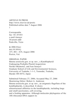
  • 9. dots). Red: Autofluorescence of the symbiont plastid. The scale bar is 10mm. 0 0.2 0.6 1 ab so rp tio n 450 500 550 600 650 700 wavelength Symbiont of Hatena arenicola Chl. b Chl. b Chl. a Chl. a Figure 3. Microspectrophotometry of the symbiont plastid. Spectrogram shows absorbance similar to
  • 10. chlorophyll a/b-containing plastids. 404 N. Okamoto and I. Inouye Fig. 1 H). The chromosomes separate (Fig. 1 I). Following nuclear division (Fig. 1 J), cytokinesis results in one green cell with the symbiont and one colorless cell without it (Fig. 1 K-L). Fluorescence Microscopy: Figure 2 A-H shows DIC and fluorescence images of the same cells. DAPI label (blue) indicates a large host nucleus in the cell center and one to four smaller symbiont nuclei (Fig. 2 B,D,F,H). Each symbiont nucleus is independent of all others and is surrounded by plastid(s), shown in red (due to autofluorescence). Interestingly, the cell with multiple symbiont nuclei has only a single eyespot at the apex of the host cell (Fig. 2 E-G; arrowheads). Microphotometry: Microphotometry of seven cells shows an absorption pattern characteristic of plastids with chlorophyll a/b. Average absorp- tion is shown in Figure 3. The prominent peaks at 435 and 678 nm represent chlorophyll a absorp- tion, while the smaller peaks at 470 and 650 nm correspond to chlorophyll b absorption. Uptake of Prey Cells and Symbiont Specifici- ty: Molecular phylogenetic analysis of 16S rDNA indicates that the symbiont is a member of Nephroselmis (Okamoto and Inouye 2005a). In feeding experiments using a Nephroselmis strain (NIES1417; different from that of the symbiont in 16S rDNA sequence; data not shown), colorless cells of H. arenicola phagocytotically engulfed the alga (Fig. 4 A-I) and tentatively maintained it.
  • 11. However, none developed fully, likely because the strain used in the experiments was not the exact symbiont of H. arenicola. This suggests that symbiont specificity is at the species or strain level. As the H. arenicola cells which engulfed Nephroselmis NIES1417 died, it is unclear whether the Nephroselmis cells were digested. Crawling Motion: Hatena arenicola displays a conspicuous crawling motion that makes it easy to recognize this organism in a crude sample. Figure 5 A-H illustrates cell motion and flagellar movement. The anterior flagellum produces most of the propulsion, while the posterior flagellum is used to change direction. To move forward, a cell casts the anterior flagellum, which adheres to the substratum by its tip (Fig. 5 A-C), and pulls itself forward (Fig. 5 D, E). It then anchors itself with the posterior flagellum (Fig. 5 F-H), and repeats the process. The tip of the flagellum is sharply bent while it is attached to the substratum, as shown in the supplemental material and in Figure 5 I. The cell bends the posterior flagellum to change direction (not shown). Electron Microscopy Electron microscopy (EM) revealed that the green plastid-like structure in the cell is a eukaryotic ARTICLE IN PRESS Figure 4. Uptake of Nephroselmis (NIES1417) by Hatena arenicola. A-H were taken at 6-s intervals. I:
  • 12. corresponds to Hatena arenicola and the symbiont of each frame. Figure 5. Crawling motion of Hatena arenicola. High-speed video images recorded at 0 ms (A), 60 ms (B), 100 ms (C), 130 ms (D), 220 ms (E), 280 ms (F), 300 ms (G), 340 ms (H), respectively. I. SEM image showing the anterior flagellum sharply bent at the distal end (arrowhead). The scale bar is 10 mm in I. 405Hatena arenicola: Halfway to a Plant? endosymbiont. A single nucleus, usually dorsiven- trally flattened, is located in the middle posterior region of the cell, and its chromatin is always condensed and electron dense (Fig. 6 A,B). Multi- ple mitochondrial profiles are present throughout the cytoplasm, though these could be different sections of a single, large reticulate mitochon- drion. Mitochondrial cristae are tubular (Fig. 6 C), and there is a single large Golgi body near the groove of flagellar insertion between the nucleus and the flagellar apparatus (Fig. 6 D). Ejectisomes: There are two types of ejecti- somes beneath the membrane: large, Type I ejectisomes sensu Vørs 1992b (Fig. 6 D-E) that are arrayed in two rows near the flagellar insertion (Figs 1 D, 6 F-G); and smaller Type II ejectisomes sensu Vørs 1992b (Fig. 6 H-I) that are arrayed in numerous rows all over the cell (Fig. 6 F-G). Each ejectisome consists of a coiled ribbon with a gradual depression (Fig. 6 D, H), and bound by a single membrane. Discharged ribbons are slightly curved (Fig. 6 J; type I ejectisome). The ribbon does not have any kink, unlike those of
  • 13. Cryptophyta. Type II ejectisomes are situated beneath the plasma membrane of the cell (Fig. 7 A,B) except for a small smooth area in the apical region, below which the eyespot is situated (asterisk in Fig. 6 G). The ejectisomes are spaced regularly between longitudinally oriented cytoskeletal microtubular bundles (arrowheads in Fig. 7 A). Hatena arenicola lacks Type III ejectisomes that are characteristic of the genus Leucocryptos (Vørs 1992b). Cellular and Flagellar Covering: The plasma membrane of each cell has a characteristic cellular covering composed of a thick inner basal layer (asterisk in Fig. 7 B) and an outer layer of electron-opaque material (arrowhead in Fig. 7 B). This structure extends to the flagellar surface. The outer layer comprises a regularly arrayed and spiraling envelope around the whole flagellum (Fig. 7 C). Endoplasmic Reticulum: The endoplasmic re- ticulum (ER) does not form a conspicuous stack but is loosely distributed throughout the cell. The rough ER extends beneath the surface of the cell (double arrow in Fig. 7 B). Flagella and Basal Bodies: The long anterior and shorter posterior flagella emerge from a ARTICLE IN PRESS
  • 14. Figure 6. Ultrastructure of Hatena arenicola. A. A longitudinal section of a Hatena arenicola cell showing a nucleus (N). Most of the nuclear content is condensed. The rest of the cytoplasm is occupied by the symbiont (Sym). The symbiont has a large plastid region with multiple pyrenoids. B. A transverse section of Hatena arenicola showing host nucleus (N) and the symbiont (Sym) with the vestigial cytoplasm (asterisk). C. Mitochondrial profiles showing tubular cristae. D. A Golgi body near the flagellar basal bodies (arrows) and Type I ejectisomes sensu Vørs 1992 (nearly longitudinal view). E. A transverse section of a Type I ejectisome (sensu Vørs 1992). F. SEM image of Hatena arenicola showing Type I ejectisomes near the flagellar insertion as well as smaller ejectisomes (Type II ejectisomes sensu Vørs 1992) regularly arrayed over the cell surface. G. Magnified SEM image of the same cell. Note that the apical region of the cell (asterisk) lacks ejectisomes. H. A longitudinal section of Type II ejectisome. I. A transverse section of Type II ejectisome. J. A whole mount TEM image of a discharged type I ejectisome. The scale bar is 10 mm in A, E; 2 mm in B, G; 50 nm in C; 500 nm in D, F; 1mm in I; 200 nm in H, I. 406 N. Okamoto and I. Inouye ARTICLE IN PRESS Figure 7. Surface structure and flagellar transition region of Hatena arenicola. A. A tangential section of the surface of Hatena arenicola. Cytoskeletal micro- tubular bundles (arrows) are arrayed longitudinally below the cell surface; Type II ejectisomes are
  • 15. situated between them. Each pore corresponds to the position of an ejectisome, where the surface sheath is thin. B. A transverse section of the cell periphery showing a bilayered surface sheath. An arrowhead indicates the outer layer. An asterisk indicates the thick basal layer. An arrow indicates the plasma membrane. A double arrowhead indi- cates a profile of rough ER. C. A tangential section of the flagellum shows the outer layer of the surface sheath enveloping the flagellum in a spiraling fashion. D-J. Flagellar transition region of Hatena arenicola. D. Diagram of flagellar transition region reconstructed based on serial ultrathin sections. Each letter in D indicates which part corresponds to one of the transverse sections (E-J). The scale bar is 500 nm in A, C; 200 nm in B, E-J. 407Hatena arenicola: Halfway to a Plant? shallow subapical furrow. The flagella are coated by a ‘‘surface sheath’’. The diagram of the basal body and the flagellar transition zone (Fig. 7 D) is based on serial sections of the flagellar transition zone (Fig. 7 E-J). At the proximal end of the basal body, there is a cartwheel structure with a central tube. The triplet microtubules of the basal body terminate below the plasma membrane (Fig. 7 E), whereas the doublet microtubules attach to the plasma membrane by a connecting fiber (Fig. 7 F). The flagellar transition region extends above the plasma membrane, and an electron-dense rod structure is present (Fig. 7 G) in the middle of this region. Surrounding the transition region are outer doublet microtubules lined in a loose, electron- dense material (Fig. 7 H). The flagellar transition region ends in a terminal plate with electron- opaque material at the center (Fig. 7 I). The
  • 16. axonemal central pair begins above the terminal plate (Fig. 7 J). Feeding Apparatus: As reported in Okamoto and Inouye (2005a), colorless cells lacking the symbiont have a complex feeding apparatus at their apex (Fig. 8 A-C), consisting of transverse tubular rings (arrowheads in Fig. 8 B-C) and longitudinal microtubules arrayed in a single layer (arrow in Fig. 8 B-C). These microtubules are different from those that form the cytoskeleton (double arrowheads in Fig. 8 A). Inside the micro- tubular skeletons of the feeding apparatus are several electron-opaque granules, some of which are large and elongate (light gray) and others that are smaller, granulated, and pigmented (Fig. 8 A, C). These granules are restricted to the feeding apparatus, never seen elsewhere in the cell. Symbiont-bearing cells do not have a feeding structure; the corresponding region is occupied by the eyespot of the symbiont (Fig. 8 D-E). Symbiont: The symbionht retains not only a plastid but also its own cytoplasm with a nucleus and mitochondrion(a) (Fig. 9 A-C). Most symbiont cells contain one nucleus, which is often attached to the membrane, and symbiont and host nuclei often face each other (Fig. 9 A-C). The symbiont is bounded by a single membrane (arrowhead in Fig. 9 D), whose origin is unknown. Mitochondria have flat, often degraded cristae, though the extent of degradation varies among individual cells (Fig. 9 E-F). The plastid, which is the largest structure in the symbiont (Fig. 6 A-B), contains multiple pyrenoids bounded by a thin starch sheath (Fig. 9 G). The pyrenoid has shallow invaginations of the thylakoid membrane. The plastid has a single conspicuous eyespot, where
  • 17. the morphological association between host and symbiont is present (see below). Free ribosomes are densely distributed through- out the symbiont cytoplasm, though no ribosome- bearing membrane (rough ER) is present. Occa- sionally there are flattened, stacked membranes next to the nucleus (Fig. 9 A). This structure would normally be a Golgi body but it is present in a degraded or inactive form, because no Golgi vesicles were seen around the structure. Some randomly shaped vacuoles of unknown origin are also present (Fig. 9 B-C). Because each cell division will result in half the population carrying ARTICLE IN PRESS Figure 8. Feeding apparatus of Hatena arenicola. A. A nearly transverse section of the feeding apparatus. Feeding apparatus is distinct from cytoskeletal microtubules (double arrowheads). B. A magnified view showing microtubules (arrow) regularly arrayed in a single layer along the external side of the tubular rings (arrowheads). C. Longitudinal section of the feeding apparatus showing numerous transverse tubular rings (arrowhead) and longitudinal microtubules arrayed in a single layer (arrows). D. An eyespot (e) of symbiont is located at the corresponding place in the symbiont bearing cell. E. A schematic illustration of the feeding apparatus and the corresponding place of the symbiont-bearing Hatena arenicola. The scale bar is 500 nm in A, C-D; 250 nm in B. 408 N. Okamoto and I. Inouye
  • 18. the symbiont, these morphological varieties likely reflect degradation (see Discussion). Cytoskeletal structures, including the flagella, basal bodies, the flagellar apparatus, and micro- tubular rootlets, are completely absent. These morphological changes must affect intracellular functions, such as protein synthesis and distribu- tion in the symbiont (see Discussion). The lysosome of the host cytosol is discontinuous with the symbiont compartment. The lysosome of some cells contains scales of Nephroselmis and Pyramimonas (Fig. 10 A,B), indicating that H. arenicola cells engulf other prey in addition to its Nephroselmis partner. Because Pyramimonas cells are digested, H. arenicola may be partly heterotrophic (see Discussion). Eyespot: The eyespot is composed of a single- layered sheet of osmiophilic granules (Fig. 11 A) that connects to the inner plastid envelope. Near the eyespot, the plastid, symbiont, and host plasma membranes are tightly layered (Fig. 11 A,B). In some cells, single microtubules are aligned longitudinally between the plasma membrane and the symbiont membrane overlying the eyespot region (Mts in Fig. 11 C,D). These single microtubules are distinct from the cytoskeletal microtubular bundles (arrow- head in Fig. 11 D), and the space between them lacks Type II ejectisomes; this is consistent with the smooth surface appearance of the eyespot region (Fig. 6 F). Molecular Phylogeny
  • 19. Partial SSU rDNA sequences of H. arenicola (AB212285) aligned with the homologues of known eukaryotes were subjected to phylogenetic analyses. The resulting maximum likelihood (ML) tree (Fig. 12) showed the typical topology of the SSU rDNA tree, and H. arenicola was included in the katablepharid clade, which was robustly supported with high bootstrap probability in ML (99%), NJ (100%), and MP (99%) analyses. Discussion Hatena arenicola has several unique features that suggest it is in the process of endosymbiosis with ARTICLE IN PRESS Figure 10. Lysosome of Hatena arenicola with scales of prasinophytes characteristic to Pyramimo- nas (A) and Nephroselmis (B) respectively. The scale bar is 500 nm. Figure 9. Ultrastructure of the symbiont. A-C. The symbiont cytoplasm, retaining a nucleus, mitochondria, and sometimes a Golgi body-like vesicle (A) or membranes of random shape (B-C), likely in an intermediate state of integration. D. The single symbiont-enveloping membrane (arrowhead) separates the symbiont compartment (Sym) and host cytoplasm (H). Double membranes of the symbiont plastid are also shown (arrows). E. Mitochondrial profiles that retain flat cristae. F. A relatively degenerated mitochondrial profile. G. A pyrenoid, surrounded by a starch sheath. Random shallow invagination of the thylakoids. The scale bar is
  • 20. 1 mm in A-C; 250 nm in D-G. 409Hatena arenicola: Halfway to a Plant? a Nephroselmis partner (Okamoto and Inouye 2005a). We will discuss the taxonomy and classification of H. arenicola first, and then focus on its unique endosymbiosis. Taxonomy Morphological and molecular analyses of H. arenicola clearly show that it belongs to the recently established division/phylum Katablephar- idophyta (International Code of Botanical Nomen- clature, ICBN)/Kathablepharida (International Code of Zoological Nomenclature, ICZN) (Oka- moto and Inouye 2005b). The katablepharids comprise an ultrastructurally well-defined and small group of heterotrophic flagellates that includes 10 species in two genera: nine species of Katablepharis Skuja (correct spelling in ICBN which will be used in this paper)/Kathablepharis Skuja (original spelling in the ICZN), and one species of Leucocryptos (Braarud) Butcher. Kata- blepharidaceae (ICBN) was originally described by Skuja (1939) based on ovate or cylindrically ovate cell shape, two flagella emerging from a subapical depression, and conspicuous ejectisomes aligned ARTICLE IN PRESS Figure 11. Eyespot of the symbiont plastid. A. A longitudinal section of the eyespot (E). B. A magnified view of another cell clearly shows the eyespot granules, the inner/outer envelop of the symbiont plastid (arrows),
  • 21. the single symbiont enveloping membrane (arrowhead), and the host plasma membrane (double arrowhead) associated with each other. C-D. Tangential sections of the eyespot region show single microtubules (Mts) distinctive from the cytoskeletal microtubules (arrowheads) longitudinally situated between the eyespot (E) and the plasma membrane. The scale bar is 250 nm in A; 100 nm in B; 1mm in C-D. 410 N. Okamoto and I. Inouye in two rows near the flagellar insertion. Vørs (1992b), and Clay and Kugrens (1999b) emended the family by adding the following ultrastructural features: the entire surface of the cell, including the flagella, is coated with a bilayered surface sheath that appears to form spiraling rows around the cell body; tubular mitochondrial cristae; a complex, truncated conical feeding apparatus and cytoskeleton; a Golgi apparatus situated anteriorly and a centrally located nucleus; and a food vacuole in the posterior part of the cell. Hatena arenicola shares all these characters, except that the cell is dorsiventrally compressed and the food vacuole is absent. We occasionally observed a vacuole containing scales of prasino- phytes, though its position was anterior to the nucleus. In addition, the flagellar transition zone containing a rod-shaped structure is fundamen- tally the same as that of K. ovalis (Lee et al. 1992). Based on these ultrastructural similarities and the molecular phylogenetic data, H. arenicola un- doubtedly belongs to the katablepharids. Currently, all the katablepharids belong to a single family, Katablepharidaceae, whose cells are
  • 22. defined as either ‘‘oblong or cylindrically ovate’’ (Katablepharis; Clay and Kugrens, 1999b; Vørs, 1992b) or ‘‘ovate, pyriform elliptical outline’’ (Leucocryptos; Butcher 1967). The most important distinguishing feature of Leucocryptos is the presence of Type III ejectisomes, which have been found only in Leucocryptos. Because H. arenicola lacks Type III ejectisomes, it does not belong to Leucocryptos. Hatena arenicola is distinct from all other katablepharids previously described in that it is dorsiventrally compressed with a flat-oval shape, which suggests that it does not belong to the genus Katablepharis. Its crawling motion and the feeding apparatus composed of single-layered microtubules are also distinctive from the other ARTICLE IN PRESS 0.1 Chlorarachnion CCMP242 Cercomonas longicauda Heteromita globosa Thaumatomonas seravini Euglypha rotunda Paulinella chromatophora Ochromonas danica Phytophthora megasperma
  • 23. Skeletonema pseudocostatum Pteridomonas danica Prorocentrum micans Prorocentrum mexicanum Gymnodinium sp. MUCC284 Pfiesteria sp. B112456 Alexandrium minutum Cryptosporidium parvum Toxoplasma gondii Prorodon teres Platyophrya vorax Emiliania huxleyi Pavlova salina Chlorokybus atmophyticus Mesostigma viride Arabidopsis thaliana Fossombronia pusilla Pyramimonas propulsa Ulothrix zonata 'Chlorella' ellipsoidea Tetraselmis striata Coleochaete scutata Closterium littorale Cyanophora paradoxa Cyanoptyche gloeocystis
  • 24. Glaucocystis nostochinearum Gloeochaete wittrockiana Goniomonas truncata Rhodomonas mariana Hanusia phi Geminigera cryophila Cryptomonas ovata Chroomonas sp. M1318 Hemiselmis brunnescens Leukocryptos marina Hatena arenicola Katablepharis japonica Heterophrys marina Chlamydaster sterni Raphidiophrys ambigua Rhodella maculata Stylonema alsidii Bangia sp. Porphyra umbilicalis Acanthamoeba castellanii Dictyostelium discoideum Leptomyxa reticulata Hartmannella vermiformis Scutellospora cerradensis
  • 25. Pneumocystis carinii Saccharomyces cerevisiae Schizosaccharomyces pombe Basidiobolus haptosporus Chytriomyces hyalinus Monosiga brevicollis Clathrina cerebrum Cirripathes lutkeni 18S rDNA (65species 1252 sites) 100/100/99 99/83/96 64/73/62 54/86/60 58/73/- 52/88/72 53/98/67 100/89/100 100/100/100 99/100/100 99/88/56
  • 27. 91/79/84 100/100/100 75/88/66 99/97/83 75/64/- 65/85/89 74/-/- 76/81/84 93/-/- -/-/54 53/-/- 50/59/- 92/77/79 ML/NJ/MP Figure 12. Unrooted eukaryotic tree based on the SSU rDNA. The best tree of the ML method is shown. Bootstrap propotions are shown at the internal branches, in the order of ML/NJ/MP methods. The length of each branch is proportional to the estimated number of substitutions. Bar denotes 10% substitutions per site. For details of the phylogenetic reconstruction methods, see text. The organisms included in the tree are listed
  • 28. in Table 1. Unambiguously aligned 1252 nucleotide positions were used for the analysis. 411Hatena arenicola: Halfway to a Plant? katablepharids (Clay and Kugrens 1999a, b; Kugrens et al. 1994; Lee and Kugrens 1992; Lee et al. 1991; Okamoto and Inouye 2005b; Vørs 1992a, b). Based on those features, we propose that the organism should be assigned to a new genus, Hatena. The family-level taxonomy of the katablepharids is still unclear, primarily because little molecular sequence data exist. Until further studies eluci- date katablepharid taxonomy, it is best to include the genus Hatena in the family Katablepharida- ceae. ARTICLE IN PRESS AH Loss of Feeding Appar atus Plastid: Enlarged Symtiont: Degraded 412 N. Okamoto and I. Inouye Endosymbiosis Endosymbiosis is a major driving force in plant evolution, and thus it is important to understand this process. Hatena arenicola may be an im- portant model of early plastid acquisition. Sym-
  • 29. biotic Nephroselmis differs from free-living individuals in having enlarged plastids with a greater number of pyrenoids, degraded subcellu- lar structures, and morphologically distinct eye- spots. Cells of known Nephroselmis species are a maximum of 20mm in length (Nephroselmis astigmatica; Inouye and Pienaar 1984) and pos- sess a single plastid with a single pyrenoid. The symbiont occupies most of the host cytoplasm, suggesting that the symbiont plastid(s) grows more than ten fold after being engulfed by the host. Pyrenoids also multiply after being engulfed. In contrast, the symbiont cytoplasm loses other major cell components including flagellar appara- tus and microtubular roots, endomembranes such as the ER and transport vesicles, Golgi-like vesicles, and amorphous membranous structures. The dramatic growth of the plastid is in stark contrast to the degradation of the other orga- nelles. Because the cytoplasm is in such a degraded state, it must be difficult to sustain the growth and maintenance of the plastid alone. It is likely that some metabolites from the host cell are used to develop and maintain the plastid. B DE F G C Predator phase Plant phase Formation of
  • 30. Feeding apparatus Uptake of the partner Figure 13. Half-plant, half-predator-hypothesis. The solid line indicates a witnessed process, and the broken line indicates a hypothetical process. A-D: A green cell with the symbiont, lacking the feeding apparatus (A) divides (B) into one green (C) and one colorless cell (D). E-G: The colorless cell should form a feeding apparatus de novo and engulfs a Ne- phroselmis cell. G-H: The symbiont plastid selec- tively grows in the host cytoplasm. Because cell division of a colorless cell or a cell with an ‘‘immature’’ symbiont (H) has never been observed, uptake and the subsequent changes in both host and symbiont apparently occur within one generation. Eyespot Morphology The morphology of the eyespot is most suggestive of a host-symbiont coordination. The tight layering of the four distinct membranes of different origin implies functional cooperation. The eyespot is an important component of a photo-sensing complex found in various algal groups (Melkonian and Robenek 1984; Gualtieri 2001). It effectively regulates the light received by a photoreceptor located on a nearby membrane, thereby allowing the alga to detect light direction. Preliminary observations have shown that lateral, and not vertical, incidence, is important for the behavior of H. arenicola. Hence, we speculate that the photoreceptor and the regulatory mechanism would be laterally aligned in the cell, so that the
  • 31. eyespot can effectively shade the photorecepter from the lateral incidence. Assuming that the eyespot is functional in H. arenicola, the photo- receptor should be situated in either of the outer or inner plastid membrane, the single endosymbiont envelope, or the plasma membrane. Their mor- phological association can be explained as a consequence of functional collaboration. Because H. arenicola crawls two-dimensionally, a photo- tactic response to laterally projected light is plausible. To test whether a functional association exists, further investigation of the threshold and the efficiency of phototaxis in both colored and colorless H. arenicola cells are required. Morphological Changes of the Host and ‘‘Half-plant, half-predator’’ Model Hatena arenicola cells without a symbiont have a complex feeding apparatus in place of an eye- spot, indicating that symbiont acquisition is accompanied by drastic morphological changes in both the symbiont and the host. ARTICLE IN PRESS 413Hatena arenicola: Halfway to a Plant? Such structural changes would necessitate life cycle changes. Based on observations presented in this paper, we propose the ‘‘half-plant, half- predator’’ hypothesis, where H. arenicola switches its lifestyle between that of a plant and a predator (Fig. 13; see also Okamoto and Inouye 2005a for detailed explanation). This proposed life cycle is also supported by the observation that the extent
  • 32. of degradation of symbiont mitochondria and membranous structures varies among individual cells. The presence of prasinophyte scales in a H. arenicola lysosome suggests that H. arenicola lives heterotrophically to some extent, perhaps until it engulfs its symbiotic partner. This repre- sents an intermediate state of trophic alteration. Although there are several assumptions in the model, it promises to lead to new insights and helps elucidate the plastid integration process. Evolutionary Implications Extra secondary and tertiary endosymbioses in dinoflagellates may represent evolutionary events that occurred during plastid acquisition (for re- views, see Hackett et al. 2004a; Morden and Sherwood 2002; Schnepf and Elbrächter 1999). The earliest stage is represented by the crypto- phyte symbiont, in which only cytoskeletal com- ponents are lost, as in Amphidinium latum Lebour (Horiguchi and Pienaar 1992), A. poecilochroum Larsen (Larsen 1988), and Gymnodinium acidotum Nygaard (Fields and Rhodes 1991; Wilcox and Wedemayer 1984). The cell cycles of the host and symbiont are not synchronized, and the host cell must repeatedly capture symbionts. The next stage is represented by the symbiont of diatom origin, in which various subcellular structures are lost, except the nucleus, mitochondrion(a) and plastid, as in Durinskia baltica (Levander) Carty et Cox (formerly Peridinium balticum; Chesnick and Cox 1987, 1989; Chesnick et al. 1997; Eschbach et al. 1990; Tippit and Pickett-Heaps 1976; Tomas and Cox 1973) and Kryptoperidinium foliaceum (Stein) Lindemann (Dodge 1971; Eschbach et al.
  • 33. 1990). At this stage, the symbiont divides syn- chronously with the host cell (Chesnick and Cox 1987, 1989; Tippit and Pickett-Heaps 1976), so that the association between the host and symbiont becomes permanent, and repeated uptake of the symbiont is no longer necessary. Finally, the symbiont cytoplasm is reduced, as seen in Lepidodinium viride (Watanabe et al. 1987; Watanabe et al. 1990), Gymnodinium chlorophor- um (Elbrächter and Schnepf 1996), both with symbionts of prasinophyte origin, and Karenia brevis (Davis) Hansen et Moestrup, Karenia mikimotoi (Miyake et Kominami ex Oda) Hansen et Moestrup, Karlodinium veneficum (Leadbeater et Dodge) Larsen with symbionts of haptophyte origin (Daugbjerg et al., 2000; Inagaki et al., 2000; Tangen and Björnland, 1981). The symbiont of Karenia and Karlodinium species has no remnant of cytoplasm, and can therefore be recognized as an integrated plastid. Based on cytoplasm reduc- tion and the lack of cell cycle synchronization, the symbiosis of H. arenicola can be placed between the cryptophyte and diatom types of symbiosis mentioned above. Nevertheless, their morpholo- gical association suggests an intimate host—sym- biont relationship. Previous studies on symbiosis in dinoflagellates have focused on symbiont degradation only. In this study, we demonstrated that major morphological changes also occur in the host, suggesting that plastid acquisition is not merely ‘‘enslavement’’ where the symbiont is degraded, but also a process during which the host itself changes to establish a new association with the symbiont. The rigid pattern of asymmetrical inheritance of
  • 34. the symbiont is also suggestive of a partly regulated association. The symbiont always comes to the left side of the host cell (ventral view) before the migration of the host nucleus, implying an interaction between the symbiont compartment and the host cytoskeleton. If the symbiont moved to the division plane, and not to one side of the cell, the symbiont would co- segregate, just as in the division of D. baltica, where the center-positioned symbiont co-segre- gates upon host cytokinesis (Tippit and Pickett- Heaps 1976). Another question is whether the association of H. arenicola and the symbiont has developed genetic modification. The process of endosym- biosis is hypothesized to include genetic changes such as lateral gene transfer (LGT) from symbiont to host, coupled with evolution of a protein transport machinery from host to symbiont (e.g. Gilson and McFadden 2002). It is unclear when those changes start and how they integrate. Considering host—symbiont intimacy, H. arenico- la would have already experienced or may be experiencing some of these changes. Therefore, the study of LGT or of a protein transport machinery in H. arenicola would be an interesting topic for future studies. Recently, Marin et al. (2005) reported another example of primary endosymbiosis in Paulinella chromatophora Lauterborn, a freshwater thecate amoeba that bears a cyanobacterium-like
  • 35. ARTICLE IN PRESS 414 N. Okamoto and I. Inouye structure. They reported that the symbiosis of P. chromatophora is a more recent event than the origin of all other plastids, based on the molecular phylogeny of ribosomal DNA operon sequences. This is consistent with a morphological feature of the symbiont, namely, a peptidoglycan layer that must have originated from the cell wall of an ancestral cyanobacterium. The symbiotic relationships of H. arenicola, P. chromatophora, and the dinoflagellates probably represent different intermediate steps in plastid acquisition via primary or secondary endosymbio- sis. Continued study and comparison of these groups should provide further insight into plastid evolution. Concluding Remarks Hatena arenicola gen. et sp. nov. is likely in the process of plastid acquisition via secondary endosymbiosis. Although it is in an early inter- mediate stage of acquisition, the two organisms have already established an intimate association in ultrastructure and likely in metabolic function. Based on behavioral and ultrastructural observa- tions, we propose a ‘‘half-plant, half-predator’’ life cycle. Because H. arenicola shows an early intermediate state of plastid acquisition, it should provide further insight into plant evolution. This study provides a foundation for future studies on the topic. Methods
  • 36. Sampling and temporary maintenance in the laboratory: Because it is not possible to culture Hatena arenicola in the laboratory, we used crude samples from the natural habitat. Cells were collected at Isonoura Beach, Wakayama Prefec- ture, Japan (Fig. 1 B,C), April—December from 2000 to 2004. Samples were maintained in the laboratory at room temperature in f/2 medium, under ca. 10mmol photons m�2 s�1, and the light—dark cycle was L:D ¼ 5:19 h. Morphological observations: Light micro- scopy (LM) and fluorescence microscopy (FM) was conducted using a Leica DMR light micro- scope (Leica Wetzlar GmbH, Wetzlar) and the LM image was taken with a Keyence VB6010 digital chilled CCD camera (Keyence, Osaka). For FM, 40,6-Diamidino-2-phenylindole (DAPI) was used to stain the nucleus. The DAPI fluorescence along with the autofluorescence of the plastid were observed using a D filter cube (Leica Wetzlar GmbH, Wetzlar). Microspectrophotometry was performed by majoring three different regions of the symbiont in each of seven cells. Each absorption spectrum was recorded in the range of 300—800 nm with a light microscope (ECLIPSE, Nikon, Tokyo) equipped with a high-resolution multichannel photodetector (MCPD 7000, Otsuka Eelectronics, Osaka) at Okazaki National Institute for Basic Biology, Japan. The average of the three measure- ments was considered the representative absor- bance of each cell. Because the absorptions obtained were almost uniform across the seven cells, their average is shown.
  • 37. A unialgal culture of Nephroselmis sp. (NIES1417) was established from the same sam- ple site by micropipette isolation and maintained in f/2 medium at 20 1C under ca. 10mE light intensity, and a light-dark cycle of L:D ¼ 14:10 h. The uptake of Nephroselmis sp. (NIES1417) was photographed under a CKX31 inversed light microscope (Olympus, Tokyo) equipped with a COOLPIX 990 digital camera (Nikon, Tokyo) at 6-s intervals. High-speed video images were recorded at 200 frames per second using an OPTIPHOT micro- scope (Nikon, Tokyo), equipped with an MHS-200 high-speed video capturing system (Nac Inc., Tokyo). The images were digitized on a Macintosh computer using an NIH imaging program (public domain, developed at the US National Institute of Health; available at http://rsb.info.nih.gov/nih-im- age/) for analysis of the cellular and flagellar motion. Preparation for transmission electron micro- scopy (TEM) and scanning electron microscopy (SEM) was performed as described elsewhere (Moriya et al. 2000; Okamoto and Inouye 2005b). The observations were made with a JEOL 100CXII electron microscope (JEOL, Tokyo) and a JSM- 6330 scanning electron microscope (JEOL, To- kyo). Molecular phylogeny: To avoid contamination of the prey genome, single cells of Hatena arenicola were isolated with micropipette into a 0.2-ml PCR tube containing 10 ml of sterilized
  • 38. double distilled water, and immediately frozen at �80 1C for more than 15 min to completely disrupt the cells. The first and the second nested PCR were performed with existing degenerate primer sets (Moriya et al. 2000) using rTaq (TOYOBO, Osaka). Thermal cycling for the first PCR con- sisted of 33 cycles. Annealing temperatures ranged from 50 to 47 1C (six cycles decreasing http://rsb.info.nih.gov/nih-image/ http://rsb.info.nih.gov/nih-image/ ARTICLE IN PRESS Table 1. Sequences used for the phylogenetic analyses Organism Accession number SSU rDNA Katablepharidophyta/Kathablepharida Hatena arenicola AB212285 Katablepharis japonica AB231617 Leucocryptos marina AB193602 Metazoa Cirripathes lutkeni AF052902 Clathrina cerebrum U42452 Monosiga brevicollis AF084618 Fungi Basidiobolus haptosporus AF113413 Chytriomyces hyalinus M59758
  • 39. Pneumocystis carinii L27658 Saccharomyces cervisiae V01335 Schizosaccharomyces pombe Z19578 Scutellospora cerradensis AB041344 Amoebae Acanthamoeba castellanii M13435 Hartmannella vermiformis M95168 Leptomyxa reticulata AF293898 Dictyostelium discoideum K02641 Cercozoa Cercomonas longicauda AF101052 Chlorarachnion CCMP242 U03479 Euglypha rotunda X77692 Heteromita globosa U42447 Paulinella chromatophora X81811 Thaumatomonas seravini AF411259 Viridiplantae Arabidopsis thaliana X16077 ‘Chlorella’ ellipsoidea D13324 Chlorokybus atmophyticus M95612 Closterium littorale AF115438 Coleochaete scutata X68825 Fossombronia pusilla X78341 Mesostigma viride AJ250108 Pyramimonas propulsa AB017123 Tetraselmis striata X70802 Ulothrix zonata Z47999 Heterokontophyta Ochromonas danica M32704 Pteridomonas danica L37204 Skeletonema pseudocostatum X85394 Phytophthora megasperma X54265
  • 40. Alveolata Cryptosporidium parvum X64340 Toxoplasma gondii X75429 Platyophrya vorax AF060454 Prorodon teres X71140 Alexandrium minutum U27499 Gymnodinium sp. MUCC284 AF022196 Pfiesteria sp. B112456 AF218805 Table 1. (continued ) Organism Accession number Prorocentrum mexicanum Y16232 Prorocentrum micans M14649 Cryptophyta Chroomonas sp. M1318 AJ007279 Cryptomonas ovata AJ421147 Geminigera cryophila U53124 Hanusia phi U53126 Hemiselmis brunnescens AJ007282 Rhodomonas mariana X81373 Goniomonas truncata U03072 Glaucophyta Cyanophora paradoxa X68483 Cyanoptyche gloeocystis AJ007275 Glaucocystis nostochinearum X70803 Gloeochaete wittrockiana X81901 Haptophyta Emiliania huxleyi L04957 Pavlova salina L34669
  • 41. Rhodophyta Bangia sp. AF043362 Porphyra umbilicalis AB013179 Rhodella maculata U21217 Stylonema alsidii L26204 Centroheliozoa Chlamydaster sterni AF534709 Heterophrys marina AF534710 Raphidiophrys ambigua AF534708 415Hatena arenicola: Halfway to a Plant? the temperature by 0.5 1C for each cycle, and 27 cycles at a constant temperature). An extension was performed at 72 1C for 1 min, and denaturing was done at 94 1C for 30 s. The final extension period was at 72 �C for 7 min. Thermal cycling for the second PCR consisted of 33 cycles with an annealing step at 53 1C for 30 s, an extension step at 72 1C for 1 min, and denaturing at 94 1C for 30 s, with a final extension period at 72 1C for 7 min. The sequences were determined by direct sequen- cing. Cycle sequencing reaction was performed using a DYEnamic ET terminator cycle sequencing kit (Amersham biosciences, Buckinghamshire), as per the manufacturer’s instructions. Sequencing was conducted with an ABI PRISM 377 DNA Sequencer (Applied Biosystems, California), and sequences were confirmed free of contaminants and not of haptophyte prey origin by comparing at least two cells or performing a BLAST search at the National Center for Biotechnology Informa- tion (NCBI) server (http://www.ncbi.nlm.nih.gov/ http://www.ncbi.nlm.nih.gov/BLAST/
  • 42. ARTICLE IN PRESS 416 N. Okamoto and I. Inouye BLAST/). The sequence (AB212285) was depos- ited to GenBank. The SSU sequence was manually aligned to the existing alignments of global eukaryotes (Okamo- to and Inouye 2005b), which include 65 taxa for SSU rDNA (Table 1). To avoid the long-branch attraction (LBA) artifact in SSU rDNA analysis, organisms with an extraordinary evolutionary rate, such as the Euglenozoa, the diplomonads, and the palabasalids, were excluded after confirming that they are not a sister group of the katablephar- ids. A total of 1252 unambiguously aligned nucleotide positions were selected for phyloge- netic analyses. The maximum likelihood (ML), neighbor joining (NJ), and maximum parsimony (MP) methods for phylogenetic analysis were applied to the data set using PAUP* ver.4.0 b 10 (Swofford 2003). In the ML analysis, the evolutionary model was selected using the Akaike information criterion (AIC) test in Modeltest ver.3 (Posada and Crandall 1998), which selected the general time reversible (GTR) model with rate variation among sites and invariant sites (GTR+I+gamma). The estimated gamma-shape parameter (alpha) of the discrete gamma-distribution was 0.5645, and the propor- tion of invariant sites was 0.3257. A further tree search with GTR+I+gamma model with eight site rate categories that approximates site rate was
  • 43. used to produce the optimal tree. Bootstrap proportion (BP) values for internal branches of the optimal tree of the ML analysis were obtained using PAUP* through 300 bootstrap resamplings for the ML method and 1000 resamplings for the NJ and MP methods. Acknowledgments We thank Dr. Masakatsu Watanabe and Dr. Shigeru Matsunaga (Graduate University for Ad- vanced Studies, Japan) for their dedication to the microspectrophotometric work and assistance in sampling, and Dr. Hiroshi Kawai (Research Center for Inland Seas, University of Kobe) for use of his laboratory equipment. We appreciate Dr. Tetsuo Hashimoto and Dr. Miako Sakaguchi for their assistance and advices in molecular phylogeny. We are grateful to Dr. Jeremy Pickett-Heaps for taking movie images of H. arenicola. This research was supported in part by Japan Society for the Promotion of Sciences (JSPS) Grants RFTF00L0162 (to I.I.) and 1612007 (to N.O.). N.O. was also supported by a JSPS Research Fellowship for Young Scientists as a JSPS Research Fellow. Appendix A. Supplementary materials Supplementary data associated with this article can be found in the online version at doi:10.1016/ j.protis.2006.05.011 References Andersson JO, Roger AJ (2002) A cyanobacterial gene in nonphotosynthetic protists—an early chlor- oplast acquisition in eukaryotes? Curr Biol 12: 115—119
  • 44. Archibald JM, Rogers MB, Toop M, Ishida K, Keeling PJ (2003) Lateral gene transfer and the evolution of plastid-targeted proteins in the second- ary plastid-containing alga Bigelowiella natans. Proc Natl Acad Sci USA 100: 7678—7683 Baldauf SL (2003) The deep roots of eukaryotes. Science 300: 1703—1706 Baldauf SL, Roger AJ, Wenk-Siefert I, Doolittle WF (2000) A kingdom-level phylogeny of eukaryotes based on combined protein data. Science 290: 972—977 Bapteste E, Brinkmann H, Lee JA, Moore DV, Sensen CW, Gordon P, Durufle L, Gaasterland T, Lopez P, Muller M, Philippe H (2002) The analysis of 100 genes supports the grouping of three highly divergent amoebae: Dictyostelium, Entamoeba, and Mastigamoeba. Proc Natl Acad Sci USA 99: 1414—1419 Bhattacharya D, Yoon HS, Hackett JD (2004) Photosynthetic eukaryotes unite: endosymbiosis connects the dots. Bioessays 26: 50—60 Butcher RW (1967) An Introductory Account of the Smaller Algae of British Coastal Waters. 4. Crypto- phyceae. Fishery Investigations, Series IV. HMSO, London Cavalier-Smith T (2003) Genomic reduction and evolution of novel genetic membranes and protein- targeting machinery in eukaryote—eukaryote chi- maeras (meta-algae). Philos Trans R Soc Lond B
  • 45. 358: 109—133 Chesnick JM, Cox ER (1987) Synchronized sexu- ality of an algal symbiont and its dinoflagellate host, Peridinium balticum (Levander) Lemmermann. Bio- Systems 21: 69—78 http://www.ncbi.nlm.nih.gov/BLAST/ dx.doi.org/10.1016/j.protis.2006.05.011 dx.doi.org/10.1016/j.protis.2006.05.011 ARTICLE IN PRESS 417Hatena arenicola: Halfway to a Plant? Chesnick JM, Cox ER (1989) Fertilization and zygote development in the binucleate dinoflagellate Peridinium balticum (Pyrrhophyta). Am J Bot 76: 1060—1072 Chesnick JM, Kooistra WHCF, Wellbrock U, Medlin LK (1997) Ribosomal RNA analysis indicates a benthic pennate diatom ancestry for the endo- symbionts of the dinoflagellates Peridinium folia- ceum and Peridinium balticum (Pyrrhophyta). J Eukaryot Microbiol 44: 314—320 Clay BL, Kugrens P (1999a) Description and ultrastructure of Kathablepharis tenuis sp. nov. and K. obesa sp. nov. — two new freshwater Kathable- pharis (Kathablepharididae) from Colorado Wyom- ing. Europ J Protistol 35: 435—447 Clay BL, Kugrens P (1999b) Systematics of the enigmatic Kathablepharids, including EM character- ization of the type species, Kathablepharis phoeni-
  • 46. koston, and new observations on K. remigera comb. nov. Protist 150: 43—59 Daugbjerg N, Hansen G, Larsen J, Moestrup Ø (2000) Phylogeny of some of the major genera of dinoflagellates based on ultrastructure and partial LSU rDNA sequence data, including the erection of three new genera of unarmoured dinoflagellates. Phycologia 39: 302—317 Dodge JD (1971) A dinoflagellate with both a mesokaryotic and a eukaryotic nucleus. Protoplas- ma 73: 145—157 Douglas S, Zauner S, Fraunholz M, Beaton M, Penny S, Deng LT, Wu XN, Reith M, Cavalier- Smith T, Maier UG (2001) The highly reduced genome of an enslaved algal nucleus. Nature 410: 1091—1096 Elbrächter M, Schnepf E (1996) Gymnodinium chlorophorum, a new, green, bloom-forming dino- flagellate (Gymnodiniales, Dinophyceae) with a ves- tigial prasinophyte endosymbiont. Phycologia 35: 381—393 Eschbach S, Speth V, Hansmann P, Sitte P (1990) Freeze-fracture study of the single membrane between host cell and endocytobiont in the dino- flagellates Glenodinium foliaceum and Peridinium balticum. J Phycol 26: 324—328 Falkowski PG, Katz ME, Knoll AH, Quigg A, Raven JA, Schofield O, Taylor FJR (2004) The evolution of modern eukaryotic phytoplankton. Science 305: 354—360
  • 47. Fields SD, Rhodes RG (1991) Ingestion and reten- tion of Chroomonas spp (Cryptophyceae) by Gym- nodinium acidotum (Dinophyceae). J Phycol 27: 525—529 Gilson PR, Su V, Slamovits CH, Reith ME, Keeling PJ, McFadden GI (2006) Complete nucleotide sequence of the chlorarachniophyte nucleomorph: Nature’s smallest nucleus. Proc Natl Acad Sci USA 103: 9566—9571 Graham LE, Wilcox LW (2000) Algae. Prentice-Hall, NJ Gualtieri P (2001) Morphology of photoreceptor systems in microalgae. Micron 32: 411—426 Hackett JD, Anderson DM, Erdner DL, Bhatta- charya D (2004a) Dinoflagellates: a remarkable evolutionary experiment. Am J Bot 91: 1523—1534 Hackett JD, Yoon HS, Soares MB, Bonaldo MF, Casavant TL, Scheetz TE, Nosenko T, Bhatta- charya D (2004b) Migration of the plastid genome to the nucleus in a peridinin dinoflagellate. Curr Biol 14: 213—218 Hashimoto H (2005) The ultrastructural features and division of secondary plastids. J Plant Res 118: 163—172 Horiguchi T, Pienaar RN (1992) Amphidinium latum (Dinophyceae), a sand-dwelling dinoflagellate feed- ing on cryptomonads. Jpn J Phycol (Sorui) 40: 353—363
  • 48. Huang CY, Ayliffe MA, Timmis JN (2003) Direct measurement of the transfer rate of chloroplast DNA into the nucleus. Nature 422: 72—76 Inagaki Y, Dacks JB, Doolittle WF, Watanabe KI, Ohama T (2000) Evolutionary relationship between dinoflagellates bearing obligate diatom endosym- bionts: insight into tertiary endosymbiosis. Int J Syst Evol Microbiol 50: 2075—2081 Inouye I, Pienaar RN (1984) Light and electron microscope observation on Nephroselmis astigmati- ca sp. nov. (Prasinophyceae). Nord J Bot 4: 409—423 Katz LA (2002) Lateral gene transfers and the evolution of eukaryotes: theories and data. Int J Syst Evol Microbiol 52: 1893—1900 Kugrens P, Lee RE, Corliss JO (1994) Ultrastruc- ture, biogenesis, and functions of extrusive orga- nelles in selected non-ciliate protist. Protoplasma 181: 164—190 Larsen J (1988) An ultrastructural study of Amphi- dinium poecilochroum (Dinophyceae), a phago- trophic dinoflagellate feeding on a small species of cryptophytes. Phycologia 27: 366—377 Lee RE, Kugrens P (1992) Relationship between the flagellates and the ciliates. Microbiol Rev 56: 529—542 ARTICLE IN PRESS
  • 49. 418 N. Okamoto and I. Inouye Lee RE, Kugrens P, Mylnikov AP (1991) Feeding apparatus of the colorless flagellate Katablepharis (Cryptophyceae). J Phycol 27: 725—733 Lee RE, Kugrens P, Mylnikov AP (1992) The structure of the flagellar apparatus of two strains of Katablepharis (Cryptophyceae). Br Phycol J 27: 369—380 Marin B, Nowack ECM, Melkonian M (2005) A plastid in the making: evidence for a second primary endosymbiosis. Protist 156: 425—432 Martin W (2003a) Gene transfer from organelles to the nucleus: frequent and in big chunks. Proc Natl Acad Sci USA 100: 8612—8614 Martin W (2003b) The smoking gun of gene transfer. Nature Genet 33: 442 Martin W, Herrmann RG (1998) Gene transfer from organelles to the nucleus: How much, what hap- pens, and why? Plant Physiol 118: 9—17 Martin W, Rujan T, Richly E, Hansen A, Cornelsen S, Lins T, Leister D, Stoebe B, Hasegawa M, Penny D (2002) Evolutionary analysis of Arabidopsis, cyanobacterial, and chloroplast genomes reveals plastid phylogeny and thousands of cyanobacterial genes in the nucleus. Proc Natl Acad Sci USA 99: 12246—12251 McFadden GI (2001) Primary and secondary en- dosymbiosis and the origin of plastids. J Phycol 37:
  • 50. 951—959 Melkonian M, Robenek H (1984) The Eyespot Apparatus of Flagellated Green Algae: a Critical Review. In Round FE, Chapman DJ (eds) Progress in Phycological Research, Vol. 3. Biopress Ltd., Bristol, pp 93—268 Morden CW, Sherwood AR (2002) Continued evolutionary surprises among dinoflagellates. Proc Natl Acad Sci USA 99: 11558—11560 Moriya M, Nakayama T, Inouye I (2000) Ultrastruc- ture and 18SrDNA sequence analysis of Wobblia lunata gen. et sp. nov., a new heterotrophic flagellate (stramenopiles, incertae sedis). Protist 150: 835—846 Nozaki H, Matsuzaki M, Misumi O, Kuroiwa H, Hasegawa M, Higashiyama T, Shin T, Kohara Y, Ogasawara N, Kuroiwa T (2004) Cyanobacterial genes transmitted to the nucleus before divergence of red algae in the Chromista. J Mol Evol 59: 103—113 Nozaki H, Matsuzaki M, Takahara M, Misumi O, Kuroiwa H, Hasegawa M, Shin-i T, Kohara Y, Ogasawara N, Kuroiwa T (2003) The phylogenetic position of red algae revealed by multiple nuclear genes from mitochondria-containing eukaryotes and an alternative hypothesis on the origin of plastids. J Mol Evol 56: 485—497 Okamoto N, Inouye I (2005a) A secondary symbio- sis in progress? Science 310: 287
  • 51. Okamoto N, Inouye I (2005b) The katablepharids are a distant sister of the Cryptophyta: a proposal of Katablepharidophyta divisio nova/Kathablepharida phylum novum based on SSU rDNA and beta tubulin phylogeny. Protist 156: 163—179 Posada D, Crandall KA (1998) Modeltest: testing the model of DNA substitution. Bioinformatics 14: 817—818 Schnepf E, Elbrächter M (1999) Dinophyte chloro- plasts and phylogeny — a review. Grana 38: 81—97 Simpson AGB, Roger AJ (2002) Eukaryotic evolu- tion: Getting to the root of the problem. Curr Biol 12: R691—R693 Skuja H (1939) Beitrag zur Algenflora Lettlands. II. Acta Hortic Bot Univ Latviensis 11/12:41—168 Stegemann S, Hartmann S, Ruf S, Bock R (2003) High-frequency gene transfer from the chloroplast genome to the nucleus. Proc Natl Acad Sci USA 100: 8828—8833 Swofford DL (2003) PAUP*: Phylogenetic Analysis Using Parsimony (* and other methods), Version 4.0b10. Sinauer Associates, Inc., Sunderland, MA Tangen K, Björnland T (1981) Observations on pigments and morphology of Gyrodinium aureolum Hulburt, a marine dinoflagellate containing 190- hexanoyloxyfucoxanthin as the main carotenoid. J Plankton Res 3: 389—401 Tippit DH, Pickett-Heaps JD (1976) Apparent
  • 52. amitosis in the binucleate dinoflagellate Peridinium balticum. J Cell Sci 21: 273—289 Tomas RN, Cox ER (1973) Peridinium balticum (Levander) Lemmermann, an unusual dinoflagellate with a mesocaryotic and an eukaryotic nucleus. J Phycol 9: 91—98 van der Giezen M, Cavalier-Smith T, Martin W, Wilkins A, Doolittle WF, Allen JF, Leaver CJ, Whatley FR, Howe CJ (2003) Genomic reduction and evolution of novel genetic membranes and protein-targeting machinery in eukaryote—eukar- yote chimaeras (meta-algae) — Discussion. Philos Trans R Soc Lond B 358: 133—134 van Dooren GG, Schwartzbach SD, Osafune T, McFadden GI (2001) Translocation of proteins across the multiple membranes of complex plastids. Biochim Biophys Acta Mol Cell Res 1541: 34—53 ARTICLE IN PRESS 419Hatena arenicola: Halfway to a Plant? Vørs N (1992a) Heterotrophic amoebae, flagellates and heliozoa from the Tvärminne area, Gulf of Finland, in 1988—1990. Ophelia 36: 1—109 Vørs N (1992b) Ultrastructure and autecology of the marine, heterotrophic flagellate Leucocryptos marina (Braarud) Butcher 1967 (Katablepharidaceae/Katha- blepharidae), with a discussion of the genera Leucocryptos and Katablepharis/Kathablepharis. Europ J Protistol 28: 369—389
  • 53. Watanabe MM, Suda S, Inouye I, Sawaguchi T, Chihara M (1990) Lepidodinium viride gen. et sp. nov. (Gymnodiniales, Dinophyta), a green dinoflagel- late with a chlorophyll a- and b-containing endo- symbiont. J Phycol 26: 741—751 Watanabe MM, Takeda Y, Sasa T, Inouye I, Suda S, Sawaguchi T, Chihara M (1987) A green dinoflagellate with chlorophylls a and b: morphology, fine structure of the chloroplast and chlorophyll composition. J Phycol 23: 1987 Wilcox LW, Wedemayer GJ (1984) Gymnodinium acidotum Nygaard (Pyrrophyta), a dinoflagellate with an endosymbiotic cryptomonad. J Phycol 20: 236—242 Hatena arenicola gen. et sp. nov., a Katablepharid Undergoing Probable Plastid AcquisitionIntroductionResultsDescriptionLatin DiagnosisLight MicroscopyGeneral MorphologyCell DivisionFluorescence MicroscopyMicrophotometryUptake of Prey Cells and Symbiont SpecificityCrawling MotionElectron MicroscopyEjectisomesCellular and Flagellar CoveringEndoplasmic ReticulumFlagella and Basal BodiesFeeding ApparatusSymbiontEyespotMolecular PhylogenyDiscussionTaxonomyEndosymbiosisEyespot MorphologyMorphological Changes of the Host and ’’Half- plant, half-predator’’ ModelEvolutionary ImplicationsConcluding RemarksMethodsAcknowledgmentsSupplementary materialsReferences