2. 6-2
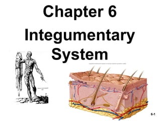
The Integumentary System
• Functions of the skin and subcutaneous
tissue
– epidermis and dermis
– hypodermis
– thick and thin skin
– skin color
– skin markings
• Hair and nails
• Cutaneous glands
• Skin disorders
3. 6-3
Skin & Subcutaneous Tissue
• The bodies largest organ and constitutes 15% of
body weight.
– i.e. 150 pound person = 22.5 pounds is skin.
• Every 24 hours, the surface of the skin sheds a layer
of dead cells, constantly renewing about every 28
days
• An average of 40 kilos of skin is shed during a
lifetime
4. 6-4
Overview
Skin consists of two layers:
• Epidermis
–Keratinized stratified squamous epithelium
• Dermis
–connective tissue layer
• Hypodermis- not part of skin but studied
• Thickness variable, normally 1-2 mm (0.03)
- Lacks hair follicles or sebaceous glands,
–Thickness due to dermis, up to 6 mm (0.24 inches)
– thin skin contains hair follicles, sebaceous & sweat
glands
6. 6-6
Functions of the Skin
1. Resistance to trauma and infection
– packed with keratin and linked by desmosomes;
– Bacteria & fungi are kept in check by the dryness &
acid mantle (pH 4-6) of skin;
1. Barrier - to ultraviolet light, H2O lose & absorption,
permeable to some drugs & poisons;
2. Vitamin D synthesis- first step
3. Sensory receptors – heat, cold, texture, pressure,
vibration, injury
4. Thermoregulation- through sweating, thermoreceptors
5. Nonverbal communication
7. 6-7
Transdermal Absorption
The ability to absorb substances through the skin:
- medicine
- ointments (hydrocortisone)
- lotions
- adhesive patches (ADHD, nitroglycerine)
- poisons
- toxic alkaloids (poison ivy)
- mercury, lead, arsenic, carbon tetrachloride
- acetone (nail cleaner)
Causes- liver, kidney failure, brain damage.
9. 6-9
Cells of the Epidermis
Most epidermis cells are keratinized,
stratified squamous, which mean:
Flat, layered cells that contain dead cells packed
with tough protein keratin
Like other epithelia the Epidermis lack a
blood supply, and must depend upon
diffusion from the underlying connective
tissue.
10. 6-10
Cells of the Epidermis
Composed of 5 types of cells:
– undifferentiated cells in deepest layers (ONLY
Stratum basale);
– Undergo mitosis to give rise to keratinocytes;
-most of the skin cells, synthesis
keratin;
-ONLY in Stratum basale, synthesize
melanin that shield UV;
– Receptors for touch,cells associated with nerve fibers
– macrophages guard against pathogens, toxins;
– Found in Stratum spinosum & stratum granulosum
layer;
1. Stem Cells
2. Keratinocytes
3. Melanocytes
4. Tactile (merkel) cells
5. Dendritic (langerhans) cells
12. 6-12
Layers of the Epidermis
• The epidermis is made up of 4-5 (thick) layers of cells.
13. 6-13
1. Stratum Basale
• Single layer cells on basement membrane
(usually cuboidal)
• Cell types in this layer
• undergo mitosis to replace epidermis
• distribute melanin through cell processes
• melanin picked up by keratinocytes
- are touch receptors
• form Merkel disc
Layers of Epidermis
1. Stem Cells
3. merkel cells
2. melanocytes
14. 6-14
2. Stratum Spinosum
• Several layers of keratinocytes (thickest layer)
– Lowest layer capable of mitosis but as they are pushed
upward, they instead produce keratin filaments that
flatten out the cells as they move to surface.
• Contains:
– macrophages from bone marrow
that migrate to the epidermis
– 800 cells/millimeter2
– help protect body against pathogens by “presenting”
them to the immune system
Dendritic (Langerhans) Cells
15. 6-15
3. Stratum Granulosum
3 to 5 layers Flat keratinocytes
• Contain keratinohyalin granules
– Produces lipid-filled vesicles that
release a glycolipid by exocytosis
to waterproof the skin
• forms a barrier between surface cells
and deeper layers of the epidermis
16. 6-16
4. Stratum Lucidum
• Thin translucent zone seen only in thick skin
• Keratinocytes are packed with eleidin, a precursor
to keratin
– does not stain well
• Cells have no nucleus or organelles
17. 6-17
5. Stratum Corneum
• Up to 30 layers of dead, scaly,
keratinized cells
– surface cells flake off (exfoliate)
– Especially resistant to abrasion,
penetration, and water loss
20. 6-20
Life History of Keratinocytes
• Produced by stem cells in stratum basale
• New cells push others toward surface
– 30 to 40 days keratinocytes make their way to the surface and
flake off.
– Accelerated growth = calus or corn
– As they get closer to the surface the form a membrane-coating
vesicle.
• Keratinocytes in the S. granulosum layer
• Undergo apopthosis
• The keratohylin granules release their substance
• Membrane coating vesicles release a lipid mixture
• Epidermal water barrier forms between S. granulosum and S.
spinosum
• Retains water in the body and prevents dehydration.
– Cells above the barrier are cut off from nutrients die.
23. 6-23
Dermis
• Is a connective tissue layer
– Composition
• collagen, elastic and reticular fibers, fibroblasts
• Well supplied with blood vessels, nerve
endings, sebaceous glands, sweat glands, hair
follicles, smooth muscles (Piloerector), skeletal
muscles allow for facial expression, smiles etc.
24. 6-24
Dermis
• Dermal papillae - extensions of the dermis into
the epidermis
• Epidermal ridges – extension of epidermis into
dermis
– forming the ridges of the fingerprints
25. 6-25
Dermis
• Dermis contains two zones (layers)
– papillary layer: loosely organized areolar
tissue allows motility of leukocytes and other
defense against breaks in epidermis.
– reticular layer: contains dense irregular
connective tissue. Excessive stretch can tear
these fibers producing Striae (stretch marks)
28. 6-28
Hypodermis
• Aka Subcutaneous tissue/ superficial
fascia
• Mostly adipose (subcutaneous fat) and
areolar, distribution is different throughout
the body and sexes.
• Functions
– energy reservoir
– thermal insulation
• Hypodermic injections (subQ)
– highly vascular
32. 6-32
Skin Colors (Pigmentation)
• Main factor is Melanin
• Produced by melanocytes, accumulates in
the keratinocytes in the S.basale and S.
Spinosum.
33. 6-33
Melanin
• 2 Forms:
1. Eumelanin: brownish – black
2. Pheomelanin: reddish yellow pigment
All races generally have the same number of melanocytes, but in darker
skinned people melanocytes produce a greater amount of melanin,
conversely lighter skinned people do not.
Location of melanin in the body may differ in regions.
Other factors in skin color:
Hemoglobin: imparts a reddish to pinkish hues to the skin.
Carotene: a yellow pigment found in egg yolks and yellow
orange vegetable.
34. 6-34
Abnormal Skin Colors
Abnormal Color for diagnosis
• Cyanosis = blueness from deficiency of
oxygen in the circulating blood (cold
weather)
• Erythema = redness due to dilated
cutaneous vessels (anger, sunburn,
embarrassment)
• Jaundice = yellowing of skin and sclera due
to excess of bilirubin in blood (liver disease)
35. 6-35
Abnormal Skin Colors
• Bronzing = golden-brown color of
Addison disease (deficiency of
glucocorticoid hormone)
• Pallor = pale color from lack of blood flow
• Albinism = a genetic lack of melanin
• Hematoma = a bruise (visible clotted
blood)
36. 6-36
• Freckles and moles = aggregations of melanocytes
– freckles are flat; moles are elevated
• Friction ridges leave oily fingerprints on touched
surfaces
– unique pattern formed during fetal development
• Flexion creases form after birth by repeated closing
of the hand
• Flexion lines form in wrist and elbow areas
Skin Markings
37. 6-37
Skin Markings
• Hemangiomas (birthmarks)
– discolored skin caused by benign tumors of dermal blood
capillaries
• Types:
– Capillary Hemangiomas (strawberry birthmarks):
bright red or purple, disappear in childhood.
– Cavernous Hemangiomas (port wine stains): flat
and duller in color last for life
39. 6-39
Characteristics of Human Hair
• Hair: composed of dead, keratinized cells
• Hair found almost everywhere
– The number of hairs on a given area does not differ much from person to
person
– differences between sexes or individuals is difference in texture and
color of hair
• 3 different body hair types
– lanugo -- fine, unpigmented fetal hair, last 3 months
– vellus -- fine, unpigmented hair of children and women
– terminal hair -- coarse, long, pigmented hair of scalp
40. 6-40
Structure of Hair and Follicle
Hair divided into 3 zones along its length:
1. Bulb:
2. Root:
3. Shaft:
Swelling at base where hair originates in dermis
Remainder of the hair within the follicle
Portion above the skin
41. 6-41
Structure of Hair and Follicle
Cross section shows 3 layer within hair:
1. Medulla:
2. Cortex:
3. Cuticle:
Loosely arranged cells, air space found in thick hair but
absent from thin hair.
Layer of keratinized cuboidal cells
Surface layer of scaly cells
42. 6-42
Hair Texture & Color
Texture is related to cross-sectional shape.
• Straight hair is round.
• Wavy hair is oval.
• Tight curly hair is relatively flat.
Color is due to pigment granules in the cells
of the cortex.
44. 6-44
Blond hair contain pheomelanin pigment, but little
eumelanin.
Hair Color and Texture, Blonde
45. 6-45
Red hair contains little eumelanin but lots of
pheomelanin.
Hair Color and Texture, Red
46. 6-46
White & Gray hair = air in medulla and lack of
pigment in cortex.
Hair Color and Texture, Gray and White
47. 6-47
Functions of Hair
1. Body hair (too thin to provide warmth)
– alert us to parasites crawling on skin (lice,fleas)
1. Scalp hair
– heat retention and sunburn cover
1. Beard, pubic and axillary hair indicate sexual
maturity and help distribute sexual scents
2. Guard hairs (vibrissae) and eyelashes
– prevent foreign objects from getting into nostrils, ear
canals or eyes
1. Expression of emotions with eyebrows
50. 6-50
Nails
• Derivative of stratum corneum
– densely packed dead cells filled with hard keratin
• Flat nails allow for fleshy, sensitive fingertips
• Growth rate is 1 mm per week
– new cells added by mitosis in the nail matrix (under skin)
– nail plate is visible part of nail
• medical diagnosis of iron deficiency = concave nails
• Finger nails can become clubbed in long term hypoxia (O2
deficiency in blood) such as congenital heart defects.
55. 6-55
Sweat (sudoriferous) Glands
Two kinds (described in Ch.5)
1. Merocrine glands is simple tubular gland
– Most abundant, millions of them help cool the body by
producing watery perspiration
– Simple tubular gland, lined with stratified cuboidal
epithelium
– Myoepithelial cells: incorporated with secretory cells,
respond to nervous stimuli thus squeezing the duct.
56. 6-56
Sweat (sudoriferous) Glands
2. Apocrine glands produce sweat
containing fatty acids (thicker)
– Opens to hair follicle rather than surface.
– Respond to stress and sexual stimulation
– No odor, unless trapped in clothing
– bromhidrosis is body odor produced by
bacterial action on fatty acids (body odor).
57. 6-57
Sweat (sudoriferous) Glands (FYI)
• Protein-free filtrate of plasma and some
waste products
– K+, urea, lactic acid, ammonia, & NaCl
– 500 ml of insensible perspiration/day, no
noticeable wetness of skin.
– sweating with visible wetness is diaphoresis
58. 6-58
Sebaceous Glands
• Oily secretion called sebum that contains
broken-down cells
– lanolin in skin creams is sheep sebum
• Flask-shaped gland with duct that opens into
hair follicle
• Sebum- Keeps skin and hair from becoming dry,
brittle and cracked.
59. 6-59
Ceruminous Glands
Found only in external ear canal
Their secretion combines with sebum to
produce earwax (cerumen).
• Cerumen Function:
1. Waterproof auditory canal
2. keeps eardrum flexible
3. bitterness repel mites and other pests
60. 6-60
Mammary Glands
• Breasts of both sexes rarely contain glands
– secondary sexual characteristic of females
• found only during lactation and pregnancy
– modified apocrine sweat gland
– thicker secretion released by ducts open on the nipple
• Anatomy and Physiology is in greater detail in
Chapter 28
64. 6-64
Skin Cancer
• Skin Cancer is one of the most common cancers
most often on head and neck where exposure is
greatest.
• Easiest one to treat and has one of the highest
survival rate when it is detected and treated
early.
• There are 3 types, named for their epidermal
cells from which they originate and distinguished
by their lesions.
65. 6-65
Skin Cancer
• Induced by UV rays of the sun
1. basal cell carcinoma (least dangerous)
• Most common
• arises from stratum basale and invades dermis
• Lesion starts as a small, shiny bump
66. 6-66
Skin Cancer
2. Squamous cell carcinoma
• arises from keratinocytes in stratum spinosum
• Lesion: raised, reddened, scaly, later an ulcer
• Lesions appear can appear on:
– Scalp, ears, lower lip, back of hand
• metastasis to the lymph nodes can be lethal
67. 6-67
Skin Cancer
3. Malignant melanoma (most deadly)
• arises from melanocytes of a preexisting mole
• Metastasizes quickly if untreated
• Greater incident in men then women
• Result of oncogene BRAF in men, in women it does not
trigger malignant melanoma but other cancers like breast
and ovarian.
68. 6-68
American Cancer Society
• “ABCD rule”
• A =
• B =
• C =
• D =
Asymmetry, one side of lesion looks different from the other.
Border irregularity, contour not uniform but wavy or scalloped
Color, mixture of brown, black, tan, sometimes red and blue
Diameter, greater than 6 mm
70. 6-70
Burns
• Hot water, sunlight, radiation, electric shock
or acids and bases
• Death from fluid loss, infection and other
toxic effects of Eschar.
• Eschar: Burned, dead tissue
71. 6-71
Degree of Burns
1st
Degree Burns
• Involve only epidermis
Characterized by:
• reddness, slight edema, pain
• Heals is a few days rarely scars
• Most sunburns
72. 6-72
Degree of Burns
2nd
Degree Burns
• involves epidermis and part of dermis
– Since it leaves some of the dermis
intact, 1st
and 2nd
degree burns are
considered PARTIAL – THICKNESS
BURNS
• Characterized by:
– red, tan or white and is blistered, very
painful
– May heal from 2 weeks to several
months, may scar
– epidermis regenerates from epithelial
cells in hair follicles and sweat glands
– Scalds and some sunburns
73. 6-73
Degree of Burns
3rd
Degree Burns (aka
Full-thickness burn)
• Epidermis, dermis and
deeper tissues are
destroyed.
• Require skin graphs
• If not taken care of,
contracture (abnormal
connective tissue fibrosis)
results = severe
disfigurement.
74. 6-74
Treatment
2 major conditions to treat:
1. fluid replacement
– Patient can lose several liters of water, electrolytes and protein
each day.
– As fluid is lost from tissues, it is replaced by the blood stream,
thus circulating blood volume declines.
– Within a few hours a person can lose 75% of blood plama
resulting in the principle cause of death from burns:
• Circulatory shock and cardiac arrest
– Intravenous fluids are given
75. 6-75
Treatment
2. Infection
– Controlled by keeping the patient in an aseptic
(sterile) environment
– Administer antibiotics
– Eschar is sterile in the first 24 hours but will quickly
become infected and have a toxic effect on digestive,
respiratory, and other systems
– To control the infection Debridement is used. This is
the removal of the eschar.
77. 6-77
UVA, UVB and Sunscreens
• UVA and UVB are improperly called
“tanning rays” and “burning rays”
• Both thought to initiate skin cancer
• As sale of sunscreens has risen so has
skin cancer
– those who use have higher incidence of
basal cell
– chemical in sunscreen damage DNA and
generate harmful free radicals
• PABA, zinc oxide and titanium dioxide
Editor's Notes
Thick and thin based on thickness of the epidermis alone. THICK skin covers palms, soles and corresponding surfaces of the fingers and toes.
Barrier to water, prevents the body from absorbing water when swimming and bathing, as well as prevents water loss.
O’Riain’s Sign (Shrivel Test): indicated when the fingers do not wrinkle after placed in warm water (30 minutes). NON wrinkling is due to denervation (sensibility) of the that particular nerve. (Return 90-120 days after injury).
Vitamin D needed for bone development and maintenance.
Extensive sense organ, can detect heat, cold, touch, texture, pressure, vibration and tissue injury. Location various areas.
The dermis has nerve endings that are called Thermoreceptors that transmit back to the brain, then back to dermal blood vessels to vasoconstrict or vasodilate.
Social function.
Melanocytes only in s. basale. Synthesize brown to black pigment melanin.
Tactile receptors for sense of touch.
Macrophages originate in the bone marrow and migrate to the epidermis of various sites.
Guard against toxins,microbes, and other pathogens that penetrate into the skin.
Named for its histological appearance. Spinosum means shiny appearance.
Dead cell constantly flake off the skin surface, forming much of the house dust.
Continuously being relaced.
Thus the S. Corneum consists of compact dead cells. Flake off in specks called DANDER.
Dermatophagoides (der-MAT-oh-fah-GOY-deez)
Reticular layer contains dense irregular connective tissue. Stretching of the skin can tear this collagen fibers producing STRIAE or stretch marks.
Dermal Papillae: Epidermal and dermal areas interweave like corrugated cardboard. Produces resistance for non slip between the epidermis and dermis layers.
Look at wrist and see delicate rectangule to rhomboid shapes. The dermal papillae for the raised areas between the furrows
The most significant factor in skin color is melanin. Produced by melanocytes but accumulates in the keratinocytes of the stratum basale and stratum spinosum.
Hard Keratin seen in hair and nails, more compact than soft keratin and is toughened by cross-linkage between keratin molecules.
Soft Keratin seen skin, pliable stratum corneum.
Difference is not the amount of hair but….
SEE FIGURE 6.8 in text
The question WHY DO HUMANS HAVE HAIR?
Hair of the trunk and limbs is vestigial, with little known function.
BODY HAIR kept our ancestors warm, but……. (go to slide).
SCALP HAIR the brain receives a rich supply of warm blood and the scalp lack an insulating fat layer so heat is easily conducted through the bone of the skull and lost to surrounding air.
GUARD HAIRS (aka vibrissae)
EXPRESSION: not protect from sweat. Specialized frontalis muscles for this purpose.
Most mammals have claws, whereas primates have flat nail ( a distinguishing feature).
Not only sensative but serve as strong keratinized “tools” for digging, grooming, picking food apart.
CLUBBING
Apocrine sweat glands in groin, axillia. Their ducts lead to nearby hair follicles rather than opening directly onto skin. Produce their secretion like merocrine glands – exocytosis.
hyperhidrosis :hands, prescriptions, botox injections and surgery 'endoscopic transthoracic sympathectomy' may cause increase sweating else where.
The sheen of well brushed hair is due to sebum, but we go through great lengths of trying to wash it out.
Mammary glands and breasts are often regarded as the one and the same.
Breasts are present in both sexes, and in both sexes rarely contain mammary glands.
Skin Cancer is one of the most common cancers most often on head and neck where exposure is greatest.
Easiest one to treat and has one of the highest survival rate when it is detected and treated early.
3 types named for the epidermal cells in which they originate.
Basal Cell Carcinoma: common type, least dangerous bec. It seldom metastasizes.
Squamous Cell Carcinoma: Usually appears on the scalp, ears, lower lip, or back of hand.
Malignant Melanoma: (only accounts for 5% of all cases), metastasizes quickly,fatal if not treated.
Men have a higher incident than women. Due to the BRAF oncogene in men. BRAF does not appear to trigger malignant melanoma, but it has been linked to some breast and ovarian cancers.
Leading cause of accidental deaths.
DEATH results from fluid loss, infection and toxic effects called ESCHAR– burned, dead tissue.
1st degree are sunburns!
2nd degree aka Partial-thickness burns! Some sunburns and scalds.
3rd degree aka Full- thickness burns! Contracture (abnormal connective tissue fibrosis), severe disfigurement
Fluid is lost from the tissues, more is transferred from the bloodstream.
A person can lose up to 75% of blood plasma within a few hours leading to circulatory shock and cardiac arrest-Principal cause of death in burn patients.
Infection is controlled by keeping the patient in an aseptic environment.