
Recommended
More Related Content
What's hot
What's hot (20)
Similar to EM-I-1-8-male-pelvis-and-perineum-Székely-A-2019.pptx
Similar to EM-I-1-8-male-pelvis-and-perineum-Székely-A-2019.pptx (20)
Recently uploaded
Recently uploaded (20)
EM-I-1-8-male-pelvis-and-perineum-Székely-A-2019.pptx
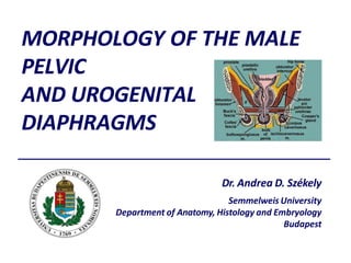
- 1. MORPHOLOGY OF THE MALE PELVIC AND UROGENITAL DIAPHRAGMS Dr. Andrea D. Székely Semmelweis University Department of Anatomy, Histology and Embryology Budapest
- 2. • It makes a fundamental contribution to movement and stability • Functions in coordination with the abdominal, back, and hip muscles • Especially important is its relationship to the Transversus Abdominis (the deepest layer of the abdominal muscles), and the Multifidus muscles in the low back, to maintain the integrity of pelvic, sacral, and spinal joints during movement • It supports the prostate, bladder, rectum, and seminal vesicles • It regulates continence, opening and closing the urethra and anus as needed • It plays an essential role in sexual function • It acts reciprocally with the respiratory diaphragm in breathing. • It is a flexor of the coccyx (tail bone) • The pelvic floor is the center of gravity in your frame, the place where movement is initiated, and is essential to your overall sense of well- being. FUNCTIONS AND ROLES OF THE PELVIC FLOOR
- 3. BONY AND LIGAMENTOUS FRAMEWORK
- 6. MORPHOLOGICAL DIMORPHISM Male and female perineal muscles
- 7. PERINEAL LAYERS OF MUSCLES AND FASCIAE
- 8. MUSCLE LAYERS OF THE PELVIC FLOOR 1. Pelvic diaphragm : levator ani and the fasciae 1. (ant) pubococcygeus 2. pubovaginalis 3. puborectalis 4. (post) iliococcygeus 5. coccygeus (ischiococcygeus) External anal sphincter 2. Urogenital Diaphragm : deep transverse perineal and the fasciae 1. urethrovaginal sphincter 2. compressor urethrae 3. urethral sphincter PERINEAL BODY 3. Urogenital trigone : - ischiocavernosus - bulbospongiosus - superficial transverse perineal m. corrugator cutis ani (smooth muscle)
- 9. FASCIAL LAYERS Fascia transversalis continues as the endopelvic fascia lining the pelvic cavity PELVIC FASCIA Lamina parietalis (obturator internus + Piriformis) Lamina visceralis (M. levator ani and M. coccygeus) = = superior fascia of the pelvic diaphragm * surrounding the pelvic viscera) * to be joined by the inferior fascial layer - see later
- 10. M. pubococcygeus (+ rectococcygeus) ORIGIN: Sup. Ramus of pubic bone INSERTION: Anococcygeal lig., Sacrum, Coccyx, M. sphincter ani ext. Fibres cross in an 8 shape limiting 2 openings Urogenital Hiatus Anal Hiatus M. iliococcygeus ORIGIN: Tendinous arch of the obturator int. + ischiac spine INSERTION: anococcygeal ligament joined by the M. coccygeus from posterior ORIGIN: Ischiac spine INSERTION: sacrotuberal lig., Coccyx, Sacrum) Function: Strengthens the pelvic floor PELVIC DIAPHRAGM LEVATOR ANI
- 11. ORIGIN: Obturator membrane + foramen INSERTION: Trochanteric Fossa Completes the lateral wall of the pelvis M. obturator internus Inferior fascia of the pelvic diaphragm FURTHER MUSCLES AND FASCIAE OF THE PELVIC WALL The piriformis or the triceps coxae will NOT contribute to the pelvic floor or wall covers the lower surface * Joins the superior fascial layer - see earlier Below the tendinous arch (of the levator ani) this fascia forms the lining of the isciorectal fossa together with the obturator fascia
- 12. PELVIC FLOOR LAYERS, LEVATOR ANI ORIGINS AND DIVISIONS Urogenital hiatus – anal hiatus iliococcygeus (ischio)coccygeus pubococcygeus piriformis puborectalis Anococcygeal ligament
- 13. PERINEAL MUSCLES, UROGENITAL DIAPHRAGM Deep transverse perineal muscle Trapezoid in shape, pulls between the inferior rami of the pubic bones. Forms the inferior cover of the pelvic diaphragm (fused to it at the perineal body) Passes the dorsal penile nerves and vessels between the arcuate pubic lig. and the transverse pelvic lig. Spalteholz M. bulbospongiosus ORIGIN: external anal sphincter + raphe of penile bulb, fused to the perineal body (erection + ejaculation) M. ischiocavernosus ORIGIN: ischiac tuberosity INSERTION: Crus penis + cavernous bodies (erection) Superficial transverse perineal muscle ORIGIN ischiac tuberosity INSERTIONperineal body)
- 14. SUPERFICIAL PERINEAL FASCIA LAMINA SUPERFICIALIS thick areolar CT, continues in the tunica dartos, fused to the skin in the midline LAMINA PROFUNDA (fascia Colles) thin, fibre rich CT membrane, continues in the tunica dartos, deep penile fascia, deep spermatic fascia, covers the muscles. Fused to the superficial lamina in the midline DEEP PERINEAL FASCIA (TRIANGULAR LIGAMENT) LAMINA INFERIOR Anteriorly - open towards the arcuate ligament, posteriorly fuses to the perineal body, covers the deep transverse perineal muscle LAMINA SUPERIOR Continuation of the obturator fascia, anterior attm to the pubic arch, posteriorly merges with the deep lamina (ligamentum transversum pelvis) UROGENITAL TRIGONE (perineal pouch) superficial transverse perineal bulbospongiosus ischiocavernosus PERINEAL FASCIAE AND SPACES
- 15. THE ISCHIORECTAL FOSSA WALLS ANTERIOR (recessus anterior) MED: levator ani LAT: obturator internus INF: deep transverse perineal POSTERIOR MED: anal fascia, sphincter ani ext. LAT: ischiac tuberosity, obturator fascia ANT: superfic and deep perineal fasciae POST: sacrotuberal lig., gluteus maximus CONTENT Adipose CT, containing : inferior hemorrhoidal aa., vv., nn. cutaneous br. of the pudendal plexus posterior scrotal aa., vv., nn. Alcock’s canal : Fascial duplication of the obturator internus CONTENT pudendal n. internal pudendal a. internal pudendal v.
- 16. DEEP AND SUPERFICIAL PERINEAL SPACES
- 17. PELVIC CONNECTIVE TISSUE SPACES Corning Prevesical space (of Reitzus) Contains the prostatic venous plexus (of Santorini) Paracysticum Retrovesical space Paraproctium Retrorectal space Contains the sup. rectal, median and lateral sacral arteries; pelvic and sacral splanchnic nerves; branches of the hypogastric & sacral plexus
- 18. Branches of the internal pudendal a./v. Branches of the pudendal n. VASCULAR AND NERVOUS SUPPLY
- 20. PELVIC PASSAGES – THE MALE URETHRA
- 21. External anal sphincter Voluntary action (striated muscle) Pars profunda Pars superficialis (between the perineal body and the anococcygeal ligament) Pars subcutanea Rauber-Kopsch Sobotta PELVIC PASSAGES – THE ANAL SPHINCTER
- 22. PELVIC PASSAGES – THE ANAL CANAL
- 24. ECTOPIC TESTICLE VARICOCELE HYDROCOCELE INGUINAL CANAL CLINICAL RELEVANCES
- 25. CLINICAL RELEVANCES - HAEMORRHOIDS
- 26. THANK YOU FOR YOUR ATTENTION!