SlideShare a Scribd company logo
1 of 93
Copyright © John Wiley & Sons, Inc. All rights reserved.
Chapter 17
The Special
Senses
Copyright © John Wiley & Sons, Inc. All rights reserved.
Special SensesRecall that a sensation is the conscious or
subconscious awareness of an internal or external stimulus
For this chapter, “external stimulus” means light rays striking
the retina of the eye, sound waves impinging on the tympanic
membrane of the ear, molecules in the air and food transmitting
smells and tastes to the chemical sensors in the nose an on the
tongue, and the force of gravity acting on equilibrium receptors
in the inner ear which sense changes in inertia
Copyright © John Wiley & Sons, Inc. All rights reserved.
Special SensesReceptors for the special senses of smell, taste,
vision, hearing, and equilibrium are anatomically distinct from
one another and are concentrated in specific locations in the
head
In addition to the stimuli and the receptors, there are specific
afferent pathways and translation sites in the brain for
information assembled from these special senses
Copyright © John Wiley & Sons, Inc. All rights reserved.
Special Senses
General Senses
Include somatic sensations (tactile, thermal, pain, and
proprioceptive) and visceral sensations
Are scattered throughout the body
Are relatively simple structures
Special Senses
Include smell, taste, vision, hearing and equilibrium
Are concentrated in specific locations in the head
Are anatomically distinct structures
Form complex neural pathwaysComparing the general senses
and the special senses
Copyright © John Wiley & Sons, Inc. All rights reserved.
Olfaction and TasteOlfaction is the process of perceiving
smells. Smell and taste are brought about through the
interpretation of chemicals present in the environment
Olfactory and gustatory (taste) impulses travel not only to the
cerebral cortex, but also to the limbic systemthis is why we can
have emotional responses and trigger strong memories to certain
smells and tastesgustation and olfaction work together but
olfaction
is much stronger/more sensitive (when someone has a
cold it is difficult to taste food)
Copyright © John Wiley & Sons, Inc. All rights reserved.
OlfactionThe olfactory epithelium is located in the superior part
of the nasal cavity covering the surface of the cribriform plate
and extending along the superior nasal concha
Copyright © John Wiley & Sons, Inc. All rights reserved.
OlfactionThe olfactory epithelium consists of 3 kinds of cells:
The olfactory receptor is a bipolar neuron with cilia (called
olfactory hairs). There are 10-100 million of these receptors in
the nose that respond to odorant molecules
Supporting cells provide
support and nourishment
Basal cells are stem cells
that replace olfactory
receptors
*
Copyright © John Wiley & Sons, Inc. All rights reserved.
OlfactionThe olfactory apparatus can detect about 10,000
different odors, often in concentrations as low as 1/25 billionth
of a milligram per milliliter of air When an odorant binds to
the
receptor of an olfactory hair it
initiates a cascade of intracellular
events through a G-protein and
Because olfaction is much more sensitive than taste, a given
concentration of a food substance may stimulate the olfactory
system thousands of times more strongly than it stimulates the
gustatory system. When you have a cold or are suffering from
allergies and cannot taste your food, it is actually olfaction that
is blocked, not taste.
A smell can be detected in a concentration as low as 1/25
billionth of a milligram per milliliter of air (the concentration
of the methyl mercaptan additive to natural gas).
*
Copyright © John Wiley & Sons, Inc. All rights reserved.
OlfactionOnce generated, nerve impulses travel through the two
olfactory area in the temporal lobe of the cortex
Olfaction is the only sensory
system that has direct
cortical projections
without first going
through relay stations
in the thalamus
*
Copyright © John Wiley & Sons, Inc. All rights reserved.
OlfactionOlfactory sensory pathways (centrally) are rapidly
adapting, decreasing activity by 50% in the first second, and
completely accommodating in 1–2 minutesOlfactory supporting
cells and glands are innervated by the facial (VII) nerve, a
component of which provides parasympathetic motor
innervation to lacrimal glands and the mucous membranes in the
nasal cavity. This is why certain odors will make our nose run
and cause us to produce tears
*
Olfactory Rc adapt very little. It’s the central pathways that
adapt.
Copyright © John Wiley & Sons, Inc. All rights reserved.
GustationGustation, or taste, is much simpler than olfaction in
that only five primary tastes can be distinguished: sour, sweet,
bitter, salty, and umami (“meaty” or “savory”)
Umami is believed to arise from taste receptors that are
stimulated by monosodium glutamate (MSG), a substance
naturally present in many foods and added to others as a flavor
enhancer
All other flavors, such as chocolate, pepper, and coffee, are
combinations of the five primary tastes, plus accompanying
olfactory and tactile (touch) sensations
Chemicals that stimulate gustatory receptor cells are known as
tastants. Once a tastant is dissolved in saliva, it can make
contact with the plasma membrane of the gustatory hairs, which
are the sites of taste transduction. The result is a receptor
potential that stimulates exocytosis of synaptic vesicles from
the gustatory receptor cell. In turn, the liberated
neurotransmitter molecules trigger nerve impulses in the first-
order sensory neurons that synapse with gustatory receptor
cells.
*
Copyright © John Wiley & Sons, Inc. All rights reserved.
We have nearly 10,000 taste buds located on the tongue, soft
Each taste bud is composed of about 50 gustatory receptor
cells, surrounded by a number of supporting cells
Basal cells located near the CT base multiply and differentiate,
first to become
the supporting cells
around the bud, then
the gustatory receptor
cells inside the taste
bud
Gustation
*
Copyright © John Wiley & Sons, Inc. All rights reserved.
A single, long microvillus, called a gustatory hair, projects from
each receptor cell to the surface through the taste pore Each
gustatory
receptor cell
has a lifespan
of about
10 days
Gustation
*
Copyright © John Wiley & Sons, Inc. All rights reserved.
GustationTaste buds are found in 3 different types of papillae
(elevations on the tongue which provide a rough texture
About 12 very large vallate papillae form a row at the back of
the tongue (each houses 100–300 taste buds)
Fungiform papillae are mushroom-shaped and are scattered over
the entire surface of the tongue (containing about 5 taste buds
each)
Foliate papillae are located in small trenches on the lateral
margins of the tongue, but most of their taste buds degenerate in
early childhood
Vallate papillae are also called circumvallate papillae (or
circular vallate papillae).
*
Copyright © John Wiley & Sons, Inc. All rights reserved.
In addition, the entire surface of the tongue has filiform
papillae that contain tactile
receptors but no
taste buds
They increase
friction between the
tongue and food,
making it easier to
move food in the oral cavity
Gustation
*
Copyright © John Wiley & Sons, Inc. All rights reserved.
GustationThree cranial nerves contain axons of the first-order
gustatory neurons that innervate the taste buds
The facial (VII) nerve serves taste buds in the anterior 2/3 of
the tongue
The glossopharyngeal (IX)
nerve serves taste buds in the
posterior 1/3 of the tongue
The vagus (X) nerve serves taste
buds in the throat and epiglottis
Copyright © John Wiley & Sons, Inc. All rights reserved.
Nerve impulses propagate along these cranial nerves to the
gustatory nucleus in the medulla oblongata. From there, axons
carrying taste signals project
to the hypothalamus, limbic
system, and thalamus
Taste is perceived consciously as
signals from the thalamus arrive
at the primary gustatory area at
the base of the somatosensory
cortex in the parietal lobe
Gustation
Copyright © John Wiley & Sons, Inc. All rights reserved.
GustationThe threshold for taste varies for each of the primary
tastes
We are most sensitive to bitter substances, such as quinine.
Because poisonous substances are often bitter, this high
sensitivity may have a protective function
The threshold for sour substances is somewhat higher, followed
by salty and sweet substancesComplete adaptation to a specific
taste can occur in
1–5 minutes of continuous stimulation
Copyright © John Wiley & Sons, Inc. All rights reserved.
VisionOur visual perception is dependent on the eye, its
accessory structures, the optic tracts, and the 1o visual cortex
and it’s association areas
Vision is possible because of
photoreceptors that are able
to “catch” photons of EM
radiation in the 400-700 nm
wavelengths – what we
perceive as visual light
Copyright © John Wiley & Sons, Inc. All rights reserved.
VisionThe eyeball is about 2.5 cm in diameter, with only about
16% of it viewable by just looking at a person
The accessory structures
of the eye are the
extraocular muscles,
palpebra, conjunctiva,
and the lacrimal glands
and ducts. The pupil is
an opening for light to
pass into the back of the eye
Copyright © John Wiley & Sons, Inc. All rights reserved.
The upper and lower palpebrae are the eyelids, with the fissure
being the space between themCN III supplies 4 of the 6
extraocular muscles, plus the levator palpebrae superioris
muscles that raise the
upper eyelidThe conjunctiva is a clear
mucous membrane that
covers the white (avascular)
part of the eye
Accessory Eye Structures
Visceral motor to parasympathetic innervation of the constrictor
pupillae and ciliary muscles. CN IV to the superior oblique.
CN VI to the lateral rectus
Production of tears superiorly and laterally, drain
inferomedially. Tears have lysozymes which help to destroy
bacteria.
*
Copyright © John Wiley & Sons, Inc. All rights reserved.
Accessory Eye StructuresThe lacrimal glands are each about the
size an almond, situated superolateral to the eyeball. Leading
from the lacrimal glands are 6 to 12 excretory lacrimal ducts
Tears (lacrimal fluid) run from the lacrimal glands, into the
excretory lacrimal ducts,
onto the surface of the
conjunctiva, over the
surface of the eyeballsome lacrimal fluid
also evaporates
*
Copyright © John Wiley & Sons, Inc. All rights reserved.
Accessory Eye StructuresTears drain into the lacrimal puncta,
which are two openings on the nasal side of the extreme edge of
the eyeball. Superior and inferior lacrimal canals empty the
tears into the nasolacrimal sac and nasolacrimal duct
The right and left sided nasolacrimal ducts empty into each side
of
the nose
*
Copyright © John Wiley & Sons, Inc. All rights reserved.
Accessory Eye StructuresWatery eyes occur when lacrimal fluid
builds up, as when something obstructs the nasolacrimal ducts
for instance
Blocked nasolacrimal ducts can be caused by an inflammation
of the nasal mucosa, such as a cold
Over production of lacrimal fluid occurs in response to
parasympathetic stimulation, caused by an emotional response
(crying), and tears spill over the edges of the eyelids and drain
into the nasal cavity (causing nasal stuffiness)
*
Copyright © John Wiley & Sons, Inc. All rights reserved.
The wall of the eyeball consists of three layers or tunics: The
fibrous tunic is the outer layer and is composed of the sclera
(“white” of the eye) and the cornea (the transparent epithelium
the protects the front of the eye)
The vascular tunic or uvea is
the middle layer and is
composed of the choroid,
the ciliary body and the iris
The nervous tunic is the
inner retinal layer
Anatomy of the Eye
Copyright © John Wiley & Sons, Inc. All rights reserved.
Anatomy of the EyeEven though you can’t easily see it, the
cornea is a very important structure in the outer avascular
fibrous tunic
It’s composed of a transparent epithelium that covers the
anterior eye and helps focus light onto the retinaLASIK is a
common visual corrective procedure that is performed on the
cornea of the eyeBecause of the amount of collagen fibers in the
sclera it forms the tough, white part of the eye
The sclera gives the eye it’s shape and protects the inner
anatomical parts
*
Copyright © John Wiley & Sons, Inc. All rights reserved.
Anatomy of the EyeOf the 3 parts of the middle tunic the
choroid forms the major vascular portion that lines the internal
surface of the sclera
The ciliary body consists of two parts:The ciliary processes that
secrete aqueous humor The ciliary muscle that changes the
shape of the lens to adapt to near and far vision
The iris is the colored portion of the eyeball consisting of
circular and radial smooth muscle fibers
*
Ciliary process is folded epithelial tissue
Iris has pigmented epithelium + two groups of smooth muscle
cells
The uvea (Lat. uva, grape), also called the uveal layer, uveal
coat, uveal tract, or vascular tunic, is the pigmented middle of
the three concentric layers that make up an eye. The name is
possibly a reference to its almost black colour, wrinkled
appearance and grape-like size and shape when stripped intact
from a cadaveric eye. Its use as a technical term in anatomy and
ophthalmology is relatively modern.
Copyright © John Wiley & Sons, Inc. All rights reserved.
The inner nervous tunic (retina) lines the posterior 2/3 of the
eye
The retina consist of a layer of melanin pigmented epithelium
that allows light to be absorbed rather than scattered. Without
the melanin, scattered
light in our eye would
cause us to always be
squinting, even in a
moderately lit room
Anatomy of the Eye
*
We don’t need the retina to cover the entire inside of the eye
because the light only strikes the back of the eye.
Copyright © John Wiley & Sons, Inc. All rights reserved.
The exact center of the retina is called the macula lutea, and in
its center is a small depression called the central fovea (or
fovea centralis)
There are no rods or nerve cells in the fovea, only a high
concentration of cones - this gives us the sharp central vision
necessary in any
activity where
detail is of
primary importance
Anatomy of the Eye
The sharp central vision is also called foveal vision.
*
Copyright © John Wiley & Sons, Inc. All rights reserved.
The retina can be viewed through the pupil using an
ophthalmoscope, allowing direct inspection of the retinal
vessels for any pathological changes. This is the only place in
the body where arterial vessels can be so viewed (without
opening
the body)
Anatomy of the Eye
*
Copyright © John Wiley & Sons, Inc. All rights reserved.
Anatomy of the EyeThe optic disc is where the optic nerve and
retinal vessels enter and exit the eyeball. Its existence creates a
necessary defect on the retina – an area where there are no
cones or rods. Bilateral vision, and
saccade (involuntary,
quick) muscle movements
allow our brain to correct
for this “blind spot”, and
most are not even aware
they have one
(try the test on the next page)
*
Copyright © John Wiley & Sons, Inc. All rights reserved.
*
The blind spot can be demonstrate using this chart
Instructions:
Situate yourself so that your nose is pointing in-between the
cross and the black circle.
Cover your LEFT eye and stare at the cross with your RIGHT
eye.
Now SLOWLY move towards the computer screen while still
staring at the cross with your RIGHT eye.
At somewhere around 10-14 inches from the computer screen –
the black circle will disappear and the area where the black
circle was…will now be all white - this is your BLIND SPOT.
If you move closer to the screen or farther away - the circle
will re-appear. At just the right distance – the circle will
disappear.
Now try the OTHER eye…but this time cover your RIGHT eye
and look at the CIRCLE with your LEFT eye…..move closer
and you will see that the CROSS now disappears!!
Copyright © John Wiley & Sons, Inc. All rights reserved.
Anatomy of the EyeThe retina consists of two types of
photoreceptor cells, rods and cones
Rods are abundant in the periphery of the retina whereas cones
are found more frequently in the central areas
*
Copyright © John Wiley & Sons, Inc. All rights reserved.
Anatomy of the EyeEach eye contains ≈ 120 million rod-shaped
photoreceptors that are adapted for a low light threshold (high
sensitivity) - they produce low resolution, black and white
images
a loss of rods with age makes it difficult to drive at night
*
Copyright © John Wiley & Sons, Inc. All rights reserved.
Anatomy of the EyeCone-shaped photoreceptors function in
bright light to produce high resolution color images
They exists in three varieties,
corresponding to the type of
pigment they contain: red, green or blue
The photopigments are concentrated in
the outer segment of the receptor, while the
inner segment contains the
nucleus and organelles
A complete loss of cones will result in legal blindness. A
relative loss, or a deficiency of one type results in color
blindness.
*
Copyright © John Wiley & Sons, Inc. All rights reserved.
Eye Cavities and ChambersThe lens is an avascular refractory
structure situated posterior to the pupil and iris. It consists of a
capsule with crystallin proteins arranged in layers, and like the
cornea, the lens is transparent
It attaches to the ciliary muscle
of the ciliary body
by suspensory
ligaments that fine
tune the focusing of
light on the retina
Copyright © John Wiley & Sons, Inc. All rights reserved.
Eye Cavities and ChambersThe lens divides the eyeball into two
cavities: An anterior cavity anterior to the lens, and a posterior
cavity (vitreous chamber) behind the lens
The anterior cavity is further
divided at the level of the
iris into anterior and
posterior chambers
(both filled with
aqueous humor)
Copyright © John Wiley & Sons, Inc. All rights reserved.
Eye Cavities and ChambersThe much larger posterior cavity of
the eyeball (vitreous chamber) lies between the lens and the
retina
Within the vitreous chamber is the vitreous body, a transparent
jellylike substance that holds the retina flush against the
choroid, giving the retina an even surface for the reception of
clear imagesoccasionally, collections of debris called vitreal
floaters cast shadows on the retina and create a spot in our field
of vision (they are usually harmless and do not require
treatment)
*
Copyright © John Wiley & Sons, Inc. All rights reserved.
Eye Cavities and ChambersThis cow eye dissection shows an
eye bisected into anterior and posterior sections along its
coronal axis. The anterior structures of the
iris and pupil are seen in
the bottom half; the
posterior retina,
choroid, and optic
disc are seen in the
top half
*
Copyright © John Wiley & Sons, Inc. All rights reserved.
Aqueous HumorThe eye requires a constant bath in a nourishing
fluid to deliver enough O2 to support the avascular lens and
cornea
It also needs fluid to help “inflate” the walls of the eyeball
(maintain a constant intraocular pressure – IOP) and support the
vitreous bodythis need is accomplished through the production
of aqueous humor, which flows through the anterior cavity of
the eye and is replaced every 90 minutes
*
Copyright © John Wiley & Sons, Inc. All rights reserved.
Aqueous HumorAqueous humor is produced at the ciliary body
and flows first through the posterior chamber (of the anterior
cavity of the eye)
Traveling along the posterior surface of the iris it passes
through the pupil to enter the anterior chamber
It proceeds along the anterior surface of the iris until it is
reabsorbed into the scleral venous sinus (canal of Schlemm) and
returned to the venous system
*
Copyright © John Wiley & Sons, Inc. All rights reserved.
Aqueous HumorAny sort of blockage to aqueous humor flow,
or overproduction at the ciliary body may result in an increase
of pressure inside the eye –
a condition called glaucoma
If not treated,
glaucoma can lead
to a degeneration
of eye function
*
Too high or too low of an IOP are both problems. IOP can be
measured with a device called an applanation tonometer that
sends a puff of air to ricochet off the cornea and back to the
machine to be measured.
Copyright © John Wiley & Sons, Inc. All rights reserved.
Retinal DetachmentThe vitreous body (humor) also contributes
to maintain proper intraocular pressure as it holds the retina
against the choroid. The vitreous humor, however, is only
formed during embryological development and is not replaced.
As we age, shrinkage of the vitreous body may lead to a
detachment of the retina from the choroid
A retinal detachment is considered a medical emergency and
needs immediate repair before vision loss becomes permanent
Copyright © John Wiley & Sons, Inc. All rights reserved.
The pupil is an opening in the center of the iris. It is composed
of a radial muscle that “radiates” away from the center, and a
circular muscle that is in the center
Contraction of the inner circular muscle fibers cause the pupil
to constrict
while contraction
of the radial fibers
cause it to dilate
The Pupillary Response
Copyright © John Wiley & Sons, Inc. All rights reserved.
Refraction and ImageNormal image formation depends on
refraction of light waves, accommodation of the lens,
constriction of the pupil, and convergence of the two eyes
Refraction is the process of bending light rays. Both the cornea
and the lens refract light rays, and both must be functioning in
order to
properly focus light onto
the right spot on the retina
to produce clear vision
Copyright © John Wiley & Sons, Inc. All rights reserved.
Refraction and ImageSince the cornea has a fixed shape, its
“focal length” is also fixed; and its ability to refract light is
likewise fixedIn order to focus light that has already been bent
by the cornea the lens must change shape – the amount
depending on the type of light rays we are trying to “see”
Copyright © John Wiley & Sons, Inc. All rights reserved.
Refraction and ImageAn increase in the curvature of the lens for
near vision is called accommodation
The near point of vision is the minimum distance from the eye
that an object
can be clearly
focused - about 4 in
(a distance that
increases with age
due to a loss of
elasticity in the lens)
Images focused on the retina are inverted and reversed, but our
brain learns to flip them around.
*
Copyright © John Wiley & Sons, Inc. All rights reserved.
Refraction and ImageConvergence is the inward movement of
the eyes so that both are directed at the object being viewed -
becoming a little cross-eyed when viewing things close up
The nearer the object, the greater the degree of convergence
needed to maintain binocular visionthe coordinated action of the
extrinsic eye muscles brings about convergence.
Convergence helps us maintain our binocular vision and see in
three dimensions
Copyright © John Wiley & Sons, Inc. All rights reserved.
Refraction and ImageWith nearsightedness (myopia), only close
objects can be seen clearly: Light rays coming in from distant
objects are naturally focused in front of the retina and appear
blurry
Correction involves the use of a concave
(negative) lensWith farsightedness (hyperopia), only distant
objects can be seen clearly: Light rays coming in from nearer
objects are naturally focused behind the retina
Correction involves the use of a convex (positive) lens
Copyright © John Wiley & Sons, Inc. All rights reserved.
Abnormal refractive capabilities of the eye are the result of a
misshapen eyeball (usually too long or too short), or because
the lens becomes stiff (usually
with age). Corrections are
accomplished using either
a positive (convex) or
negative (concave) lens
(eyeglasses, contacts, or
lens replacements)
Refraction and Image
Astigmatism is an irregular curvature of the lens or cornea that
causes portions of objects to be out of focus.
*
Copyright © John Wiley & Sons, Inc. All rights reserved.
Visual TransductionOnce light waves have been successfully
focused on the retina, the information “stored” in that
electromagnetic energy must be changed by photopigments in
the photoreceptors into signals our brain can interpret - a
process called visual transductionThe single type of
photopigment in rods is rhodopsin, whereas there are 3 different
cone photopigments
Color vision results from different colors of light selectively
activating the different cone photopigments
Copyright © John Wiley & Sons, Inc. All rights reserved.
Visual TransductionThe first step in visual transduction is
absorption of light by a photopigment, a colored protein that
undergoes structural changes when it
absorbs light in the outer
segment of a photoreceptor
Light absorption initiates
a series of events that
lead to the production
of a receptor potential
(number 4 in the diagram)
Copyright © John Wiley & Sons, Inc. All rights reserved.
Visual TransductionAll photopigments associated with vision
contain two parts: a glycoprotein known as opsin and a
derivative of vitamin A called retinal
Although there are 4 different opsins, retinal is the light-
absorbing part of all visual photopigmentsTo simplify the
process we can say that there is a cyclical bleaching and
regeneration of photopigment
Bleaching is a term describing a conformational change in the
retinal molecule in response to light
Copyright © John Wiley & Sons, Inc. All rights reserved.
Visual TransductionIn darkness, retinal has a bent shape called
cis-retinal
Absorption of a photon of light causes it to straighten into the
trans-retinal form in a process called isomerization
Trans-retinal completely separates from the opsin; since the
final products look colorless, this part of the cycle is called
bleaching of photopigment
An enzyme converts trans-retinal→ cis-retinal
The cis-retinal regenerates the photopigment
In darkness, the neurotransmitter glutamate is released keeping
Na+ channels open and inhibiting the bipolar cell: This inflow
of Na+is called the “dark current”. Photons cause Na+ channels
to close, and the rod hyperpolarizes. It’s strange that when your
eyes are closed and you are asleep the rods are the most active.
*
Copyright © John Wiley & Sons, Inc. All rights reserved.
Bleaching and regeneration of photopigments are summarized
here
Horizontal cells transmit inhibitory signals to bipolar cells in
the areas lateral to excited rods and cones.
*
Copyright © John Wiley & Sons, Inc. All rights reserved.
Visual TransductionIn daylight, regeneration of rhodopsin
cannot keep up with the bleaching process, so rods contribute
little to daylight vision. In contrast, cone photopigments
regenerate rapidly enough that some of the cis form is always
present, even in very bright light
light conditions) happens in seconds; dark adaptation (from
adapt)
After complete bleaching, regeneration of half of the rhodopsin
takes 5 minutes; half of the cone photopigments regenerate in
only 90 seconds. Full regeneration of bleached rhodopsin takes
30 to 40 minutes.
*
Copyright © John Wiley & Sons, Inc. All rights reserved.
Visual TransductionMost forms of color blindness, an inherited
inability to distinguish between certain colors, result from the
absence or deficiency of one of the three types of cones
Most common type is red-green color blindness in which red
cones or green cones are missingProlonged vitamin A
deficiency and the resulting below-normal amount of rhodopsin
may cause night blindness or nyctalopia, an inability to see well
at low light levels
Copyright © John Wiley & Sons, Inc. All rights reserved.
The graded potentials generated by the photoreceptors undergo
considerable processing at synapses among the various types of
neurons in the retina (horizontal cells, bipolar cells, and
amacrine cells)- certain features of visual
input are enhanced while others
are discarded
Overall, convergence pre-
dominates as 126 million
photo-receptors impinge on only
1 million ganglion cells
The Visual Pathway
Horizontal cells transmit inhibitory signals to bipolar cells in
the areas lateral to excited rods and cones. Horizontal cells also
assist in the differentiation of various colors. Amacrine cells,
which are excited by bipolar cells, synapse with ganglion cells
and transmit information to them that signals a change in the
level of illumination of the retina. When bipolar or amacrine
cells transmit excitatory signals to ganglion cells, the ganglion
cells become depolarized and initiate nerve impulses.
*
Copyright © John Wiley & Sons, Inc. All rights reserved.
The Visual PathwayThe axons of retinal ganglion cells provide
output that travels back “towards the light”, exiting the eyeball
as the
optic nerve, which emerges from the vitreous surface of the
retina
The axons then pass
through a crossover point
called the optic chiasm
Copyright © John Wiley & Sons, Inc. All rights reserved.
Some axons cross to the opposite side, while others remain
uncrossed. Once through the
optic chiasm the axons enter the
brain matter as the optic tracts
(most terminate in thalamus)
Here they synapse with
neurons that project to the
1o visual cortex in the
occipital lobes
The Visual Pathway
Copyright © John Wiley & Sons, Inc. All rights reserved.
Visual field of
left eye
Temporal
half
Visual field of
right eye
Temporal
half
Nasal
half
Midbrain
Left eye
Temporal
retina
Optic
radiations
Left eye and its pathways
Primary visual area of cerebral
cortex (area 17) in occipital lobe
Lateral geniculate nucleus
of the thalamus
Optic
radiations
Midbrain
Temporal
retina
Nasal
retina
Right eye
Right eye and its pathways
Nasal
half
Nasal retina
1
1
Visual field of
left eye
Temporal
half
Visual field of
right eye
Temporal
half
Nasal
half
Midbrain
Left eye
Temporal
retina
Optic
radiations
Left eye and its pathways
Primary visual area of cerebral
cortex (area 17) in occipital lobe
Lateral geniculate nucleus
of the thalamus
Optic
radiations
Midbrain
Temporal
retina
Nasal
retina
Right eye
Right eye and its pathways
Nasal
half
Nasal retina
1
1
2
2
Visual field of
left eye
Temporal
half
Visual field of
right eye
Temporal
half
Nasal
half
Midbrain
Left eye
Temporal
retina
Optic
radiations
Left eye and its pathways
Primary visual area of cerebral
cortex (area 17) in occipital lobe
Lateral geniculate nucleus
of the thalamus
Optic
radiations
Midbrain
Temporal
retina
Nasal
retina
Right eye
Right eye and its pathways
Nasal
half
Nasal retina
1
1
2
2
3
3
Visual field of
left eye
Temporal
half
Visual field of
right eye
Temporal
half
Nasal
half
Midbrain
Left eye
Temporal
retina
Optic
radiations
Left eye and its pathways
Optic
tract
Primary visual area of cerebral
cortex (area 17) in occipital lobe
Lateral geniculate nucleus
of the thalamus
Optic
radiations
Midbrain
Temporal
retina
Nasal
retina
Right eye
Right eye and its pathways
Nasal
half
Nasal retina
1
1
2
2
4
4
3
3
Visual field of
left eye
Temporal
half
Visual field of
right eye
Temporal
half
Nasal
half
Midbrain
Left eye
Temporal
retina
Optic
radiations
Left eye and its pathways
Optic
tract
Primary visual area of cerebral
cortex (area 17) in occipital lobe
Lateral geniculate nucleus
of the thalamus
Optic
radiations
Midbrain
Temporal
retina
Nasal
retina
Right eye
Right eye and its pathways
Nasal
half
Nasal retina
1
1
2
2
4
4
5
5
3
3
Visual field of
left eye
Temporal
half
Visual field of
right eye
Temporal
half
Nasal
half
Midbrain
Left eye
Temporal
retina
Optic
radiations
Left eye and its pathways
Optic
tract
Primary visual area of cerebral
cortex (area 17) in occipital lobe
Lateral geniculate nucleus
of the thalamus
Optic
radiations
Midbrain
Temporal
retina
Nasal
retina
Right eye
Right eye and its pathways
Nasal
half
Nasal retina
1
1
2
3
2
4
3
4
5
5
6
6
Copyright © John Wiley & Sons, Inc. All rights reserved.
The EarAudition, the process of hearing, is accomplished by the
organs of the ear. The ear is an engineering marvel because its
sensory receptors can transduce sound vibrations with
amplitudes as small as the diameter of an atom of gold into
electrical
signals 1000 times faster than
the eye can respond to light
The ear also contains
receptors for equilibrium
Copyright © John Wiley & Sons, Inc. All rights reserved.
The EarThe ear has 3 principle regions
The external ear, which uses air to collect and channel sound
waves
The middle ear, which uses a
bony system to amplify
sound vibrations
The internal ear, which
generates action potentials to transmit
sound and balance information to the brain
*
The pathways for sound transmission starts in the air, the to the
solid bone of the middle ear, then to the endolymph of the inner
ear.
Copyright © John Wiley & Sons, Inc. All rights reserved.
The External EarThe anatomy of the external ear includes
The auricle (pinna), a flap of elastic cartilage covered by skin
and containing ceruminous glands
A curved 1” long external auditory canal situated in the
temporal bone leading from
the meatus to the tympanic
membrane (TM – or ear
drum) which separates the
outer ear from the cavity
of the middle ear
*
Copyright © John Wiley & Sons, Inc. All rights reserved.
The middle ear is an air-filled cavity in the temporal bone. It is
lined with epithelium and contains 3 auditory ossicles (bones)
The stapes (stirrup)
The incus (anvil)
The handle of
the malleus
(hammer) attaches
to the TM
The Middle Ear
*
Copyright © John Wiley & Sons, Inc. All rights reserved.
The Middle EarTwo small skeletal muscles (the tensor tympani
and stapedius) attach to
the ossicle and
dampen vibrations
to prevent damage
from sudden,
loud sounds
*
Copyright © John Wiley & Sons, Inc. All rights reserved.
The Middle Ear
*
Copyright © John Wiley & Sons, Inc. All rights reserved.
The Middle EarThe Eustachian (auditory) tube connects the
middle ear with the nasopharynx (upper portion of the throat)
It consists of bone and hyaline
cartilage and is normally
passively collapsed. It opens to
equalize pressures on each side
of the TM
(allowing it
to vibrate freely)
The chamber of the middle ear is continuous through the
eustachian tube with the nasopharynx, but also with the mastoid
antrum and mastoid air cells. Infection of the mucosa lining the
middle ear will extend to the mastoid air cells in the bone
behind the ear.
*
Copyright © John Wiley & Sons, Inc. All rights reserved.
The Inner EarThe internal ear (inner ear) is also called the
labyrinth because of its complicated series of canals
Structurally, it consists of two main divisions: an outer bony
labyrinth that encloses an inner membranous labyrinththe bony
labyrinth is sculpted out of the petrous part of the temporal
bone, and divided into three areas: (1) the semicircular canals,
(2) the vestibule,
and (3) the cochlea
*
The oval window starts the inner ear. As the stapes rocks back
and forth the oval window and round window oscillates (like
pushing on the end of a waterbed).
Copyright © John Wiley & Sons, Inc. All rights reserved.
The Inner EarThe vestibule is the middle part of the bony
labyrinth
The membranous labyrinth in the vestibule consists of two sacs
called the utricle and the saccule (both contain rc for static
equilibrium)The cochlea , located anterior to the
vestibule, contains rc for hearingThe three semicircular canals
are above the vestibule, each
ending in a swollen
enlargement called the ampulla
(for dynamic equilibrium)
*
Copyright © John Wiley & Sons, Inc. All rights reserved.
The Inner EarThe snail shaped cochlea contains the hearing
apparatus
Two types of fluid (perilymph and endolymph) fill its 3
different internal channels: The scala vestibuli, scala tympani,
and cochlear duct
A section
through one
turn of the
cochlea is shown
*
Copyright © John Wiley & Sons, Inc. All rights reserved.
The Inner EarPerilymph transmits the vibrations coming from
the stapes in the oval window up and around the scala vestibuli,
and then back down and around the scala tympani – causing the
endolymph in the cochlear duct to vibrate
Pressure waves in the endolymph cause the basilar membrane of
the cochlear duct to vibrate, moving the hair cells of the spiral
organ of Corti against an overhanging flexible gelatinous
membrane called the tectorial membrane
*
Copyright © John Wiley & Sons, Inc. All rights reserved.
The Inner EarNote how the sound waves between the number 1
and number 2 in this diagram are shown impacting different
parts of membranous labyrinth. This is a representation of
sounds waves of different frequencies being transduced at the
segment of the basilar membrane that
is “tuned” for
a particular
pitch
*
Copyright © John Wiley & Sons, Inc. All rights reserved.
The Inner EarMovements of the hair cells in contact with the
tectorial membrane transduce mechanical vibrations into
electrical signals which generate nerve impulses along the
cochlear branch of CN VIII
Copyright © John Wiley & Sons, Inc. All rights reserved.
The Auditory PathwayThis graphic depicts the events in the
stimulation of auditory receptors, from channeling sound waves
into the external ear and onto the TM, to the transduction of
those vibrations
into local
receptor
potentials
Sound waves enter the external auditory canal and strike the
eardrum. The vibrations of the eardrum cause the ossicles to
vibrate and the stapes pushes the membrane of the oval window
in and out. The movement of the oval window sends fluid
pressure waves into the perilymph of the scala vestibuli which
then transmit them to the scala tympani and eventually to the
round window (causing it to bulge outward into the middle ear).
The pressure waves move into the endolymph of the cochlear
duct and cause the basilar membrane to vibrate which moves the
hair cells of the spiral organ against the tectorial membrane.
This leads to bending of the stereocilia and ultimately to the
generation of nerve impulses in first-order neurons in cochlear
nerve fibers. Sound waves of various frequencies cause certain
regions of the basilar membrane to vibrate more intensely than
other regions.
Each segment of the basilar membrane is “tuned” for a
*
Copyright © John Wiley & Sons, Inc. All rights reserved.
Scala
vestibuli
Cochlear duct
(contains endolymph)
Scala
tympani
Perilymph
Basilar
membrane
Cochlea
Sound waves
Helicotrema
Stapes vibrating
in oval window
Malleus
Incus
External auditory
canal
Tympanic
membrane
Secondary tympanic
membrane vibrating
in round window
Auditory tube
Vestibular membrane
Middle ear
Tectorial membrane
Spiral organ
(organ of Corti)
1
Scala
vestibuli
Cochlear duct
(contains endolymph)
Scala
tympani
Perilymph
Basilar
membrane
Cochlea
Sound waves
Helicotrema
Stapes vibrating
in oval window
Malleus
Incus
External auditory
canal
Tympanic
membrane
Secondary tympanic
membrane vibrating
in round window
Auditory tube
Vestibular membrane
Middle ear
Tectorial membrane
Spiral organ
(organ of Corti)
1
2
Scala
vestibuli
Cochlear duct
(contains endolymph)
Scala
tympani
Perilymph
Basilar
membrane
Cochlea
Sound waves
Helicotrema
Stapes vibrating
in oval window
Malleus
Incus
External auditory
canal
Tympanic
membrane
Secondary tympanic
membrane vibrating
in round window
Auditory tube
Vestibular membrane
Middle ear
Tectorial membrane
Spiral organ
(organ of Corti)
1
2
3
Scala
vestibuli
Cochlear duct
(contains endolymph)
Scala
tympani
Perilymph
Basilar
membrane
Cochlea
Sound waves
Helicotrema
Stapes vibrating
in oval window
Malleus
Incus
External auditory
canal
Tympanic
membrane
Secondary tympanic
membrane vibrating
in round window
Auditory tube
Vestibular membrane
Middle ear
Tectorial membrane
Spiral organ
(organ of Corti)
1
2
3
4
Scala
vestibuli
Cochlear duct
(contains endolymph)
Scala
tympani
Perilymph
Basilar
membrane
Cochlea
Sound waves
Helicotrema
Stapes vibrating
in oval window
Malleus
Incus
External auditory
canal
Tympanic
membrane
Secondary tympanic
membrane vibrating
in round window
Auditory tube
Vestibular membrane
Middle ear
Tectorial membrane
Spiral organ
(organ of Corti)
1
2
3
4
5
Scala
vestibuli
Cochlear duct
(contains endolymph)
Scala
tympani
Perilymph
Basilar
membrane
Cochlea
Sound waves
Helicotrema
Stapes vibrating
in oval window
Malleus
Incus
External auditory
canal
Tympanic
membrane
Secondary tympanic
membrane vibrating
in round window
Auditory tube
Vestibular membrane
Middle ear
Tectorial membrane
Spiral organ
(organ of Corti)
1
2
3
4
5
6
Scala
vestibuli
Cochlear duct
(contains endolymph)
Scala
tympani
Perilymph
Basilar
membrane
Cochlea
Sound waves
Helicotrema
Stapes vibrating
in oval window
Malleus
Incus
External auditory
canal
Tympanic
membrane
Secondary tympanic
membrane vibrating
in round window
Auditory tube
Vestibular membrane
Middle ear
Tectorial membrane
Spiral organ
(organ of Corti)
1
2
3
4
5
6
7
Scala
vestibuli
Cochlear duct
(contains endolymph)
Scala
tympani
Perilymph
Basilar
membrane
Cochlea
Sound waves
Helicotrema
Stapes vibrating
in oval window
Malleus
Incus
External auditory
canal
Tympanic
membrane
Secondary tympanic
membrane vibrating
in round window
Auditory tube
Vestibular membrane
Middle ear
Tectorial membrane
Spiral organ
(organ of Corti)
1
2
3
4
5
6
7
8
8
Scala
vestibuli
Cochlear duct
(contains endolymph)
Scala
tympani
Perilymph
Basilar
membrane
Cochlea
Sound waves
Helicotrema
Stapes vibrating
in oval window
Malleus
Incus
External auditory
canal
Tympanic
membrane
Secondary tympanic
membrane vibrating
in round window
Auditory tube
Vestibular membrane
Middle ear
Tectorial membrane
Spiral organ
(organ of Corti)
1
2
3
4
5
6
7
8
8
9
Copyright © John Wiley & Sons, Inc. All rights reserved.
The Auditory PathwayThe cell bodies of the sensory neurons
are located in the spiral ganglia. Nerve impulses pass along the
axons of these neurons, which
form the cochlear
branch of the
vestibulo-
cochlear (VIII)
nerve
Copyright © John Wiley & Sons, Inc. All rights reserved.
The Auditory PathwayThe nerve impulses follow CN VIII en
route to the medulla, pons, midbrain, and thalamus, and finally
to the primary auditory cortex in the temporal lobe. Slight
differences in the timing of nerve
impulses arriving from the
two ears at the superior
olivary nuclei in the
pons allow us to
locate the source
of a sound
Copyright © John Wiley & Sons, Inc. All rights reserved.
EquilibriumEquilibrium is another function of the inner ear -
controlled by the vestibular apparatus (the saccule and utricle of
the vestibule, and the 3 semicircular canals)
Static equilibrium refers to a
state of balance relative to
the force of gravity
Dynamic equilibrium
involves the maintenance
of balance during sudden movements
*
Copyright © John Wiley & Sons, Inc. All rights reserved.
Static EquilibriumStatic equilibrium is controlled by the
sensory hairs within the macula of the utricle
and saccule
*
Copyright © John Wiley & Sons, Inc. All rights reserved.
Static EquilibriumAn otolithic membrane, studded with dense
calcium carbonate crystals (otoliths), responds to gravity when
head position is changed
This movement opens
transduction channels
in the hair cells,
producing local potentials
which summate to
form nerve AP
*
Copyright © John Wiley & Sons, Inc. All rights reserved.
Dynamic EquilibriumDynamic equilibrium is controlled by the
sensory hairs within the ampulla of the
semicircular canals
Within each ampulla
is a small elevation
called the crista
*
Copyright © John Wiley & Sons, Inc. All rights reserved.
Dynamic EquilibriumEach crista contains hair cells and
supporting cells covered by gelatinous material called the
cupula
With movement, the endolymph within the ampulla lags behind
the moving cupola, causing a difference in the inertial forces –
the hair bundle
of the cupola
bends and nerve
impulses are generated
Copyright © John Wiley & Sons, Inc. All rights reserved.
Equilibrium PathwayOnce generated, nerve impulse travel up
the vestibular branch of CN VIII. Most of these axons synapse
in the major integrating centers for equilibrium, in the medulla
and pons, which also receive input from the eyes and
proprioceptors
parietal lobe to provide us with conscious awareness of the
position and movements of the head and limbs
*
Copyright © John Wiley & Sons, Inc. All rights reserved.
Homeostatic ImbalancesA cataract is an opaque defect in the
cornea or lens of the eye – most cataracts are in the lens
Cataracts are causes by injury, medications, and diseases like
diabetes. They are common in old ageConjunctivitis is an
inflammation of the conjunctival membrane which covers part
of the front of the eye
Conjunctivitis is caused most frequently by viral infections
(pink eye) and allergy. It can also result from bacterial
infections and many other irritants
Copyright © John Wiley & Sons, Inc. All rights reserved.
Homeostatic ImbalancesAge Related Macular Degeneration
results in a loss of vision in the center of the visual field (the
macula) because of damage to the retina. It is a major cause of
visual impairment in older adults (>50 years)
It can become impossible
to recognize faces, yet
enough peripheral vision
remains to allow other
activities of daily life
Copyright © John Wiley & Sons, Inc. All rights reserved.
Homeostatic ImbalancesMyringitis is an inflammation of the ear
drum
Infections of the middle ear cavity (otitis media) are common in
children between 6 mo. – 5 yrs. old, and usually presents with a
crying child and a TM (viewed through an otoscope), that looks
angry, red, and bulgingOtitis externa (commonly called
“swimmer’s ear”) is a dermatitis of the epithelium of the outer
ear (infectious and noninfectious). The chlorine, water, and ear
plugs associated with swimming can result in irritated, inflamed
tissues of the outer ear and ear canal
Copyright © John Wiley & Sons, Inc. All rights reserved.
Distended Eardrum Caused by
Otitis Media
Dr. P. Marazzi/Photo Researchers, Inc.
Copyright © John Wiley & Sons, Inc. All rights reserved.
Homeostatic ImbalancesMeniere’s disease is a disorder of the
inner ear that can affect hearing and balance, and is thought to
be due to increased pressure in the cochlea and semicircular
canals (extra endolymph)
Episodes of vertigo (the room spinning) and ringing in the ears
(tinnitus) can be a mild annoyance, or a chronic, disabling
disability
Copyright © John Wiley & Sons, Inc. All rights reserved.
End of Chapter 17
Copyright 2012 John Wiley & Sons, Inc. All rights reserved.
Reproduction or translation of this work beyond that permitted
in section 117 of the 1976 United States Copyright Act without
express permission of the copyright owner is unlawful. Request
for further information should be addressed to the Permission
Department, John Wiley & Sons, Inc. The purchaser may make
back-up copies for his/her own use only and not for distribution
or resale. The Publisher assumes no responsibility for errors,
omissions, or damages caused by the use of these programs or
from the use of the information herein.
Copyright © John Wiley & Sons, Inc. All rights reserved.
Chapter 16
Sensory, Motor, and Integrative Systems
Copyright © John Wiley & Sons, Inc. All rights reserved.
In this chapter we explore the levels and components of
pathways that convey sensory nerve impulses from the body to
the brain, and the general sensations (somatic and visceral) that
result. We will
also examine the activation of
motor pathways and movements
In chapter 17 we will look at
the special senses of sight,
hearing, taste, and smell
General Sensations
Copyright © John Wiley & Sons, Inc. All rights reserved.
General SensationsAs sensory impulses reach the CNS, they
become part of a large pool of sensory input
(though not every one
will elicit a response)
Each piece of
incoming information
is combined with other arriving
and previously stored information
in a process called integration
Copyright © John Wiley & Sons, Inc. All rights reserved.
General Sensations
Integration occurs at many places along pathways in the spinal
cord, brain stem, cerebellum, basal nuclei,
and cerebral cortex. Sensations result in
and evoke a conscious perception
or subconscious awareness that
changes have occurred in the
external or internal environment.
The motor responses are also
modified at several of these levels.
Copyright © John Wiley & Sons, Inc. All rights reserved.
General SensationsExamples of complex integrative functions
of the brain include wakefulness and sleep, and learning and
memory
Copyright © John Wiley & Sons, Inc. All rights reserved.
Sensory ModalitiesEach unique type of sensation is called a
sensory modality, and a given sensory neuron carries
information for only one modality, be it somatic, visceral, or
“special”
Somatic senses include tactile sensations (touch, pressure,
vibration, itch, and tickle), thermal sensations (warm and cold),
pain sensations, and proprioception (awareness of limb and joint
position in space)
Visceral senses provide information about conditions within
internal organs
Copyright © John Wiley & Sons, Inc. All rights reserved.
Sensory ModalitiesThe process of sensation begins in a sensory
receptor, which can be either a specialized cell or the dendrites
of a sensory neuron
A particular kind of stimulus (a change in the environment)
activates certain sensory receptors, while other sensory
receptors respond only weakly or not at all – a characteristic
known as selectivity
Copyright © John Wiley & Sons, Inc. All rights reserved.
Sensory ModalitiesFor a sensation to arise, four events typically
occur:
Stimulation of the sensory receptor - an appropriate stimulus
must occur within the receptor’s receptive field
Transduction of the stimulus - a sensory receptor converts
energy in a stimulus into a graded potential. Recall that graded
potentials (but not APs) vary in amplitude depending on the
strength of the stimulus that causes them, and are not
propagated
Sensory receptors produce two different kinds of graded
potentials—generator potentials and receptor potentials—in
response to a stimulus. When stimulated, the dendrites of free
nerve endings, encapsulated nerve endings, and the receptive
part of olfactory receptors produce a generator potential (Figure
16.1a, b). When a generator potential is large enough to reach
threshold, it triggers one or more nerve impulses in the axon of
a first-order sensory neuron. The resulting nerve impulse
propagates along the axon into the CNS. Thus, generator
potentials generate action potentials.
By contrast, sensory receptors that are separate cells produce
graded potentials termed receptor potentials.
*
Copyright © John Wiley & Sons, Inc. All rights reserved.
Sensory ModalitiesThe four events that bring about a sensation,
cont’d…
Generation of nerve impulses – occurs when the sum of graded
potentials reach threshold in first-order neurons (the first
neuron in a specific tract – in this case from the PNS into the
CNS)
Integration of sensory input – occurs when a particular region
of the CNS integrates a number (and even a variety) of sensory
nerve impulses and results in a conscious sensations or
perceptions
Copyright © John Wiley & Sons, Inc. All rights reserved.
Sensory Modalities
Copyright © John Wiley & Sons, Inc. All rights reserved.
Sensory ReceptorsSensory receptors can be grouped into several
classes
based on structural and functional characteristics:
Microscopic structure – free nerve endings vs encapsulated
endings, for example
Location…of the receptors and the origin of the stimuli that
activate them
The type of stimulus detected (nociceptors for pain,
mechanoreceptors for pressure, etc.)
Interoceptors (visceroceptors) are located in blood vessels,
visceral organs, muscles, and the nervous system and monitor
conditions in the internal environment. The nerve impulses
produced by interoceptors usually are not consciously
perceived; occasionally, however, activation of interoceptors by
strong stimuli may be felt as pain or pressure.
*
Copyright © John Wiley & Sons, Inc. All rights reserved.
Sensory ReceptorsSensory receptors can be grouped into several
classes
based on structural and functional characteristics:
Microscopic structure – free nerve endings vs encapsulated
endings, for example
Location…of the receptors and the origin of the stimuli that
activate them – exteroceptors near the external surface vs
interoceptors (visceroceptors), for example
The type of stimulus detected (nociceptors for pain,
mechanoreceptors for pressure, etc.)
Interoceptors (visceroceptors) are located in blood vessels,
visceral organs, muscles, and the nervous system and monitor
conditions in the internal environment. The nerve impulses
produced by interoceptors usually are not consciously
perceived; occasionally, however, activation of interoceptors by
strong stimuli may be felt as pain or pressure.
*
Copyright © John Wiley & Sons, Inc. All rights reserved.
Sensory ReceptorsReceptors named according to their location
include:
Exteroceptors, which are located at or near the external surface
of the body and respond to external stimuli
Interoceptors (visceroceptors), which are located in blood
vessels, organs, and muscles and produce impulses which
usually are not consciously perceived
Proprioceptors, which are located in muscles, tendons, joints,
and the inner ear. They provide information about body
position and movement of joints
Exteroceptors provide information about the external
environment.
Interoceptors monitor the internal environment. Occasionally
activation of interoceptors by strong stimuli may be felt as pain
or pressure.
*
Copyright © John Wiley & Sons, Inc. All rights reserved.
Sensory ReceptorsReceptors can also denote the type of
stimulus that excites them
*
Copyright © John Wiley & Sons, Inc. All rights reserved.
Sensory ReceptorsReceptors named according to mode of
activation are:
Mechanoreceptors, which are sensitive to deformation
Thermoreceptors, which detect changes in temperature
Nociceptors, which respond to painful stimuli
Photoreceptors, which are activated by photons of light
Chemoreceptors, which detect chemicals in the mouth (taste),
nose (smell) and body fluids
Osmoreceptors, which detect the osmotic pressure of body
fluids
Mechanoreceptors provide sensations of touch, pressure,
vibration, proprioception, and hearing and equilibrium. They
also monitor the stretching of blood vessels and internal organs.
*
Copyright © John Wiley & Sons, Inc. All rights reserved.
Sensory ReceptorsA characteristic feature of most sensory
receptors is adaptation, in which the generator potential or
receptor potential decreases in amplitude during a sustained or
constant stimulus
Because there is an accommodation response at the receptor
level, the frequency of nerve impulses traveling to the cerebral
cortex decreases and the perception of the sensation fades even
though the stimulus persistsreceptors vary in how quickly they
adapt (rapidly adapting and slowly adapting receptors)
Receptors associated with pressure, touch, and smell are rapidly
adapting.
*
Copyright © John Wiley & Sons, Inc. All rights reserved.
MechanoreceptionMany of the mechanoreceptors and
nociceptors previously described are
located in
the skin
*
Copyright © John Wiley & Sons, Inc. All rights reserved.
MechanoreceptionThis graphic illustrates some representative
examples of general somatic mechanoreceptors and the first-
order neurons to which they belong. Receptors for special
senses are not shown.
*
Copyright © John Wiley & Sons, Inc. All rights reserved.
All of our sensory modalities are important, but pain serves a
protective function and is indispensable for survival
Nociceptors are chemoreceptive free nerve
endings activated by tissue damage from intense thermal,
mechanical, or chemical stimuli - they’re found in every tissue
of the body except the brain
Nociception
Copyright © John Wiley & Sons, Inc. All rights reserved.
NociceptionThere are two types of pain: fast and slow
The perception of fast pain (acute, well localized) occurs
rapidly because the nerve impulses propagate along medium-
diameter, myelinated A fibers
By contrast, slow pain begins after a stimulus is applied and
gradually increases in intensity over a period of several seconds
or minutes. Impulses for slow pain conduct along small-
diameter, unmyelinated C fibers. This type of pain may be
excruciating and often has a burning, aching, or throbbing
quality
An example of slow pain is the pain associated with a
toothache.
*
Copyright © John Wiley & Sons, Inc. All rights reserved.
NociceptionPain that arises from stimulation of receptors in the
skin is called superficial somatic pain; stimulation of receptors
in skeletal muscles, joints, tendons, and fascia causes deep
somatic painVisceral pain results from stimulation of
nociceptors in visceral organs
In many instances of visceral pain, the pain is felt in or just
deep to the skin that overlies the stimulated organ, or in a
surface area far from the stimulated organ. This phenomenon is
called referred pain
*
Copyright © John Wiley & Sons, Inc. All rights reserved.
NociceptionCommon patterns of referred visceral pain are
shown in this graphic
*
Copyright © John Wiley & Sons, Inc. All rights reserved.
ProprioceptionMuscle spindles are the proprioceptors in skeletal
muscles that monitor changes in the muscle length and
participate in stretch reflexes
By adjusting how vigorously a muscle spindle responds to
stretching of a skeletal muscle, the brain sets an overall level of
muscle tone (the small degree of contraction that is present
while the muscle is
at rest)
Copyright © John Wiley & Sons, Inc. All rights reserved.
Each muscle spindle consists of several slowly adapting sensory
nerve endings that wrap around 3 to 10 specialized muscle
fibers. A connective tissue capsule encloses the sensory nerve
endings and anchors the spindle to the endomysium
and perimysium Muscle spindles are plentiful
in muscles that control fine
movements and much more
sparse in those that control
course or forceful movements
Proprioception
Free nerve endings and Ruffini corpuscles in the capsules of
joints respond to pressure.
Pacinian corpuscles respond to acceleration and deceleration of
joints during movement.
*
Copyright © John Wiley & Sons, Inc. All rights reserved.
Somatic Sensory PathwaysNo matter the type of receptor on the
receiving end (where the generator potential is set up), first-
order somatosensory neurons are unipolar in structure
This means that their cell body is located in the dorsal root
ganglia (DRG) just outside the CNS
Their other end terminates
nearby in the posterior
gray horns of the
cord, usually at the
level where they enter
Mechanoreceptors provide sensations of touch, pressure,
vibration, proprioception, and hearing and equilibrium. They
also monitor the stretching of blood vessels and internal organs.
*
Copyright © John Wiley & Sons, Inc. All rights reserved.
Second-order neurons conduct ascending impulses from the
brain stem where their
axons decussate (cross over to
the opposite side) before
ascending to the thalamus
Thus, all somatic sensory
information from one side
of the body reaches the
thalamus on the opposite side
Somatic Sensory Pathways
*
Copyright © John Wiley & Sons, Inc. All rights reserved.
Somatic Sensory PathwaysThird-order neurons conduct
impulses from the thalamus to the primary somatosensory area
of the cortex on the same side.
Copyright © John Wiley & Sons, Inc. All rights reserved.
Somatic Sensory PathwaysSomatic sensory neurons (and their
axons that convey somatic sensations) are not distributed evenly
in the body
The peripheral areas with the highest density are represented in
the brain with the
largest amount of gray matter in the
sensory homunculus. The most
sensitive areas in the body are
therefore the tip of the tongue,
lips, and fingertips
*
Copyright © John Wiley & Sons, Inc. All rights reserved.
There are two major spinocerebellar tracts in the spinal cord
that carry proprioceptive impulses to the cerebellum
Although they are not
consciously perceived,
sensory impulses sent
to the cerebellum
along these two pathways
are critical for
posture, balance, and
coordination of skilled movements
Somatic Sensory Pathways
*
Copyright © John Wiley & Sons, Inc. All rights reserved.
Motor activity begins in the primary motor areas of the
precentral gyrus and other cerebral integrative centers
Any motor neuron that is not directly
responsible for stimulating target
muscles is called an upper
motor neuron (UMN)UMNs connect the brain to
the appropriate level in the
spinal cord
Somatic Motor Pathways
The basal nuclei and cerebellum influence movement through
their effects on upper motor neurons.
*
Copyright © John Wiley & Sons, Inc. All rights reserved.
From there, all excitatory and inhibitory signals that control
movement converge on second-order motor neurons known as
lower motor
neurons (LMNs) that descend
to innervate skeletal muscle
Since only LMNs provide
output from the CNS to
skeletal muscle fibers they are also
called the final common pathway
Somatic Motor Pathways
*
Copyright © John Wiley & Sons, Inc. All rights reserved.
Axons of LMNs extend through cranial nerves to the skeletal
muscles of the face and head, and through spinal nerves to
innervate
skeletal muscles
of the limbs and trunk
Two of the major
LMN tracts are the
lateral and anterior
corticospinal tracts
Somatic Motor Pathways
Only LMNs provide output from the CNS to skeletal muscle
fibers. For this reason, they are also called the final common
pathway.
*
Copyright © John Wiley & Sons, Inc. All rights reserved.
Sensory and Motor Pathways
(Interactions Animation)Somatic Sensory Pathways
You must be connected to the internet to run this animation
*
Copyright © John Wiley & Sons, Inc. All rights reserved.
End of Chapter 16
Copyright 2011 John Wiley & Sons, Inc. All rights reserved.
Reproduction or translation of this work beyond that permitted
in section 117 of the 1976 United States Copyright Act without
express permission of the copyright owner is unlawful. Request
for further information should be addressed to the Permission
Department, John Wiley & Sons, Inc. The purchaser may make
back-up copies for his/her own use only and not for distribution
or resale. The Publisher assumes no responsibility for errors,
omissions, or damages caused by the use of these programs or
from the use of the information herein.
Chapter 15
The Autonomic Nervous System
Lecture slides prepared by Curtis DeFriez, Weber State
University
Copyright © John Wiley & Sons, Inc. All rights reserved.
Introduction to the ANS
In this chapter, we examine the structural and functional
features of the autonomic nervous system (ANS) and compare
the organization and actions of its two major parts, the
sympathetic and parasympathetic divisions.
The autonomic nervous system contributes to homeostasis by
responding to subconscious visceral sensations and exciting or
inhibiting smooth muscle, cardiac muscle, and many glands.
Copyright © John Wiley & Sons, Inc. All rights reserved.
Introduction to the ANS
Structurally, the ANS includes autonomic sensory neurons,
integrating centers in the CNS, and autonomic motor neurons.
The enteric division is a specialized network of nerves and
ganglia forming an independent nerve network within the wall
of the gastrointestinal (GI) tract. The enteric division will not
be further discussed in this chapter, but we will return to it in
Chapter 24.
Copyright © John Wiley & Sons, Inc. All rights reserved.
Introduction to the ANS
While both the ANS and the somatic nervous system (SNS)
include sensory and motor neurons, the ANS has many
distinctive features which set it apart.
Perhaps the biggest difference between these two systems is the
involvement of conscious control.
In the SNS, feedback via tactile, thermal, pain, and
proprioceptive sensations are consciously perceived, and
skeletal muscle is the main tool used to provide reflexive and
voluntary movement.
Copyright © John Wiley & Sons, Inc. All rights reserved.
Introduction to the ANS
If a somatic motor neuron ceases to stimulate a muscle, the
result is a paralyzed, limp muscle that has no tone.
Although we are generally not conscious of breathing, the
muscles that generate respiratory movements are skeletal
muscles controlled by somatic motor neurons.
If the respiratory motor neurons become inactive, breathing
stops.
Copyright © John Wiley & Sons, Inc. All rights reserved.
5
Introduction to the ANS
The ANS usually operates without conscious control, though
centers in the hypothalamus and brain stem do provide
regulation for ANS reflexes.
Sensory receptors called interoceptors located in blood vessels,
visceral organs, muscles, and the nervous system monitor
conditions in the internal environment.
Examples of interoceptors are chemoreceptors that monitor
blood CO2 level and mechanoreceptors that detect the degree of
stretch in the walls of organs or blood vessels.
Copyright © John Wiley & Sons, Inc. All rights reserved.
ANS Motor Pathways
Autonomic motor neurons regulate visceral activities by either
increasing (exciting) or decreasing (inhibiting) ongoing
activities in their effector tissues.
Because autonomic responses cannot be consciously altered to
any great degree, some autonomic responses are the basis for
polygraph (“lie detector”) tests.
However, practitioners of yoga and biofeedback techniques may
learn how to regulate at least some of their autonomic activities
through long practice.
Copyright © John Wiley & Sons, Inc. All rights reserved.
7
Introduction to the ANS
The anatomy of all autonomic pathways can best be understood
by picturing a double-barrelled neuronal construct consisting of
a preganglionic neuron
leading to an intermediate
ganglion that contains
the cell bodies of post-
ganglionic neurons
(that innervate an effector).
Copyright © John Wiley & Sons, Inc. All rights reserved.
8
Introduction to the ANS
Copyright © John Wiley & Sons, Inc. All rights reserved.
Introduction to the ANS
Interactions Animation
The ANS: An Introduction Animation
You must be connected to the internet to run this animation
Copyright © John Wiley & Sons, Inc. All rights reserved.
10
Most body organs have dual ANS innervation; that is, they
receive impulses from both sympathetic and parasympathetic
neurons.
Usually the nerve impulses from one division stimulate an
organ, while impulses from the other division decrease activity.
Divisions of the ANS
Copyright © John Wiley & Sons, Inc. All rights reserved.
Divisions of the ANS
Copyright © John Wiley & Sons, Inc. All rights reserved.
Divisions of the ANS
Copyright © John Wiley & Sons, Inc. All rights reserved.
Divisions of the ANS
Furthermore, the responses of the various organs to ANS
stimulation neatly group into two functional categories :
Like children on a teeter-totter, the sympathetic divisions “fight
or flight” response is balanced against the “rest and relax”
(or rest and digest)
activities of the
parasympathetic
division.
sympathetic
parasympathetic
replace graphic
Copyright © John Wiley & Sons, Inc. All rights reserved.
The Sympathetic Division
The cell bodies of neurons which participate in motor responses
of the sympathetic nervous system are located
in the lateral horns of the gray matter in the 12 thoracic
segments and the first two lumbar segments of the cord.
Sympathetic preganglionic neurons exit the spinal cord only
between levels T1-L2 (hence the name thoracolumbar division),
though sympathetic ganglia extend in the vicinity of the cord
from the cervical to the sacral region.
Copyright © John Wiley & Sons, Inc. All rights reserved.
Some of the major groups of sympathetic ganglia include:
The sympathetic trunk
(vertebral chain) ganglia
Prevertebral ganglia
The celiac, superior
mesenteric, inferior
mesenteric, aorticorenal
and renal ganglia
The Sympathetic Division
Copyright © John Wiley & Sons, Inc. All rights reserved.
The Sympathetic Division
Axons leave the sympathetic trunk in four possible ways:
They can enter and travel with spinal nerves.
They can form fine networks of periarterial preganglionic
traveling cephalad to synapse in the cervical ganglia.
Postganglionic axons exiting the sympathetic trunk can form
sympathetic nerves to the heart and lungs.
Preganglionic axons can leave the sympathetic trunk without
synapsing and form splanchnic nerves.
Copyright © John Wiley & Sons, Inc. All rights reserved.
Gray ramus: Axons of some postganglionic neurons leave the
sympathetic trunk by entering a short pathway called a gray
ramus and merge with the anterior ramus of a spinal nerve.
Gray rami communicantes: structures containing sympathetic
postganglionic axons that connect the ganglia of the
sympathetic trunk to spinal nerves.
17
The Sympathetic Division
Major groups of sympathetic ganglia.
Copyright © John Wiley & Sons, Inc. All rights reserved.
The Sympathetic Division
A truism of the sympathetic division is that a single sympathetic
preganglionic fiber synapses with many postganglionic branches
(with 20 or more) to create a diverging circuit.
The postganglionic axons typically terminate in several
different visceral effectors, making the effects of sympathetic
stimulation a widespread massive response.
This is why anger can be hard to control – it is such a diffuse
response.
Copyright © John Wiley & Sons, Inc. All rights reserved.
The Sympathetic Division
This schematic
illustrates the outflow of the sympathetic division of the ANS
via thoracolumbar pathways to the
many organs of the
body.
Copyright © John Wiley & Sons, Inc. All rights reserved.
The Parasympathetic Division
The cell bodies of preganglionic neurons which participate in
motor responses of the parasympathetic nervous system are
located in nuclei of 4 cranial nerves in the brainstem (III, VII,
IX and X) and in the lateral gray matter of sacral areas of the
spinal cord (S2-S4).
The vagus nerve (CN X) carries nearly 80% of the total
parasympathetic flow to the organs of the thorax and upper
abdomen. Lower abdominal and pelvic organs are innervated by
the sacral output.
Copyright © John Wiley & Sons, Inc. All rights reserved.
The Parasympathetic Division
Parasympathetic ganglia are called terminal ganglia because
they are located far from their origin at the “terminal” ends of
the pathways (near the target organs).
Four pairs of cranial parasympathetic ganglia innervate
structures in the head: The ciliary, pterygopalatine,
submandibular, and otic ganglia.
The cranial-sacral division also has the ganglia associated with
the vagus (X) nerve and the sacral nerves.
Copyright © John Wiley & Sons, Inc. All rights reserved.
Most of the parasympathetic ganglia are located very close to
the organs or intended action.
Copyright © John Wiley & Sons, Inc. All rights reserved.
The sacral preganglionic axons branch off of sacral spinal
nerves to form pelvic splanchnic nerves which synapse with
parasympathetic postganglionic neurons located in terminal
ganglia in the walls
of the innervated viscera.
From the terminal ganglia,
postganglionic axons innervate
smooth muscle and glands in
the walls of the colon, ureters,
urinary bladder, and
reproductive organs
The Parasympathetic Division
Copyright © John Wiley & Sons, Inc. All rights reserved.
The Parasympathetic Division
In contrast to the sympathetic system, the parasympathetic
response is more controlled.
Presynaptic parasympathetic neurons usually synapse with only
4–5 postsynaptic neurons, all of which supply a single visceral
effector. Parasympathetic stimulation leads to a narrow, focused
action on specific organs.
This is why it is possible to walk and chew gum at the same
time (not really!)
Copyright © John Wiley & Sons, Inc. All rights reserved.
The division of the sympathetic and parasympathetic divisions
of the ANS are compared in Table 15.3
Copyright © John Wiley & Sons, Inc. All rights reserved.
ANS Neurotransmitters
The total number of neurotransmitters used in the entire nervous
system is not known, but is well over 100.
Despite the variety of possible chemicals that could be used to
transmit chemical messages in the ANS, only 2, acetylcholine
and norepinephrine, are used to any great degree.
Synapses at which ACh is used are termed cholinergic.
Synapses at which norepinephrine or epinephrine are used are
termed adrenergic.
Copyright © John Wiley & Sons, Inc. All rights reserved.
ANS Neurotransmitters
The neurotransmitter used in all of the synapses of sympathetic
and parasympathetic ganglia (between the synapses of the
preganglionic and postganglionic fibers) is acetylcholine.
Receptors that respond to Ach released by these cholinergic
neurons are called cholinergic receptors and there are 2
subtypes: nicotinic receptors (found in the ganglia) and
muscarinic receptors (found in the synapses with the effector
organs).
Copyright © John Wiley & Sons, Inc. All rights reserved.
There are also two subtypes of receptors that respond to
norepinephrine: alpha and beta receptors that are widely
scattered throughout the body. Even these subtypes of
adrenergic receptors can be further subtyped.
28
ANS Neurotransmitters
Acetylcholine acts on a sub-type of cholinergic receptor (called
nicotinic receptors) at ganglia of the ANS.
Copyright © John Wiley & Sons, Inc. All rights reserved.
ANS Neurotransmitters
The neurotransmitter used at most sympathetic postganglionic
synapses is norepinephrine.
The exception to this rule is that ACh is used at sympathetic
postganglionic synapses for sweat glands.
Copyright © John Wiley & Sons, Inc. All rights reserved.
ANS Neurotransmitters
The neurotransmitter used at all parasympathetic postganglionic
synapses
is Ach.
These are all a variety
of cholinergic receptors
called muscarinic.
Copyright © John Wiley & Sons, Inc. All rights reserved.
ANS NeurotransmittersNeurons and Neurotransmitters of the
Parasympathetic Nervous
SystemPreganglionicPostganglionicCell body in brain or spinal
cordCell body in intramural ganglionAcetylcholine
(ACh)Acetylcholine (ACh)
Copyright © John Wiley & Sons, Inc. All rights reserved.
ANS NeurotransmittersNeurons and Neurotransmitters of
the Sympathetic Nervous
SystemPreganglionicPostganglionicoCell body in lateral horn of
ospinal cordCell body in sympathetico chain
ganglionooAcetylcholine (ACh)(norepinephrine, NE) ol
except sweat glands (Ach) o
Copyright © John Wiley & Sons, Inc. All rights reserved.
ANS Neurotransmitters
Interactions Animation
ANS Neurotransmitters and Neurons Animation
You must be connected to the internet to run this animation
Copyright © John Wiley & Sons, Inc. All rights reserved.
34
Physiology of the ANS
Sympathetic stimulation leads to secretion of norepinephrine by
the adrenal glands, an increase in the rate and strength of the
heartbeat, constriction of blood vessels of non-essential organs,
dilation of vessels of essential organs (skeletal muscle and the
cerebral cortex), an increase in the rate and depth of breathing,
hepatic conversion of glycogen to glucose, and decrease in GI
activity.
Copyright © John Wiley & Sons, Inc. All rights reserved.
Physiology of the ANS
Copyright © John Wiley & Sons, Inc. All rights reserved.
Physiology of the ANS
Copyright © John Wiley & Sons, Inc. All rights reserved.
Physiology of the ANS
Copyright © John Wiley & Sons, Inc. All rights reserved.
Physiology of the ANS
SLUDD is as an acronym used to describe the responses of the
parasympathetic nervous system:
Salivation (increased)
Lacrimation (increased)
Urination (increased)
Digestion (increased)
Defecation (increased)
… and 3 decreases (in the rate and force of the heart beat,
airway size and rate of breathing, and pupil size)
Copyright © John Wiley & Sons, Inc. All rights reserved.
Physiology of the ANS
Interactions Animation
The balance of autonomic sympathetic-parasympathetic tone is
regulated by feedback loops between the spinal cord and
brainstem, with input from the limbic system and oversight by
the hypothalamus.
Physiological Effects of the ANS Animation
You must be connected to the internet to run this animation
Copyright © John Wiley & Sons, Inc. All rights reserved.
End of Chapter 15
Copyright 2012 John Wiley & Sons, Inc. All rights reserved.
Reproduction or translation of this work beyond that permitted
in section 117 of the 1976 United States Copyright Act without
express permission of the copyright owner is unlawful. Request
for further information should be addressed to the Permission
Department, John Wiley & Sons, Inc. The purchaser may make
back-up copies for his/her own use only and not for distribution
or resale. The Publisher assumes no responsibility for errors,
omissions, or damages caused by the use of these programs or
from the use of the information herein.
Copyright © John Wiley & Sons, Inc. All rights reserved.

More Related Content

Similar to Copyright © John Wiley & Sons, Inc. All rights reserved..docx

Did i do that artifact1
Did i do that artifact1Did i do that artifact1
Did i do that artifact1Monkey_Toes
 
Did i do that artifact1
Did i do that artifact1Did i do that artifact1
Did i do that artifact1Monkey_Toes
 
Physiological_and_Psychological_Foundations_of_Sensory_Function.pptx
Physiological_and_Psychological_Foundations_of_Sensory_Function.pptxPhysiological_and_Psychological_Foundations_of_Sensory_Function.pptx
Physiological_and_Psychological_Foundations_of_Sensory_Function.pptxFarahsalinaHussin
 
Unit iv (sense organs)
Unit iv (sense organs)Unit iv (sense organs)
Unit iv (sense organs)QORYANI
 
Olfactory system
Olfactory system Olfactory system
Olfactory system Usra Hasan
 
Physio chemical senses
Physio   chemical sensesPhysio   chemical senses
Physio chemical sensesMBBS IMS MSU
 
human body and health.pptx
human body and health.pptxhuman body and health.pptx
human body and health.pptxCHETNASONI13
 
physiology of smell and applied aspects of smell
physiology of smell and applied aspects of smellphysiology of smell and applied aspects of smell
physiology of smell and applied aspects of smellshama praveen
 
Olfaction 111029153241-phpapp02
Olfaction 111029153241-phpapp02Olfaction 111029153241-phpapp02
Olfaction 111029153241-phpapp02Arvinder Singh
 
The Olfactory System Smell
The Olfactory System   SmellThe Olfactory System   Smell
The Olfactory System Smelll nickerson
 
Special Senses
Special SensesSpecial Senses
Special Senseslmembers
 

Similar to Copyright © John Wiley & Sons, Inc. All rights reserved..docx (20)

Olfactory and gustatory receptors
Olfactory and gustatory receptorsOlfactory and gustatory receptors
Olfactory and gustatory receptors
 
SensoryOrgansIV.ppt
SensoryOrgansIV.pptSensoryOrgansIV.ppt
SensoryOrgansIV.ppt
 
Did i do that artifact1
Did i do that artifact1Did i do that artifact1
Did i do that artifact1
 
Did i do that artifact1
Did i do that artifact1Did i do that artifact1
Did i do that artifact1
 
Physiological_and_Psychological_Foundations_of_Sensory_Function.pptx
Physiological_and_Psychological_Foundations_of_Sensory_Function.pptxPhysiological_and_Psychological_Foundations_of_Sensory_Function.pptx
Physiological_and_Psychological_Foundations_of_Sensory_Function.pptx
 
Olfaction
OlfactionOlfaction
Olfaction
 
Unit iv (sense organs)
Unit iv (sense organs)Unit iv (sense organs)
Unit iv (sense organs)
 
Olfactory system
Olfactory system Olfactory system
Olfactory system
 
Physio chemical senses
Physio   chemical sensesPhysio   chemical senses
Physio chemical senses
 
physiology of smell
physiology of smell physiology of smell
physiology of smell
 
Olfaction ppt
Olfaction pptOlfaction ppt
Olfaction ppt
 
Olfaction
OlfactionOlfaction
Olfaction
 
10. Anatomy_Sense Organs.pptx
10. Anatomy_Sense Organs.pptx10. Anatomy_Sense Organs.pptx
10. Anatomy_Sense Organs.pptx
 
human body and health.pptx
human body and health.pptxhuman body and health.pptx
human body and health.pptx
 
olfaction.pptx
olfaction.pptxolfaction.pptx
olfaction.pptx
 
physiology of smell and applied aspects of smell
physiology of smell and applied aspects of smellphysiology of smell and applied aspects of smell
physiology of smell and applied aspects of smell
 
Olfaction 111029153241-phpapp02
Olfaction 111029153241-phpapp02Olfaction 111029153241-phpapp02
Olfaction 111029153241-phpapp02
 
Olfaction
OlfactionOlfaction
Olfaction
 
The Olfactory System Smell
The Olfactory System   SmellThe Olfactory System   Smell
The Olfactory System Smell
 
Special Senses
Special SensesSpecial Senses
Special Senses
 

More from dickonsondorris

Copyright © eContent Management Pty Ltd. Health Sociology Revi.docx
Copyright © eContent Management Pty Ltd. Health Sociology Revi.docxCopyright © eContent Management Pty Ltd. Health Sociology Revi.docx
Copyright © eContent Management Pty Ltd. Health Sociology Revi.docxdickonsondorris
 
Copyright © Pearson Education 2010 Digital Tools in Toda.docx
Copyright © Pearson Education 2010 Digital Tools in Toda.docxCopyright © Pearson Education 2010 Digital Tools in Toda.docx
Copyright © Pearson Education 2010 Digital Tools in Toda.docxdickonsondorris
 
Copyright © Jen-Wen Lin 2018 1 STA457 Time series .docx
Copyright © Jen-Wen Lin 2018   1 STA457 Time series .docxCopyright © Jen-Wen Lin 2018   1 STA457 Time series .docx
Copyright © Jen-Wen Lin 2018 1 STA457 Time series .docxdickonsondorris
 
Copyright © by The McGraw-Hill Companies, Inc. The Aztec Accou.docx
Copyright © by The McGraw-Hill Companies, Inc. The Aztec Accou.docxCopyright © by The McGraw-Hill Companies, Inc. The Aztec Accou.docx
Copyright © by The McGraw-Hill Companies, Inc. The Aztec Accou.docxdickonsondorris
 
Copyright © Cengage Learning. All rights reserved. CHAPTE.docx
Copyright © Cengage Learning.  All rights reserved. CHAPTE.docxCopyright © Cengage Learning.  All rights reserved. CHAPTE.docx
Copyright © Cengage Learning. All rights reserved. CHAPTE.docxdickonsondorris
 
Copyright © by Holt, Rinehart and Winston. All rights reserved.docx
Copyright © by Holt, Rinehart and Winston. All rights reserved.docxCopyright © by Holt, Rinehart and Winston. All rights reserved.docx
Copyright © by Holt, Rinehart and Winston. All rights reserved.docxdickonsondorris
 
Copyright © 2020 by Jones & Bartlett Learning, LLC, an Ascend .docx
Copyright © 2020 by Jones & Bartlett Learning, LLC, an Ascend .docxCopyright © 2020 by Jones & Bartlett Learning, LLC, an Ascend .docx
Copyright © 2020 by Jones & Bartlett Learning, LLC, an Ascend .docxdickonsondorris
 
Copyright © 2019, American Institute of Certified Public Accou.docx
Copyright © 2019, American Institute of Certified Public Accou.docxCopyright © 2019, American Institute of Certified Public Accou.docx
Copyright © 2019, American Institute of Certified Public Accou.docxdickonsondorris
 
Copyright © 2018 Pearson Education, Inc. All Rights ReservedChild .docx
Copyright © 2018 Pearson Education, Inc. All Rights ReservedChild .docxCopyright © 2018 Pearson Education, Inc. All Rights ReservedChild .docx
Copyright © 2018 Pearson Education, Inc. All Rights ReservedChild .docxdickonsondorris
 
Copyright © 2018 Pearson Education, Inc. C H A P T E R 6.docx
Copyright © 2018 Pearson Education, Inc. C H A P T E R  6.docxCopyright © 2018 Pearson Education, Inc. C H A P T E R  6.docx
Copyright © 2018 Pearson Education, Inc. C H A P T E R 6.docxdickonsondorris
 
Copyright © 2018 Capella University. Copy and distribution o.docx
Copyright © 2018 Capella University. Copy and distribution o.docxCopyright © 2018 Capella University. Copy and distribution o.docx
Copyright © 2018 Capella University. Copy and distribution o.docxdickonsondorris
 
Copyright © 2018 Pearson Education, Inc.C H A P T E R 3.docx
Copyright © 2018 Pearson Education, Inc.C H A P T E R  3.docxCopyright © 2018 Pearson Education, Inc.C H A P T E R  3.docx
Copyright © 2018 Pearson Education, Inc.C H A P T E R 3.docxdickonsondorris
 
Copyright © 2018 by Steven Levitsky and Daniel.docx
Copyright © 2018 by Steven Levitsky and Daniel.docxCopyright © 2018 by Steven Levitsky and Daniel.docx
Copyright © 2018 by Steven Levitsky and Daniel.docxdickonsondorris
 
Copyright © 2017, 2014, 2011 Pearson Education, Inc. All Right.docx
Copyright © 2017, 2014, 2011 Pearson Education, Inc. All Right.docxCopyright © 2017, 2014, 2011 Pearson Education, Inc. All Right.docx
Copyright © 2017, 2014, 2011 Pearson Education, Inc. All Right.docxdickonsondorris
 
Copyright © 2017 Wolters Kluwer Health Lippincott Williams.docx
Copyright © 2017 Wolters Kluwer Health  Lippincott Williams.docxCopyright © 2017 Wolters Kluwer Health  Lippincott Williams.docx
Copyright © 2017 Wolters Kluwer Health Lippincott Williams.docxdickonsondorris
 
Copyright © 2016, 2013, 2010 Pearson Education, Inc. All Right.docx
Copyright © 2016, 2013, 2010 Pearson Education, Inc. All Right.docxCopyright © 2016, 2013, 2010 Pearson Education, Inc. All Right.docx
Copyright © 2016, 2013, 2010 Pearson Education, Inc. All Right.docxdickonsondorris
 
Copyright © 2017 by University of Phoenix. All rights rese.docx
Copyright © 2017 by University of Phoenix. All rights rese.docxCopyright © 2017 by University of Phoenix. All rights rese.docx
Copyright © 2017 by University of Phoenix. All rights rese.docxdickonsondorris
 
Copyright © 2016 John Wiley & Sons, Inc.Copyright © 20.docx
Copyright © 2016 John Wiley & Sons, Inc.Copyright © 20.docxCopyright © 2016 John Wiley & Sons, Inc.Copyright © 20.docx
Copyright © 2016 John Wiley & Sons, Inc.Copyright © 20.docxdickonsondorris
 
Copyright © 2016 Pearson Education, Inc. .docx
Copyright © 2016 Pearson Education, Inc.                    .docxCopyright © 2016 Pearson Education, Inc.                    .docx
Copyright © 2016 Pearson Education, Inc. .docxdickonsondorris
 
Copyright © 2015 McGraw-Hill Education. All rights reserved. N.docx
Copyright © 2015 McGraw-Hill Education. All rights reserved. N.docxCopyright © 2015 McGraw-Hill Education. All rights reserved. N.docx
Copyright © 2015 McGraw-Hill Education. All rights reserved. N.docxdickonsondorris
 

More from dickonsondorris (20)

Copyright © eContent Management Pty Ltd. Health Sociology Revi.docx
Copyright © eContent Management Pty Ltd. Health Sociology Revi.docxCopyright © eContent Management Pty Ltd. Health Sociology Revi.docx
Copyright © eContent Management Pty Ltd. Health Sociology Revi.docx
 
Copyright © Pearson Education 2010 Digital Tools in Toda.docx
Copyright © Pearson Education 2010 Digital Tools in Toda.docxCopyright © Pearson Education 2010 Digital Tools in Toda.docx
Copyright © Pearson Education 2010 Digital Tools in Toda.docx
 
Copyright © Jen-Wen Lin 2018 1 STA457 Time series .docx
Copyright © Jen-Wen Lin 2018   1 STA457 Time series .docxCopyright © Jen-Wen Lin 2018   1 STA457 Time series .docx
Copyright © Jen-Wen Lin 2018 1 STA457 Time series .docx
 
Copyright © by The McGraw-Hill Companies, Inc. The Aztec Accou.docx
Copyright © by The McGraw-Hill Companies, Inc. The Aztec Accou.docxCopyright © by The McGraw-Hill Companies, Inc. The Aztec Accou.docx
Copyright © by The McGraw-Hill Companies, Inc. The Aztec Accou.docx
 
Copyright © Cengage Learning. All rights reserved. CHAPTE.docx
Copyright © Cengage Learning.  All rights reserved. CHAPTE.docxCopyright © Cengage Learning.  All rights reserved. CHAPTE.docx
Copyright © Cengage Learning. All rights reserved. CHAPTE.docx
 
Copyright © by Holt, Rinehart and Winston. All rights reserved.docx
Copyright © by Holt, Rinehart and Winston. All rights reserved.docxCopyright © by Holt, Rinehart and Winston. All rights reserved.docx
Copyright © by Holt, Rinehart and Winston. All rights reserved.docx
 
Copyright © 2020 by Jones & Bartlett Learning, LLC, an Ascend .docx
Copyright © 2020 by Jones & Bartlett Learning, LLC, an Ascend .docxCopyright © 2020 by Jones & Bartlett Learning, LLC, an Ascend .docx
Copyright © 2020 by Jones & Bartlett Learning, LLC, an Ascend .docx
 
Copyright © 2019, American Institute of Certified Public Accou.docx
Copyright © 2019, American Institute of Certified Public Accou.docxCopyright © 2019, American Institute of Certified Public Accou.docx
Copyright © 2019, American Institute of Certified Public Accou.docx
 
Copyright © 2018 Pearson Education, Inc. All Rights ReservedChild .docx
Copyright © 2018 Pearson Education, Inc. All Rights ReservedChild .docxCopyright © 2018 Pearson Education, Inc. All Rights ReservedChild .docx
Copyright © 2018 Pearson Education, Inc. All Rights ReservedChild .docx
 
Copyright © 2018 Pearson Education, Inc. C H A P T E R 6.docx
Copyright © 2018 Pearson Education, Inc. C H A P T E R  6.docxCopyright © 2018 Pearson Education, Inc. C H A P T E R  6.docx
Copyright © 2018 Pearson Education, Inc. C H A P T E R 6.docx
 
Copyright © 2018 Capella University. Copy and distribution o.docx
Copyright © 2018 Capella University. Copy and distribution o.docxCopyright © 2018 Capella University. Copy and distribution o.docx
Copyright © 2018 Capella University. Copy and distribution o.docx
 
Copyright © 2018 Pearson Education, Inc.C H A P T E R 3.docx
Copyright © 2018 Pearson Education, Inc.C H A P T E R  3.docxCopyright © 2018 Pearson Education, Inc.C H A P T E R  3.docx
Copyright © 2018 Pearson Education, Inc.C H A P T E R 3.docx
 
Copyright © 2018 by Steven Levitsky and Daniel.docx
Copyright © 2018 by Steven Levitsky and Daniel.docxCopyright © 2018 by Steven Levitsky and Daniel.docx
Copyright © 2018 by Steven Levitsky and Daniel.docx
 
Copyright © 2017, 2014, 2011 Pearson Education, Inc. All Right.docx
Copyright © 2017, 2014, 2011 Pearson Education, Inc. All Right.docxCopyright © 2017, 2014, 2011 Pearson Education, Inc. All Right.docx
Copyright © 2017, 2014, 2011 Pearson Education, Inc. All Right.docx
 
Copyright © 2017 Wolters Kluwer Health Lippincott Williams.docx
Copyright © 2017 Wolters Kluwer Health  Lippincott Williams.docxCopyright © 2017 Wolters Kluwer Health  Lippincott Williams.docx
Copyright © 2017 Wolters Kluwer Health Lippincott Williams.docx
 
Copyright © 2016, 2013, 2010 Pearson Education, Inc. All Right.docx
Copyright © 2016, 2013, 2010 Pearson Education, Inc. All Right.docxCopyright © 2016, 2013, 2010 Pearson Education, Inc. All Right.docx
Copyright © 2016, 2013, 2010 Pearson Education, Inc. All Right.docx
 
Copyright © 2017 by University of Phoenix. All rights rese.docx
Copyright © 2017 by University of Phoenix. All rights rese.docxCopyright © 2017 by University of Phoenix. All rights rese.docx
Copyright © 2017 by University of Phoenix. All rights rese.docx
 
Copyright © 2016 John Wiley & Sons, Inc.Copyright © 20.docx
Copyright © 2016 John Wiley & Sons, Inc.Copyright © 20.docxCopyright © 2016 John Wiley & Sons, Inc.Copyright © 20.docx
Copyright © 2016 John Wiley & Sons, Inc.Copyright © 20.docx
 
Copyright © 2016 Pearson Education, Inc. .docx
Copyright © 2016 Pearson Education, Inc.                    .docxCopyright © 2016 Pearson Education, Inc.                    .docx
Copyright © 2016 Pearson Education, Inc. .docx
 
Copyright © 2015 McGraw-Hill Education. All rights reserved. N.docx
Copyright © 2015 McGraw-Hill Education. All rights reserved. N.docxCopyright © 2015 McGraw-Hill Education. All rights reserved. N.docx
Copyright © 2015 McGraw-Hill Education. All rights reserved. N.docx
 

Recently uploaded

How to Make a Pirate ship Primary Education.pptx
How to Make a Pirate ship Primary Education.pptxHow to Make a Pirate ship Primary Education.pptx
How to Make a Pirate ship Primary Education.pptxmanuelaromero2013
 
CELL CYCLE Division Science 8 quarter IV.pptx
CELL CYCLE Division Science 8 quarter IV.pptxCELL CYCLE Division Science 8 quarter IV.pptx
CELL CYCLE Division Science 8 quarter IV.pptxJiesonDelaCerna
 
Roles & Responsibilities in Pharmacovigilance
Roles & Responsibilities in PharmacovigilanceRoles & Responsibilities in Pharmacovigilance
Roles & Responsibilities in PharmacovigilanceSamikshaHamane
 
ENGLISH 7_Q4_LESSON 2_ Employing a Variety of Strategies for Effective Interp...
ENGLISH 7_Q4_LESSON 2_ Employing a Variety of Strategies for Effective Interp...ENGLISH 7_Q4_LESSON 2_ Employing a Variety of Strategies for Effective Interp...
ENGLISH 7_Q4_LESSON 2_ Employing a Variety of Strategies for Effective Interp...JhezDiaz1
 
18-04-UA_REPORT_MEDIALITERAСY_INDEX-DM_23-1-final-eng.pdf
18-04-UA_REPORT_MEDIALITERAСY_INDEX-DM_23-1-final-eng.pdf18-04-UA_REPORT_MEDIALITERAСY_INDEX-DM_23-1-final-eng.pdf
18-04-UA_REPORT_MEDIALITERAСY_INDEX-DM_23-1-final-eng.pdfssuser54595a
 
POINT- BIOCHEMISTRY SEM 2 ENZYMES UNIT 5.pptx
POINT- BIOCHEMISTRY SEM 2 ENZYMES UNIT 5.pptxPOINT- BIOCHEMISTRY SEM 2 ENZYMES UNIT 5.pptx
POINT- BIOCHEMISTRY SEM 2 ENZYMES UNIT 5.pptxSayali Powar
 
“Oh GOSH! Reflecting on Hackteria's Collaborative Practices in a Global Do-It...
“Oh GOSH! Reflecting on Hackteria's Collaborative Practices in a Global Do-It...“Oh GOSH! Reflecting on Hackteria's Collaborative Practices in a Global Do-It...
“Oh GOSH! Reflecting on Hackteria's Collaborative Practices in a Global Do-It...Marc Dusseiller Dusjagr
 
What is Model Inheritance in Odoo 17 ERP
What is Model Inheritance in Odoo 17 ERPWhat is Model Inheritance in Odoo 17 ERP
What is Model Inheritance in Odoo 17 ERPCeline George
 
Types of Journalistic Writing Grade 8.pptx
Types of Journalistic Writing Grade 8.pptxTypes of Journalistic Writing Grade 8.pptx
Types of Journalistic Writing Grade 8.pptxEyham Joco
 
Painted Grey Ware.pptx, PGW Culture of India
Painted Grey Ware.pptx, PGW Culture of IndiaPainted Grey Ware.pptx, PGW Culture of India
Painted Grey Ware.pptx, PGW Culture of IndiaVirag Sontakke
 
Enzyme, Pharmaceutical Aids, Miscellaneous Last Part of Chapter no 5th.pdf
Enzyme, Pharmaceutical Aids, Miscellaneous Last Part of Chapter no 5th.pdfEnzyme, Pharmaceutical Aids, Miscellaneous Last Part of Chapter no 5th.pdf
Enzyme, Pharmaceutical Aids, Miscellaneous Last Part of Chapter no 5th.pdfSumit Tiwari
 
Procuring digital preservation CAN be quick and painless with our new dynamic...
Procuring digital preservation CAN be quick and painless with our new dynamic...Procuring digital preservation CAN be quick and painless with our new dynamic...
Procuring digital preservation CAN be quick and painless with our new dynamic...Jisc
 
Final demo Grade 9 for demo Plan dessert.pptx
Final demo Grade 9 for demo Plan dessert.pptxFinal demo Grade 9 for demo Plan dessert.pptx
Final demo Grade 9 for demo Plan dessert.pptxAvyJaneVismanos
 
Introduction to AI in Higher Education_draft.pptx
Introduction to AI in Higher Education_draft.pptxIntroduction to AI in Higher Education_draft.pptx
Introduction to AI in Higher Education_draft.pptxpboyjonauth
 
Gas measurement O2,Co2,& ph) 04/2024.pptx
Gas measurement O2,Co2,& ph) 04/2024.pptxGas measurement O2,Co2,& ph) 04/2024.pptx
Gas measurement O2,Co2,& ph) 04/2024.pptxDr.Ibrahim Hassaan
 
Earth Day Presentation wow hello nice great
Earth Day Presentation wow hello nice greatEarth Day Presentation wow hello nice great
Earth Day Presentation wow hello nice greatYousafMalik24
 
Historical philosophical, theoretical, and legal foundations of special and i...
Historical philosophical, theoretical, and legal foundations of special and i...Historical philosophical, theoretical, and legal foundations of special and i...
Historical philosophical, theoretical, and legal foundations of special and i...jaredbarbolino94
 
Organic Name Reactions for the students and aspirants of Chemistry12th.pptx
Organic Name Reactions  for the students and aspirants of Chemistry12th.pptxOrganic Name Reactions  for the students and aspirants of Chemistry12th.pptx
Organic Name Reactions for the students and aspirants of Chemistry12th.pptxVS Mahajan Coaching Centre
 

Recently uploaded (20)

How to Make a Pirate ship Primary Education.pptx
How to Make a Pirate ship Primary Education.pptxHow to Make a Pirate ship Primary Education.pptx
How to Make a Pirate ship Primary Education.pptx
 
CELL CYCLE Division Science 8 quarter IV.pptx
CELL CYCLE Division Science 8 quarter IV.pptxCELL CYCLE Division Science 8 quarter IV.pptx
CELL CYCLE Division Science 8 quarter IV.pptx
 
ESSENTIAL of (CS/IT/IS) class 06 (database)
ESSENTIAL of (CS/IT/IS) class 06 (database)ESSENTIAL of (CS/IT/IS) class 06 (database)
ESSENTIAL of (CS/IT/IS) class 06 (database)
 
Roles & Responsibilities in Pharmacovigilance
Roles & Responsibilities in PharmacovigilanceRoles & Responsibilities in Pharmacovigilance
Roles & Responsibilities in Pharmacovigilance
 
ENGLISH 7_Q4_LESSON 2_ Employing a Variety of Strategies for Effective Interp...
ENGLISH 7_Q4_LESSON 2_ Employing a Variety of Strategies for Effective Interp...ENGLISH 7_Q4_LESSON 2_ Employing a Variety of Strategies for Effective Interp...
ENGLISH 7_Q4_LESSON 2_ Employing a Variety of Strategies for Effective Interp...
 
18-04-UA_REPORT_MEDIALITERAСY_INDEX-DM_23-1-final-eng.pdf
18-04-UA_REPORT_MEDIALITERAСY_INDEX-DM_23-1-final-eng.pdf18-04-UA_REPORT_MEDIALITERAСY_INDEX-DM_23-1-final-eng.pdf
18-04-UA_REPORT_MEDIALITERAСY_INDEX-DM_23-1-final-eng.pdf
 
POINT- BIOCHEMISTRY SEM 2 ENZYMES UNIT 5.pptx
POINT- BIOCHEMISTRY SEM 2 ENZYMES UNIT 5.pptxPOINT- BIOCHEMISTRY SEM 2 ENZYMES UNIT 5.pptx
POINT- BIOCHEMISTRY SEM 2 ENZYMES UNIT 5.pptx
 
Model Call Girl in Bikash Puri Delhi reach out to us at 🔝9953056974🔝
Model Call Girl in Bikash Puri  Delhi reach out to us at 🔝9953056974🔝Model Call Girl in Bikash Puri  Delhi reach out to us at 🔝9953056974🔝
Model Call Girl in Bikash Puri Delhi reach out to us at 🔝9953056974🔝
 
“Oh GOSH! Reflecting on Hackteria's Collaborative Practices in a Global Do-It...
“Oh GOSH! Reflecting on Hackteria's Collaborative Practices in a Global Do-It...“Oh GOSH! Reflecting on Hackteria's Collaborative Practices in a Global Do-It...
“Oh GOSH! Reflecting on Hackteria's Collaborative Practices in a Global Do-It...
 
What is Model Inheritance in Odoo 17 ERP
What is Model Inheritance in Odoo 17 ERPWhat is Model Inheritance in Odoo 17 ERP
What is Model Inheritance in Odoo 17 ERP
 
Types of Journalistic Writing Grade 8.pptx
Types of Journalistic Writing Grade 8.pptxTypes of Journalistic Writing Grade 8.pptx
Types of Journalistic Writing Grade 8.pptx
 
Painted Grey Ware.pptx, PGW Culture of India
Painted Grey Ware.pptx, PGW Culture of IndiaPainted Grey Ware.pptx, PGW Culture of India
Painted Grey Ware.pptx, PGW Culture of India
 
Enzyme, Pharmaceutical Aids, Miscellaneous Last Part of Chapter no 5th.pdf
Enzyme, Pharmaceutical Aids, Miscellaneous Last Part of Chapter no 5th.pdfEnzyme, Pharmaceutical Aids, Miscellaneous Last Part of Chapter no 5th.pdf
Enzyme, Pharmaceutical Aids, Miscellaneous Last Part of Chapter no 5th.pdf
 
Procuring digital preservation CAN be quick and painless with our new dynamic...
Procuring digital preservation CAN be quick and painless with our new dynamic...Procuring digital preservation CAN be quick and painless with our new dynamic...
Procuring digital preservation CAN be quick and painless with our new dynamic...
 
Final demo Grade 9 for demo Plan dessert.pptx
Final demo Grade 9 for demo Plan dessert.pptxFinal demo Grade 9 for demo Plan dessert.pptx
Final demo Grade 9 for demo Plan dessert.pptx
 
Introduction to AI in Higher Education_draft.pptx
Introduction to AI in Higher Education_draft.pptxIntroduction to AI in Higher Education_draft.pptx
Introduction to AI in Higher Education_draft.pptx
 
Gas measurement O2,Co2,& ph) 04/2024.pptx
Gas measurement O2,Co2,& ph) 04/2024.pptxGas measurement O2,Co2,& ph) 04/2024.pptx
Gas measurement O2,Co2,& ph) 04/2024.pptx
 
Earth Day Presentation wow hello nice great
Earth Day Presentation wow hello nice greatEarth Day Presentation wow hello nice great
Earth Day Presentation wow hello nice great
 
Historical philosophical, theoretical, and legal foundations of special and i...
Historical philosophical, theoretical, and legal foundations of special and i...Historical philosophical, theoretical, and legal foundations of special and i...
Historical philosophical, theoretical, and legal foundations of special and i...
 
Organic Name Reactions for the students and aspirants of Chemistry12th.pptx
Organic Name Reactions  for the students and aspirants of Chemistry12th.pptxOrganic Name Reactions  for the students and aspirants of Chemistry12th.pptx
Organic Name Reactions for the students and aspirants of Chemistry12th.pptx
 

Copyright © John Wiley & Sons, Inc. All rights reserved..docx

  • 1. Copyright © John Wiley & Sons, Inc. All rights reserved. Chapter 17 The Special Senses Copyright © John Wiley & Sons, Inc. All rights reserved. Special SensesRecall that a sensation is the conscious or subconscious awareness of an internal or external stimulus For this chapter, “external stimulus” means light rays striking the retina of the eye, sound waves impinging on the tympanic membrane of the ear, molecules in the air and food transmitting smells and tastes to the chemical sensors in the nose an on the tongue, and the force of gravity acting on equilibrium receptors in the inner ear which sense changes in inertia Copyright © John Wiley & Sons, Inc. All rights reserved. Special SensesReceptors for the special senses of smell, taste, vision, hearing, and equilibrium are anatomically distinct from one another and are concentrated in specific locations in the head In addition to the stimuli and the receptors, there are specific afferent pathways and translation sites in the brain for information assembled from these special senses Copyright © John Wiley & Sons, Inc. All rights reserved.
  • 2. Special Senses General Senses Include somatic sensations (tactile, thermal, pain, and proprioceptive) and visceral sensations Are scattered throughout the body Are relatively simple structures Special Senses Include smell, taste, vision, hearing and equilibrium Are concentrated in specific locations in the head Are anatomically distinct structures Form complex neural pathwaysComparing the general senses and the special senses Copyright © John Wiley & Sons, Inc. All rights reserved. Olfaction and TasteOlfaction is the process of perceiving smells. Smell and taste are brought about through the interpretation of chemicals present in the environment Olfactory and gustatory (taste) impulses travel not only to the cerebral cortex, but also to the limbic systemthis is why we can have emotional responses and trigger strong memories to certain smells and tastesgustation and olfaction work together but olfaction is much stronger/more sensitive (when someone has a cold it is difficult to taste food) Copyright © John Wiley & Sons, Inc. All rights reserved. OlfactionThe olfactory epithelium is located in the superior part of the nasal cavity covering the surface of the cribriform plate and extending along the superior nasal concha
  • 3. Copyright © John Wiley & Sons, Inc. All rights reserved. OlfactionThe olfactory epithelium consists of 3 kinds of cells: The olfactory receptor is a bipolar neuron with cilia (called olfactory hairs). There are 10-100 million of these receptors in the nose that respond to odorant molecules Supporting cells provide support and nourishment Basal cells are stem cells that replace olfactory receptors * Copyright © John Wiley & Sons, Inc. All rights reserved. OlfactionThe olfactory apparatus can detect about 10,000 different odors, often in concentrations as low as 1/25 billionth of a milligram per milliliter of air When an odorant binds to the receptor of an olfactory hair it initiates a cascade of intracellular events through a G-protein and Because olfaction is much more sensitive than taste, a given concentration of a food substance may stimulate the olfactory system thousands of times more strongly than it stimulates the gustatory system. When you have a cold or are suffering from allergies and cannot taste your food, it is actually olfaction that
  • 4. is blocked, not taste. A smell can be detected in a concentration as low as 1/25 billionth of a milligram per milliliter of air (the concentration of the methyl mercaptan additive to natural gas). * Copyright © John Wiley & Sons, Inc. All rights reserved. OlfactionOnce generated, nerve impulses travel through the two olfactory area in the temporal lobe of the cortex Olfaction is the only sensory system that has direct cortical projections without first going through relay stations in the thalamus * Copyright © John Wiley & Sons, Inc. All rights reserved. OlfactionOlfactory sensory pathways (centrally) are rapidly adapting, decreasing activity by 50% in the first second, and completely accommodating in 1–2 minutesOlfactory supporting cells and glands are innervated by the facial (VII) nerve, a component of which provides parasympathetic motor innervation to lacrimal glands and the mucous membranes in the nasal cavity. This is why certain odors will make our nose run and cause us to produce tears
  • 5. * Olfactory Rc adapt very little. It’s the central pathways that adapt. Copyright © John Wiley & Sons, Inc. All rights reserved. GustationGustation, or taste, is much simpler than olfaction in that only five primary tastes can be distinguished: sour, sweet, bitter, salty, and umami (“meaty” or “savory”) Umami is believed to arise from taste receptors that are stimulated by monosodium glutamate (MSG), a substance naturally present in many foods and added to others as a flavor enhancer All other flavors, such as chocolate, pepper, and coffee, are combinations of the five primary tastes, plus accompanying olfactory and tactile (touch) sensations Chemicals that stimulate gustatory receptor cells are known as tastants. Once a tastant is dissolved in saliva, it can make contact with the plasma membrane of the gustatory hairs, which are the sites of taste transduction. The result is a receptor potential that stimulates exocytosis of synaptic vesicles from the gustatory receptor cell. In turn, the liberated neurotransmitter molecules trigger nerve impulses in the first- order sensory neurons that synapse with gustatory receptor cells. * Copyright © John Wiley & Sons, Inc. All rights reserved. We have nearly 10,000 taste buds located on the tongue, soft Each taste bud is composed of about 50 gustatory receptor cells, surrounded by a number of supporting cells
  • 6. Basal cells located near the CT base multiply and differentiate, first to become the supporting cells around the bud, then the gustatory receptor cells inside the taste bud Gustation * Copyright © John Wiley & Sons, Inc. All rights reserved. A single, long microvillus, called a gustatory hair, projects from each receptor cell to the surface through the taste pore Each gustatory receptor cell has a lifespan of about 10 days Gustation * Copyright © John Wiley & Sons, Inc. All rights reserved. GustationTaste buds are found in 3 different types of papillae (elevations on the tongue which provide a rough texture About 12 very large vallate papillae form a row at the back of the tongue (each houses 100–300 taste buds)
  • 7. Fungiform papillae are mushroom-shaped and are scattered over the entire surface of the tongue (containing about 5 taste buds each) Foliate papillae are located in small trenches on the lateral margins of the tongue, but most of their taste buds degenerate in early childhood Vallate papillae are also called circumvallate papillae (or circular vallate papillae). * Copyright © John Wiley & Sons, Inc. All rights reserved. In addition, the entire surface of the tongue has filiform papillae that contain tactile receptors but no taste buds They increase friction between the tongue and food, making it easier to move food in the oral cavity Gustation * Copyright © John Wiley & Sons, Inc. All rights reserved. GustationThree cranial nerves contain axons of the first-order gustatory neurons that innervate the taste buds
  • 8. The facial (VII) nerve serves taste buds in the anterior 2/3 of the tongue The glossopharyngeal (IX) nerve serves taste buds in the posterior 1/3 of the tongue The vagus (X) nerve serves taste buds in the throat and epiglottis Copyright © John Wiley & Sons, Inc. All rights reserved. Nerve impulses propagate along these cranial nerves to the gustatory nucleus in the medulla oblongata. From there, axons carrying taste signals project to the hypothalamus, limbic system, and thalamus Taste is perceived consciously as signals from the thalamus arrive at the primary gustatory area at the base of the somatosensory cortex in the parietal lobe Gustation Copyright © John Wiley & Sons, Inc. All rights reserved. GustationThe threshold for taste varies for each of the primary tastes We are most sensitive to bitter substances, such as quinine. Because poisonous substances are often bitter, this high sensitivity may have a protective function The threshold for sour substances is somewhat higher, followed by salty and sweet substancesComplete adaptation to a specific taste can occur in 1–5 minutes of continuous stimulation
  • 9. Copyright © John Wiley & Sons, Inc. All rights reserved. VisionOur visual perception is dependent on the eye, its accessory structures, the optic tracts, and the 1o visual cortex and it’s association areas Vision is possible because of photoreceptors that are able to “catch” photons of EM radiation in the 400-700 nm wavelengths – what we perceive as visual light Copyright © John Wiley & Sons, Inc. All rights reserved. VisionThe eyeball is about 2.5 cm in diameter, with only about 16% of it viewable by just looking at a person The accessory structures of the eye are the extraocular muscles, palpebra, conjunctiva, and the lacrimal glands and ducts. The pupil is an opening for light to pass into the back of the eye Copyright © John Wiley & Sons, Inc. All rights reserved. The upper and lower palpebrae are the eyelids, with the fissure being the space between themCN III supplies 4 of the 6 extraocular muscles, plus the levator palpebrae superioris muscles that raise the upper eyelidThe conjunctiva is a clear mucous membrane that covers the white (avascular) part of the eye
  • 10. Accessory Eye Structures Visceral motor to parasympathetic innervation of the constrictor pupillae and ciliary muscles. CN IV to the superior oblique. CN VI to the lateral rectus Production of tears superiorly and laterally, drain inferomedially. Tears have lysozymes which help to destroy bacteria. * Copyright © John Wiley & Sons, Inc. All rights reserved. Accessory Eye StructuresThe lacrimal glands are each about the size an almond, situated superolateral to the eyeball. Leading from the lacrimal glands are 6 to 12 excretory lacrimal ducts Tears (lacrimal fluid) run from the lacrimal glands, into the excretory lacrimal ducts, onto the surface of the conjunctiva, over the surface of the eyeballsome lacrimal fluid also evaporates * Copyright © John Wiley & Sons, Inc. All rights reserved. Accessory Eye StructuresTears drain into the lacrimal puncta, which are two openings on the nasal side of the extreme edge of the eyeball. Superior and inferior lacrimal canals empty the tears into the nasolacrimal sac and nasolacrimal duct The right and left sided nasolacrimal ducts empty into each side
  • 11. of the nose * Copyright © John Wiley & Sons, Inc. All rights reserved. Accessory Eye StructuresWatery eyes occur when lacrimal fluid builds up, as when something obstructs the nasolacrimal ducts for instance Blocked nasolacrimal ducts can be caused by an inflammation of the nasal mucosa, such as a cold Over production of lacrimal fluid occurs in response to parasympathetic stimulation, caused by an emotional response (crying), and tears spill over the edges of the eyelids and drain into the nasal cavity (causing nasal stuffiness) * Copyright © John Wiley & Sons, Inc. All rights reserved. The wall of the eyeball consists of three layers or tunics: The fibrous tunic is the outer layer and is composed of the sclera (“white” of the eye) and the cornea (the transparent epithelium the protects the front of the eye) The vascular tunic or uvea is the middle layer and is composed of the choroid, the ciliary body and the iris The nervous tunic is the inner retinal layer
  • 12. Anatomy of the Eye Copyright © John Wiley & Sons, Inc. All rights reserved. Anatomy of the EyeEven though you can’t easily see it, the cornea is a very important structure in the outer avascular fibrous tunic It’s composed of a transparent epithelium that covers the anterior eye and helps focus light onto the retinaLASIK is a common visual corrective procedure that is performed on the cornea of the eyeBecause of the amount of collagen fibers in the sclera it forms the tough, white part of the eye The sclera gives the eye it’s shape and protects the inner anatomical parts * Copyright © John Wiley & Sons, Inc. All rights reserved. Anatomy of the EyeOf the 3 parts of the middle tunic the choroid forms the major vascular portion that lines the internal surface of the sclera The ciliary body consists of two parts:The ciliary processes that secrete aqueous humor The ciliary muscle that changes the shape of the lens to adapt to near and far vision The iris is the colored portion of the eyeball consisting of circular and radial smooth muscle fibers * Ciliary process is folded epithelial tissue Iris has pigmented epithelium + two groups of smooth muscle
  • 13. cells The uvea (Lat. uva, grape), also called the uveal layer, uveal coat, uveal tract, or vascular tunic, is the pigmented middle of the three concentric layers that make up an eye. The name is possibly a reference to its almost black colour, wrinkled appearance and grape-like size and shape when stripped intact from a cadaveric eye. Its use as a technical term in anatomy and ophthalmology is relatively modern. Copyright © John Wiley & Sons, Inc. All rights reserved. The inner nervous tunic (retina) lines the posterior 2/3 of the eye The retina consist of a layer of melanin pigmented epithelium that allows light to be absorbed rather than scattered. Without the melanin, scattered light in our eye would cause us to always be squinting, even in a moderately lit room Anatomy of the Eye * We don’t need the retina to cover the entire inside of the eye because the light only strikes the back of the eye. Copyright © John Wiley & Sons, Inc. All rights reserved. The exact center of the retina is called the macula lutea, and in its center is a small depression called the central fovea (or fovea centralis) There are no rods or nerve cells in the fovea, only a high concentration of cones - this gives us the sharp central vision
  • 14. necessary in any activity where detail is of primary importance Anatomy of the Eye The sharp central vision is also called foveal vision. * Copyright © John Wiley & Sons, Inc. All rights reserved. The retina can be viewed through the pupil using an ophthalmoscope, allowing direct inspection of the retinal vessels for any pathological changes. This is the only place in the body where arterial vessels can be so viewed (without opening the body) Anatomy of the Eye * Copyright © John Wiley & Sons, Inc. All rights reserved. Anatomy of the EyeThe optic disc is where the optic nerve and retinal vessels enter and exit the eyeball. Its existence creates a necessary defect on the retina – an area where there are no cones or rods. Bilateral vision, and saccade (involuntary, quick) muscle movements allow our brain to correct for this “blind spot”, and
  • 15. most are not even aware they have one (try the test on the next page) * Copyright © John Wiley & Sons, Inc. All rights reserved. * The blind spot can be demonstrate using this chart Instructions: Situate yourself so that your nose is pointing in-between the cross and the black circle. Cover your LEFT eye and stare at the cross with your RIGHT eye. Now SLOWLY move towards the computer screen while still staring at the cross with your RIGHT eye. At somewhere around 10-14 inches from the computer screen – the black circle will disappear and the area where the black circle was…will now be all white - this is your BLIND SPOT. If you move closer to the screen or farther away - the circle will re-appear. At just the right distance – the circle will disappear. Now try the OTHER eye…but this time cover your RIGHT eye and look at the CIRCLE with your LEFT eye…..move closer
  • 16. and you will see that the CROSS now disappears!! Copyright © John Wiley & Sons, Inc. All rights reserved. Anatomy of the EyeThe retina consists of two types of photoreceptor cells, rods and cones Rods are abundant in the periphery of the retina whereas cones are found more frequently in the central areas * Copyright © John Wiley & Sons, Inc. All rights reserved. Anatomy of the EyeEach eye contains ≈ 120 million rod-shaped photoreceptors that are adapted for a low light threshold (high sensitivity) - they produce low resolution, black and white images a loss of rods with age makes it difficult to drive at night * Copyright © John Wiley & Sons, Inc. All rights reserved. Anatomy of the EyeCone-shaped photoreceptors function in bright light to produce high resolution color images They exists in three varieties, corresponding to the type of pigment they contain: red, green or blue The photopigments are concentrated in
  • 17. the outer segment of the receptor, while the inner segment contains the nucleus and organelles A complete loss of cones will result in legal blindness. A relative loss, or a deficiency of one type results in color blindness. * Copyright © John Wiley & Sons, Inc. All rights reserved. Eye Cavities and ChambersThe lens is an avascular refractory structure situated posterior to the pupil and iris. It consists of a capsule with crystallin proteins arranged in layers, and like the cornea, the lens is transparent It attaches to the ciliary muscle of the ciliary body by suspensory ligaments that fine tune the focusing of light on the retina Copyright © John Wiley & Sons, Inc. All rights reserved. Eye Cavities and ChambersThe lens divides the eyeball into two cavities: An anterior cavity anterior to the lens, and a posterior cavity (vitreous chamber) behind the lens The anterior cavity is further divided at the level of the iris into anterior and posterior chambers (both filled with aqueous humor)
  • 18. Copyright © John Wiley & Sons, Inc. All rights reserved. Eye Cavities and ChambersThe much larger posterior cavity of the eyeball (vitreous chamber) lies between the lens and the retina Within the vitreous chamber is the vitreous body, a transparent jellylike substance that holds the retina flush against the choroid, giving the retina an even surface for the reception of clear imagesoccasionally, collections of debris called vitreal floaters cast shadows on the retina and create a spot in our field of vision (they are usually harmless and do not require treatment) * Copyright © John Wiley & Sons, Inc. All rights reserved. Eye Cavities and ChambersThis cow eye dissection shows an eye bisected into anterior and posterior sections along its coronal axis. The anterior structures of the iris and pupil are seen in the bottom half; the posterior retina, choroid, and optic disc are seen in the top half *
  • 19. Copyright © John Wiley & Sons, Inc. All rights reserved. Aqueous HumorThe eye requires a constant bath in a nourishing fluid to deliver enough O2 to support the avascular lens and cornea It also needs fluid to help “inflate” the walls of the eyeball (maintain a constant intraocular pressure – IOP) and support the vitreous bodythis need is accomplished through the production of aqueous humor, which flows through the anterior cavity of the eye and is replaced every 90 minutes * Copyright © John Wiley & Sons, Inc. All rights reserved. Aqueous HumorAqueous humor is produced at the ciliary body and flows first through the posterior chamber (of the anterior cavity of the eye) Traveling along the posterior surface of the iris it passes through the pupil to enter the anterior chamber It proceeds along the anterior surface of the iris until it is reabsorbed into the scleral venous sinus (canal of Schlemm) and returned to the venous system * Copyright © John Wiley & Sons, Inc. All rights reserved. Aqueous HumorAny sort of blockage to aqueous humor flow,
  • 20. or overproduction at the ciliary body may result in an increase of pressure inside the eye – a condition called glaucoma If not treated, glaucoma can lead to a degeneration of eye function * Too high or too low of an IOP are both problems. IOP can be measured with a device called an applanation tonometer that sends a puff of air to ricochet off the cornea and back to the machine to be measured. Copyright © John Wiley & Sons, Inc. All rights reserved. Retinal DetachmentThe vitreous body (humor) also contributes to maintain proper intraocular pressure as it holds the retina against the choroid. The vitreous humor, however, is only formed during embryological development and is not replaced. As we age, shrinkage of the vitreous body may lead to a detachment of the retina from the choroid A retinal detachment is considered a medical emergency and needs immediate repair before vision loss becomes permanent Copyright © John Wiley & Sons, Inc. All rights reserved. The pupil is an opening in the center of the iris. It is composed of a radial muscle that “radiates” away from the center, and a circular muscle that is in the center Contraction of the inner circular muscle fibers cause the pupil to constrict while contraction of the radial fibers
  • 21. cause it to dilate The Pupillary Response Copyright © John Wiley & Sons, Inc. All rights reserved. Refraction and ImageNormal image formation depends on refraction of light waves, accommodation of the lens, constriction of the pupil, and convergence of the two eyes Refraction is the process of bending light rays. Both the cornea and the lens refract light rays, and both must be functioning in order to properly focus light onto the right spot on the retina to produce clear vision Copyright © John Wiley & Sons, Inc. All rights reserved. Refraction and ImageSince the cornea has a fixed shape, its “focal length” is also fixed; and its ability to refract light is likewise fixedIn order to focus light that has already been bent by the cornea the lens must change shape – the amount depending on the type of light rays we are trying to “see” Copyright © John Wiley & Sons, Inc. All rights reserved. Refraction and ImageAn increase in the curvature of the lens for near vision is called accommodation The near point of vision is the minimum distance from the eye that an object can be clearly focused - about 4 in (a distance that increases with age
  • 22. due to a loss of elasticity in the lens) Images focused on the retina are inverted and reversed, but our brain learns to flip them around. * Copyright © John Wiley & Sons, Inc. All rights reserved. Refraction and ImageConvergence is the inward movement of the eyes so that both are directed at the object being viewed - becoming a little cross-eyed when viewing things close up The nearer the object, the greater the degree of convergence needed to maintain binocular visionthe coordinated action of the extrinsic eye muscles brings about convergence. Convergence helps us maintain our binocular vision and see in three dimensions Copyright © John Wiley & Sons, Inc. All rights reserved. Refraction and ImageWith nearsightedness (myopia), only close objects can be seen clearly: Light rays coming in from distant objects are naturally focused in front of the retina and appear blurry Correction involves the use of a concave (negative) lensWith farsightedness (hyperopia), only distant objects can be seen clearly: Light rays coming in from nearer objects are naturally focused behind the retina Correction involves the use of a convex (positive) lens Copyright © John Wiley & Sons, Inc. All rights reserved. Abnormal refractive capabilities of the eye are the result of a
  • 23. misshapen eyeball (usually too long or too short), or because the lens becomes stiff (usually with age). Corrections are accomplished using either a positive (convex) or negative (concave) lens (eyeglasses, contacts, or lens replacements) Refraction and Image Astigmatism is an irregular curvature of the lens or cornea that causes portions of objects to be out of focus. * Copyright © John Wiley & Sons, Inc. All rights reserved. Visual TransductionOnce light waves have been successfully focused on the retina, the information “stored” in that electromagnetic energy must be changed by photopigments in the photoreceptors into signals our brain can interpret - a process called visual transductionThe single type of photopigment in rods is rhodopsin, whereas there are 3 different cone photopigments Color vision results from different colors of light selectively activating the different cone photopigments Copyright © John Wiley & Sons, Inc. All rights reserved. Visual TransductionThe first step in visual transduction is absorption of light by a photopigment, a colored protein that undergoes structural changes when it absorbs light in the outer segment of a photoreceptor
  • 24. Light absorption initiates a series of events that lead to the production of a receptor potential (number 4 in the diagram) Copyright © John Wiley & Sons, Inc. All rights reserved. Visual TransductionAll photopigments associated with vision contain two parts: a glycoprotein known as opsin and a derivative of vitamin A called retinal Although there are 4 different opsins, retinal is the light- absorbing part of all visual photopigmentsTo simplify the process we can say that there is a cyclical bleaching and regeneration of photopigment Bleaching is a term describing a conformational change in the retinal molecule in response to light Copyright © John Wiley & Sons, Inc. All rights reserved. Visual TransductionIn darkness, retinal has a bent shape called cis-retinal Absorption of a photon of light causes it to straighten into the trans-retinal form in a process called isomerization Trans-retinal completely separates from the opsin; since the final products look colorless, this part of the cycle is called bleaching of photopigment An enzyme converts trans-retinal→ cis-retinal The cis-retinal regenerates the photopigment In darkness, the neurotransmitter glutamate is released keeping Na+ channels open and inhibiting the bipolar cell: This inflow of Na+is called the “dark current”. Photons cause Na+ channels
  • 25. to close, and the rod hyperpolarizes. It’s strange that when your eyes are closed and you are asleep the rods are the most active. * Copyright © John Wiley & Sons, Inc. All rights reserved. Bleaching and regeneration of photopigments are summarized here Horizontal cells transmit inhibitory signals to bipolar cells in the areas lateral to excited rods and cones. * Copyright © John Wiley & Sons, Inc. All rights reserved. Visual TransductionIn daylight, regeneration of rhodopsin cannot keep up with the bleaching process, so rods contribute little to daylight vision. In contrast, cone photopigments regenerate rapidly enough that some of the cis form is always present, even in very bright light light conditions) happens in seconds; dark adaptation (from adapt) After complete bleaching, regeneration of half of the rhodopsin takes 5 minutes; half of the cone photopigments regenerate in only 90 seconds. Full regeneration of bleached rhodopsin takes 30 to 40 minutes. *
  • 26. Copyright © John Wiley & Sons, Inc. All rights reserved. Visual TransductionMost forms of color blindness, an inherited inability to distinguish between certain colors, result from the absence or deficiency of one of the three types of cones Most common type is red-green color blindness in which red cones or green cones are missingProlonged vitamin A deficiency and the resulting below-normal amount of rhodopsin may cause night blindness or nyctalopia, an inability to see well at low light levels Copyright © John Wiley & Sons, Inc. All rights reserved. The graded potentials generated by the photoreceptors undergo considerable processing at synapses among the various types of neurons in the retina (horizontal cells, bipolar cells, and amacrine cells)- certain features of visual input are enhanced while others are discarded Overall, convergence pre- dominates as 126 million photo-receptors impinge on only 1 million ganglion cells The Visual Pathway Horizontal cells transmit inhibitory signals to bipolar cells in the areas lateral to excited rods and cones. Horizontal cells also assist in the differentiation of various colors. Amacrine cells, which are excited by bipolar cells, synapse with ganglion cells and transmit information to them that signals a change in the level of illumination of the retina. When bipolar or amacrine cells transmit excitatory signals to ganglion cells, the ganglion cells become depolarized and initiate nerve impulses. *
  • 27. Copyright © John Wiley & Sons, Inc. All rights reserved. The Visual PathwayThe axons of retinal ganglion cells provide output that travels back “towards the light”, exiting the eyeball as the optic nerve, which emerges from the vitreous surface of the retina The axons then pass through a crossover point called the optic chiasm Copyright © John Wiley & Sons, Inc. All rights reserved. Some axons cross to the opposite side, while others remain uncrossed. Once through the optic chiasm the axons enter the brain matter as the optic tracts (most terminate in thalamus) Here they synapse with neurons that project to the 1o visual cortex in the occipital lobes The Visual Pathway Copyright © John Wiley & Sons, Inc. All rights reserved.
  • 28. Visual field of left eye Temporal half Visual field of right eye Temporal half Nasal half Midbrain Left eye Temporal retina Optic radiations Left eye and its pathways Primary visual area of cerebral cortex (area 17) in occipital lobe Lateral geniculate nucleus of the thalamus Optic radiations Midbrain Temporal
  • 29. retina Nasal retina Right eye Right eye and its pathways Nasal half Nasal retina 1 1 Visual field of left eye Temporal half Visual field of right eye
  • 30. Temporal half Nasal half Midbrain Left eye Temporal retina Optic radiations Left eye and its pathways Primary visual area of cerebral cortex (area 17) in occipital lobe Lateral geniculate nucleus of the thalamus Optic radiations Midbrain Temporal retina Nasal retina Right eye Right eye and its pathways Nasal half Nasal retina 1 1 2 2
  • 31. Visual field of left eye Temporal half Visual field of right eye Temporal half Nasal half Midbrain Left eye Temporal retina Optic radiations Left eye and its pathways Primary visual area of cerebral cortex (area 17) in occipital lobe Lateral geniculate nucleus of the thalamus
  • 32. Optic radiations Midbrain Temporal retina Nasal retina Right eye Right eye and its pathways Nasal half Nasal retina 1 1 2 2 3 3
  • 33. Visual field of left eye Temporal half Visual field of right eye Temporal half Nasal half Midbrain Left eye Temporal retina Optic radiations Left eye and its pathways Optic tract Primary visual area of cerebral cortex (area 17) in occipital lobe Lateral geniculate nucleus of the thalamus Optic radiations Midbrain Temporal retina
  • 34. Nasal retina Right eye Right eye and its pathways Nasal half Nasal retina 1 1 2 2 4 4 3 3
  • 35. Visual field of left eye Temporal half Visual field of right eye Temporal half Nasal half Midbrain Left eye Temporal retina Optic radiations Left eye and its pathways Optic tract Primary visual area of cerebral cortex (area 17) in occipital lobe Lateral geniculate nucleus of the thalamus Optic radiations Midbrain Temporal retina Nasal retina Right eye
  • 36. Right eye and its pathways Nasal half Nasal retina 1 1 2 2 4 4 5 5 3 3
  • 37. Visual field of left eye Temporal half Visual field of right eye Temporal half Nasal half Midbrain Left eye Temporal retina Optic radiations Left eye and its pathways Optic tract Primary visual area of cerebral cortex (area 17) in occipital lobe Lateral geniculate nucleus of the thalamus Optic radiations Midbrain Temporal retina Nasal retina
  • 38. Right eye Right eye and its pathways Nasal half Nasal retina 1 1 2 3 2 4 3 4 5 5 6 6 Copyright © John Wiley & Sons, Inc. All rights reserved. The EarAudition, the process of hearing, is accomplished by the organs of the ear. The ear is an engineering marvel because its sensory receptors can transduce sound vibrations with amplitudes as small as the diameter of an atom of gold into electrical signals 1000 times faster than the eye can respond to light The ear also contains receptors for equilibrium Copyright © John Wiley & Sons, Inc. All rights reserved. The EarThe ear has 3 principle regions The external ear, which uses air to collect and channel sound
  • 39. waves The middle ear, which uses a bony system to amplify sound vibrations The internal ear, which generates action potentials to transmit sound and balance information to the brain * The pathways for sound transmission starts in the air, the to the solid bone of the middle ear, then to the endolymph of the inner ear. Copyright © John Wiley & Sons, Inc. All rights reserved. The External EarThe anatomy of the external ear includes The auricle (pinna), a flap of elastic cartilage covered by skin and containing ceruminous glands A curved 1” long external auditory canal situated in the temporal bone leading from the meatus to the tympanic membrane (TM – or ear drum) which separates the outer ear from the cavity of the middle ear * Copyright © John Wiley & Sons, Inc. All rights reserved. The middle ear is an air-filled cavity in the temporal bone. It is lined with epithelium and contains 3 auditory ossicles (bones)
  • 40. The stapes (stirrup) The incus (anvil) The handle of the malleus (hammer) attaches to the TM The Middle Ear * Copyright © John Wiley & Sons, Inc. All rights reserved. The Middle EarTwo small skeletal muscles (the tensor tympani and stapedius) attach to the ossicle and dampen vibrations to prevent damage from sudden, loud sounds * Copyright © John Wiley & Sons, Inc. All rights reserved. The Middle Ear *
  • 41. Copyright © John Wiley & Sons, Inc. All rights reserved. The Middle EarThe Eustachian (auditory) tube connects the middle ear with the nasopharynx (upper portion of the throat) It consists of bone and hyaline cartilage and is normally passively collapsed. It opens to equalize pressures on each side of the TM (allowing it to vibrate freely) The chamber of the middle ear is continuous through the eustachian tube with the nasopharynx, but also with the mastoid antrum and mastoid air cells. Infection of the mucosa lining the middle ear will extend to the mastoid air cells in the bone behind the ear. * Copyright © John Wiley & Sons, Inc. All rights reserved. The Inner EarThe internal ear (inner ear) is also called the labyrinth because of its complicated series of canals Structurally, it consists of two main divisions: an outer bony labyrinth that encloses an inner membranous labyrinththe bony labyrinth is sculpted out of the petrous part of the temporal bone, and divided into three areas: (1) the semicircular canals, (2) the vestibule, and (3) the cochlea * The oval window starts the inner ear. As the stapes rocks back
  • 42. and forth the oval window and round window oscillates (like pushing on the end of a waterbed). Copyright © John Wiley & Sons, Inc. All rights reserved. The Inner EarThe vestibule is the middle part of the bony labyrinth The membranous labyrinth in the vestibule consists of two sacs called the utricle and the saccule (both contain rc for static equilibrium)The cochlea , located anterior to the vestibule, contains rc for hearingThe three semicircular canals are above the vestibule, each ending in a swollen enlargement called the ampulla (for dynamic equilibrium) * Copyright © John Wiley & Sons, Inc. All rights reserved. The Inner EarThe snail shaped cochlea contains the hearing apparatus Two types of fluid (perilymph and endolymph) fill its 3 different internal channels: The scala vestibuli, scala tympani, and cochlear duct A section through one turn of the cochlea is shown *
  • 43. Copyright © John Wiley & Sons, Inc. All rights reserved. The Inner EarPerilymph transmits the vibrations coming from the stapes in the oval window up and around the scala vestibuli, and then back down and around the scala tympani – causing the endolymph in the cochlear duct to vibrate Pressure waves in the endolymph cause the basilar membrane of the cochlear duct to vibrate, moving the hair cells of the spiral organ of Corti against an overhanging flexible gelatinous membrane called the tectorial membrane * Copyright © John Wiley & Sons, Inc. All rights reserved. The Inner EarNote how the sound waves between the number 1 and number 2 in this diagram are shown impacting different parts of membranous labyrinth. This is a representation of sounds waves of different frequencies being transduced at the segment of the basilar membrane that is “tuned” for a particular pitch * Copyright © John Wiley & Sons, Inc. All rights reserved.
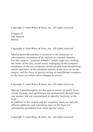
  • 44. The Inner EarMovements of the hair cells in contact with the tectorial membrane transduce mechanical vibrations into electrical signals which generate nerve impulses along the cochlear branch of CN VIII Copyright © John Wiley & Sons, Inc. All rights reserved. The Auditory PathwayThis graphic depicts the events in the stimulation of auditory receptors, from channeling sound waves into the external ear and onto the TM, to the transduction of those vibrations into local receptor potentials Sound waves enter the external auditory canal and strike the eardrum. The vibrations of the eardrum cause the ossicles to vibrate and the stapes pushes the membrane of the oval window in and out. The movement of the oval window sends fluid pressure waves into the perilymph of the scala vestibuli which then transmit them to the scala tympani and eventually to the round window (causing it to bulge outward into the middle ear). The pressure waves move into the endolymph of the cochlear duct and cause the basilar membrane to vibrate which moves the hair cells of the spiral organ against the tectorial membrane. This leads to bending of the stereocilia and ultimately to the generation of nerve impulses in first-order neurons in cochlear nerve fibers. Sound waves of various frequencies cause certain regions of the basilar membrane to vibrate more intensely than other regions. Each segment of the basilar membrane is “tuned” for a *
  • 45. Copyright © John Wiley & Sons, Inc. All rights reserved. Scala vestibuli Cochlear duct (contains endolymph) Scala tympani Perilymph Basilar membrane Cochlea Sound waves Helicotrema Stapes vibrating in oval window Malleus Incus External auditory canal Tympanic membrane Secondary tympanic membrane vibrating in round window Auditory tube Vestibular membrane Middle ear Tectorial membrane Spiral organ (organ of Corti)
  • 46. 1 Scala vestibuli Cochlear duct (contains endolymph) Scala tympani Perilymph Basilar membrane Cochlea Sound waves Helicotrema Stapes vibrating in oval window Malleus Incus External auditory canal
  • 47. Tympanic membrane Secondary tympanic membrane vibrating in round window Auditory tube Vestibular membrane Middle ear Tectorial membrane Spiral organ (organ of Corti) 1 2 Scala vestibuli Cochlear duct (contains endolymph)
  • 48. Scala tympani Perilymph Basilar membrane Cochlea Sound waves Helicotrema Stapes vibrating in oval window Malleus Incus External auditory canal Tympanic membrane Secondary tympanic membrane vibrating in round window Auditory tube Vestibular membrane Middle ear Tectorial membrane Spiral organ (organ of Corti)
  • 49. 1 2 3 Scala vestibuli Cochlear duct (contains endolymph) Scala tympani Perilymph Basilar membrane Cochlea Sound waves Helicotrema Stapes vibrating in oval window Malleus Incus External auditory canal Tympanic membrane Secondary tympanic membrane vibrating in round window Auditory tube Vestibular membrane
  • 50. Middle ear Tectorial membrane Spiral organ (organ of Corti) 1 2 3 4 Scala vestibuli Cochlear duct (contains endolymph) Scala tympani Perilymph Basilar membrane
  • 51. Cochlea Sound waves Helicotrema Stapes vibrating in oval window Malleus Incus External auditory canal Tympanic membrane Secondary tympanic membrane vibrating in round window Auditory tube Vestibular membrane Middle ear Tectorial membrane Spiral organ (organ of Corti)
  • 52. 1 2 3 4 5 Scala vestibuli Cochlear duct (contains endolymph) Scala tympani Perilymph Basilar membrane Cochlea Sound waves Helicotrema Stapes vibrating in oval window Malleus Incus External auditory canal Tympanic membrane Secondary tympanic membrane vibrating in round window Auditory tube Vestibular membrane Middle ear Tectorial membrane Spiral organ
  • 53. (organ of Corti) 1 2 3 4 5 6 Scala vestibuli Cochlear duct (contains endolymph) Scala tympani Perilymph Basilar membrane Cochlea
  • 54. Sound waves Helicotrema Stapes vibrating in oval window Malleus Incus External auditory canal Tympanic membrane Secondary tympanic membrane vibrating in round window Auditory tube Vestibular membrane Middle ear Tectorial membrane Spiral organ (organ of Corti) 1
  • 55. 2 3 4 5 6 7 Scala vestibuli Cochlear duct (contains endolymph) Scala tympani Perilymph Basilar membrane Cochlea Sound waves Helicotrema Stapes vibrating in oval window Malleus Incus External auditory canal Tympanic membrane Secondary tympanic membrane vibrating in round window Auditory tube Vestibular membrane Middle ear Tectorial membrane
  • 56. Spiral organ (organ of Corti) 1 2 3 4 5 6 7 8 8 Scala vestibuli Cochlear duct (contains endolymph) Scala tympani
  • 57. Perilymph Basilar membrane Cochlea Sound waves Helicotrema Stapes vibrating in oval window Malleus Incus External auditory canal Tympanic membrane Secondary tympanic membrane vibrating in round window Auditory tube Vestibular membrane Middle ear Tectorial membrane Spiral organ (organ of Corti)
  • 58. 1 2 3 4 5 6 7 8 8 9 Copyright © John Wiley & Sons, Inc. All rights reserved. The Auditory PathwayThe cell bodies of the sensory neurons are located in the spiral ganglia. Nerve impulses pass along the axons of these neurons, which form the cochlear branch of the vestibulo- cochlear (VIII) nerve Copyright © John Wiley & Sons, Inc. All rights reserved. The Auditory PathwayThe nerve impulses follow CN VIII en route to the medulla, pons, midbrain, and thalamus, and finally to the primary auditory cortex in the temporal lobe. Slight differences in the timing of nerve impulses arriving from the two ears at the superior
  • 59. olivary nuclei in the pons allow us to locate the source of a sound Copyright © John Wiley & Sons, Inc. All rights reserved. EquilibriumEquilibrium is another function of the inner ear - controlled by the vestibular apparatus (the saccule and utricle of the vestibule, and the 3 semicircular canals) Static equilibrium refers to a state of balance relative to the force of gravity Dynamic equilibrium involves the maintenance of balance during sudden movements * Copyright © John Wiley & Sons, Inc. All rights reserved. Static EquilibriumStatic equilibrium is controlled by the sensory hairs within the macula of the utricle and saccule * Copyright © John Wiley & Sons, Inc. All rights reserved.
  • 60. Static EquilibriumAn otolithic membrane, studded with dense calcium carbonate crystals (otoliths), responds to gravity when head position is changed This movement opens transduction channels in the hair cells, producing local potentials which summate to form nerve AP * Copyright © John Wiley & Sons, Inc. All rights reserved. Dynamic EquilibriumDynamic equilibrium is controlled by the sensory hairs within the ampulla of the semicircular canals Within each ampulla is a small elevation called the crista * Copyright © John Wiley & Sons, Inc. All rights reserved. Dynamic EquilibriumEach crista contains hair cells and supporting cells covered by gelatinous material called the cupula With movement, the endolymph within the ampulla lags behind the moving cupola, causing a difference in the inertial forces –
  • 61. the hair bundle of the cupola bends and nerve impulses are generated Copyright © John Wiley & Sons, Inc. All rights reserved. Equilibrium PathwayOnce generated, nerve impulse travel up the vestibular branch of CN VIII. Most of these axons synapse in the major integrating centers for equilibrium, in the medulla and pons, which also receive input from the eyes and proprioceptors parietal lobe to provide us with conscious awareness of the position and movements of the head and limbs * Copyright © John Wiley & Sons, Inc. All rights reserved. Homeostatic ImbalancesA cataract is an opaque defect in the cornea or lens of the eye – most cataracts are in the lens Cataracts are causes by injury, medications, and diseases like diabetes. They are common in old ageConjunctivitis is an inflammation of the conjunctival membrane which covers part of the front of the eye Conjunctivitis is caused most frequently by viral infections (pink eye) and allergy. It can also result from bacterial infections and many other irritants Copyright © John Wiley & Sons, Inc. All rights reserved.
  • 62. Homeostatic ImbalancesAge Related Macular Degeneration results in a loss of vision in the center of the visual field (the macula) because of damage to the retina. It is a major cause of visual impairment in older adults (>50 years) It can become impossible to recognize faces, yet enough peripheral vision remains to allow other activities of daily life Copyright © John Wiley & Sons, Inc. All rights reserved. Homeostatic ImbalancesMyringitis is an inflammation of the ear drum Infections of the middle ear cavity (otitis media) are common in children between 6 mo. – 5 yrs. old, and usually presents with a crying child and a TM (viewed through an otoscope), that looks angry, red, and bulgingOtitis externa (commonly called “swimmer’s ear”) is a dermatitis of the epithelium of the outer ear (infectious and noninfectious). The chlorine, water, and ear plugs associated with swimming can result in irritated, inflamed tissues of the outer ear and ear canal Copyright © John Wiley & Sons, Inc. All rights reserved. Distended Eardrum Caused by Otitis Media Dr. P. Marazzi/Photo Researchers, Inc. Copyright © John Wiley & Sons, Inc. All rights reserved.
  • 63. Homeostatic ImbalancesMeniere’s disease is a disorder of the inner ear that can affect hearing and balance, and is thought to be due to increased pressure in the cochlea and semicircular canals (extra endolymph) Episodes of vertigo (the room spinning) and ringing in the ears (tinnitus) can be a mild annoyance, or a chronic, disabling disability Copyright © John Wiley & Sons, Inc. All rights reserved. End of Chapter 17 Copyright 2012 John Wiley & Sons, Inc. All rights reserved. Reproduction or translation of this work beyond that permitted in section 117 of the 1976 United States Copyright Act without express permission of the copyright owner is unlawful. Request for further information should be addressed to the Permission Department, John Wiley & Sons, Inc. The purchaser may make back-up copies for his/her own use only and not for distribution or resale. The Publisher assumes no responsibility for errors, omissions, or damages caused by the use of these programs or from the use of the information herein. Copyright © John Wiley & Sons, Inc. All rights reserved. Chapter 16 Sensory, Motor, and Integrative Systems Copyright © John Wiley & Sons, Inc. All rights reserved. In this chapter we explore the levels and components of pathways that convey sensory nerve impulses from the body to the brain, and the general sensations (somatic and visceral) that
  • 64. result. We will also examine the activation of motor pathways and movements In chapter 17 we will look at the special senses of sight, hearing, taste, and smell General Sensations Copyright © John Wiley & Sons, Inc. All rights reserved. General SensationsAs sensory impulses reach the CNS, they become part of a large pool of sensory input (though not every one will elicit a response) Each piece of incoming information is combined with other arriving and previously stored information in a process called integration Copyright © John Wiley & Sons, Inc. All rights reserved. General Sensations Integration occurs at many places along pathways in the spinal cord, brain stem, cerebellum, basal nuclei, and cerebral cortex. Sensations result in and evoke a conscious perception or subconscious awareness that changes have occurred in the external or internal environment. The motor responses are also modified at several of these levels.
  • 65. Copyright © John Wiley & Sons, Inc. All rights reserved. General SensationsExamples of complex integrative functions of the brain include wakefulness and sleep, and learning and memory Copyright © John Wiley & Sons, Inc. All rights reserved. Sensory ModalitiesEach unique type of sensation is called a sensory modality, and a given sensory neuron carries information for only one modality, be it somatic, visceral, or “special” Somatic senses include tactile sensations (touch, pressure, vibration, itch, and tickle), thermal sensations (warm and cold), pain sensations, and proprioception (awareness of limb and joint position in space) Visceral senses provide information about conditions within internal organs Copyright © John Wiley & Sons, Inc. All rights reserved. Sensory ModalitiesThe process of sensation begins in a sensory receptor, which can be either a specialized cell or the dendrites of a sensory neuron A particular kind of stimulus (a change in the environment) activates certain sensory receptors, while other sensory receptors respond only weakly or not at all – a characteristic known as selectivity Copyright © John Wiley & Sons, Inc. All rights reserved. Sensory ModalitiesFor a sensation to arise, four events typically
  • 66. occur: Stimulation of the sensory receptor - an appropriate stimulus must occur within the receptor’s receptive field Transduction of the stimulus - a sensory receptor converts energy in a stimulus into a graded potential. Recall that graded potentials (but not APs) vary in amplitude depending on the strength of the stimulus that causes them, and are not propagated Sensory receptors produce two different kinds of graded potentials—generator potentials and receptor potentials—in response to a stimulus. When stimulated, the dendrites of free nerve endings, encapsulated nerve endings, and the receptive part of olfactory receptors produce a generator potential (Figure 16.1a, b). When a generator potential is large enough to reach threshold, it triggers one or more nerve impulses in the axon of a first-order sensory neuron. The resulting nerve impulse propagates along the axon into the CNS. Thus, generator potentials generate action potentials. By contrast, sensory receptors that are separate cells produce graded potentials termed receptor potentials. * Copyright © John Wiley & Sons, Inc. All rights reserved. Sensory ModalitiesThe four events that bring about a sensation, cont’d… Generation of nerve impulses – occurs when the sum of graded potentials reach threshold in first-order neurons (the first neuron in a specific tract – in this case from the PNS into the CNS) Integration of sensory input – occurs when a particular region of the CNS integrates a number (and even a variety) of sensory nerve impulses and results in a conscious sensations or
  • 67. perceptions Copyright © John Wiley & Sons, Inc. All rights reserved. Sensory Modalities Copyright © John Wiley & Sons, Inc. All rights reserved. Sensory ReceptorsSensory receptors can be grouped into several classes based on structural and functional characteristics: Microscopic structure – free nerve endings vs encapsulated endings, for example Location…of the receptors and the origin of the stimuli that activate them The type of stimulus detected (nociceptors for pain, mechanoreceptors for pressure, etc.) Interoceptors (visceroceptors) are located in blood vessels, visceral organs, muscles, and the nervous system and monitor conditions in the internal environment. The nerve impulses produced by interoceptors usually are not consciously perceived; occasionally, however, activation of interoceptors by strong stimuli may be felt as pain or pressure. * Copyright © John Wiley & Sons, Inc. All rights reserved. Sensory ReceptorsSensory receptors can be grouped into several classes based on structural and functional characteristics: Microscopic structure – free nerve endings vs encapsulated
  • 68. endings, for example Location…of the receptors and the origin of the stimuli that activate them – exteroceptors near the external surface vs interoceptors (visceroceptors), for example The type of stimulus detected (nociceptors for pain, mechanoreceptors for pressure, etc.) Interoceptors (visceroceptors) are located in blood vessels, visceral organs, muscles, and the nervous system and monitor conditions in the internal environment. The nerve impulses produced by interoceptors usually are not consciously perceived; occasionally, however, activation of interoceptors by strong stimuli may be felt as pain or pressure. * Copyright © John Wiley & Sons, Inc. All rights reserved. Sensory ReceptorsReceptors named according to their location include: Exteroceptors, which are located at or near the external surface of the body and respond to external stimuli Interoceptors (visceroceptors), which are located in blood vessels, organs, and muscles and produce impulses which usually are not consciously perceived Proprioceptors, which are located in muscles, tendons, joints, and the inner ear. They provide information about body position and movement of joints Exteroceptors provide information about the external environment. Interoceptors monitor the internal environment. Occasionally
  • 69. activation of interoceptors by strong stimuli may be felt as pain or pressure. * Copyright © John Wiley & Sons, Inc. All rights reserved. Sensory ReceptorsReceptors can also denote the type of stimulus that excites them * Copyright © John Wiley & Sons, Inc. All rights reserved. Sensory ReceptorsReceptors named according to mode of activation are: Mechanoreceptors, which are sensitive to deformation Thermoreceptors, which detect changes in temperature Nociceptors, which respond to painful stimuli Photoreceptors, which are activated by photons of light Chemoreceptors, which detect chemicals in the mouth (taste), nose (smell) and body fluids Osmoreceptors, which detect the osmotic pressure of body fluids Mechanoreceptors provide sensations of touch, pressure, vibration, proprioception, and hearing and equilibrium. They also monitor the stretching of blood vessels and internal organs. * Copyright © John Wiley & Sons, Inc. All rights reserved.
  • 70. Sensory ReceptorsA characteristic feature of most sensory receptors is adaptation, in which the generator potential or receptor potential decreases in amplitude during a sustained or constant stimulus Because there is an accommodation response at the receptor level, the frequency of nerve impulses traveling to the cerebral cortex decreases and the perception of the sensation fades even though the stimulus persistsreceptors vary in how quickly they adapt (rapidly adapting and slowly adapting receptors) Receptors associated with pressure, touch, and smell are rapidly adapting. * Copyright © John Wiley & Sons, Inc. All rights reserved. MechanoreceptionMany of the mechanoreceptors and nociceptors previously described are located in the skin * Copyright © John Wiley & Sons, Inc. All rights reserved. MechanoreceptionThis graphic illustrates some representative examples of general somatic mechanoreceptors and the first- order neurons to which they belong. Receptors for special senses are not shown.
  • 71. * Copyright © John Wiley & Sons, Inc. All rights reserved. All of our sensory modalities are important, but pain serves a protective function and is indispensable for survival Nociceptors are chemoreceptive free nerve endings activated by tissue damage from intense thermal, mechanical, or chemical stimuli - they’re found in every tissue of the body except the brain Nociception Copyright © John Wiley & Sons, Inc. All rights reserved. NociceptionThere are two types of pain: fast and slow The perception of fast pain (acute, well localized) occurs rapidly because the nerve impulses propagate along medium- diameter, myelinated A fibers By contrast, slow pain begins after a stimulus is applied and gradually increases in intensity over a period of several seconds or minutes. Impulses for slow pain conduct along small- diameter, unmyelinated C fibers. This type of pain may be excruciating and often has a burning, aching, or throbbing quality An example of slow pain is the pain associated with a toothache. * Copyright © John Wiley & Sons, Inc. All rights reserved.
  • 72. NociceptionPain that arises from stimulation of receptors in the skin is called superficial somatic pain; stimulation of receptors in skeletal muscles, joints, tendons, and fascia causes deep somatic painVisceral pain results from stimulation of nociceptors in visceral organs In many instances of visceral pain, the pain is felt in or just deep to the skin that overlies the stimulated organ, or in a surface area far from the stimulated organ. This phenomenon is called referred pain * Copyright © John Wiley & Sons, Inc. All rights reserved. NociceptionCommon patterns of referred visceral pain are shown in this graphic * Copyright © John Wiley & Sons, Inc. All rights reserved. ProprioceptionMuscle spindles are the proprioceptors in skeletal muscles that monitor changes in the muscle length and participate in stretch reflexes By adjusting how vigorously a muscle spindle responds to stretching of a skeletal muscle, the brain sets an overall level of muscle tone (the small degree of contraction that is present while the muscle is at rest)
  • 73. Copyright © John Wiley & Sons, Inc. All rights reserved. Each muscle spindle consists of several slowly adapting sensory nerve endings that wrap around 3 to 10 specialized muscle fibers. A connective tissue capsule encloses the sensory nerve endings and anchors the spindle to the endomysium and perimysium Muscle spindles are plentiful in muscles that control fine movements and much more sparse in those that control course or forceful movements Proprioception Free nerve endings and Ruffini corpuscles in the capsules of joints respond to pressure. Pacinian corpuscles respond to acceleration and deceleration of joints during movement. * Copyright © John Wiley & Sons, Inc. All rights reserved. Somatic Sensory PathwaysNo matter the type of receptor on the receiving end (where the generator potential is set up), first- order somatosensory neurons are unipolar in structure This means that their cell body is located in the dorsal root ganglia (DRG) just outside the CNS Their other end terminates nearby in the posterior gray horns of the cord, usually at the level where they enter
  • 74. Mechanoreceptors provide sensations of touch, pressure, vibration, proprioception, and hearing and equilibrium. They also monitor the stretching of blood vessels and internal organs. * Copyright © John Wiley & Sons, Inc. All rights reserved. Second-order neurons conduct ascending impulses from the brain stem where their axons decussate (cross over to the opposite side) before ascending to the thalamus Thus, all somatic sensory information from one side of the body reaches the thalamus on the opposite side Somatic Sensory Pathways * Copyright © John Wiley & Sons, Inc. All rights reserved. Somatic Sensory PathwaysThird-order neurons conduct impulses from the thalamus to the primary somatosensory area of the cortex on the same side. Copyright © John Wiley & Sons, Inc. All rights reserved. Somatic Sensory PathwaysSomatic sensory neurons (and their axons that convey somatic sensations) are not distributed evenly in the body
  • 75. The peripheral areas with the highest density are represented in the brain with the largest amount of gray matter in the sensory homunculus. The most sensitive areas in the body are therefore the tip of the tongue, lips, and fingertips * Copyright © John Wiley & Sons, Inc. All rights reserved. There are two major spinocerebellar tracts in the spinal cord that carry proprioceptive impulses to the cerebellum Although they are not consciously perceived, sensory impulses sent to the cerebellum along these two pathways are critical for posture, balance, and coordination of skilled movements Somatic Sensory Pathways * Copyright © John Wiley & Sons, Inc. All rights reserved. Motor activity begins in the primary motor areas of the precentral gyrus and other cerebral integrative centers Any motor neuron that is not directly responsible for stimulating target
  • 76. muscles is called an upper motor neuron (UMN)UMNs connect the brain to the appropriate level in the spinal cord Somatic Motor Pathways The basal nuclei and cerebellum influence movement through their effects on upper motor neurons. * Copyright © John Wiley & Sons, Inc. All rights reserved. From there, all excitatory and inhibitory signals that control movement converge on second-order motor neurons known as lower motor neurons (LMNs) that descend to innervate skeletal muscle Since only LMNs provide output from the CNS to skeletal muscle fibers they are also called the final common pathway Somatic Motor Pathways * Copyright © John Wiley & Sons, Inc. All rights reserved. Axons of LMNs extend through cranial nerves to the skeletal muscles of the face and head, and through spinal nerves to innervate skeletal muscles of the limbs and trunk Two of the major
  • 77. LMN tracts are the lateral and anterior corticospinal tracts Somatic Motor Pathways Only LMNs provide output from the CNS to skeletal muscle fibers. For this reason, they are also called the final common pathway. * Copyright © John Wiley & Sons, Inc. All rights reserved. Sensory and Motor Pathways (Interactions Animation)Somatic Sensory Pathways You must be connected to the internet to run this animation * Copyright © John Wiley & Sons, Inc. All rights reserved. End of Chapter 16 Copyright 2011 John Wiley & Sons, Inc. All rights reserved. Reproduction or translation of this work beyond that permitted in section 117 of the 1976 United States Copyright Act without express permission of the copyright owner is unlawful. Request for further information should be addressed to the Permission Department, John Wiley & Sons, Inc. The purchaser may make back-up copies for his/her own use only and not for distribution or resale. The Publisher assumes no responsibility for errors, omissions, or damages caused by the use of these programs or
  • 78. from the use of the information herein. Chapter 15 The Autonomic Nervous System Lecture slides prepared by Curtis DeFriez, Weber State University Copyright © John Wiley & Sons, Inc. All rights reserved. Introduction to the ANS In this chapter, we examine the structural and functional features of the autonomic nervous system (ANS) and compare the organization and actions of its two major parts, the sympathetic and parasympathetic divisions. The autonomic nervous system contributes to homeostasis by responding to subconscious visceral sensations and exciting or inhibiting smooth muscle, cardiac muscle, and many glands. Copyright © John Wiley & Sons, Inc. All rights reserved. Introduction to the ANS Structurally, the ANS includes autonomic sensory neurons, integrating centers in the CNS, and autonomic motor neurons. The enteric division is a specialized network of nerves and ganglia forming an independent nerve network within the wall of the gastrointestinal (GI) tract. The enteric division will not be further discussed in this chapter, but we will return to it in Chapter 24.
  • 79. Copyright © John Wiley & Sons, Inc. All rights reserved. Introduction to the ANS While both the ANS and the somatic nervous system (SNS) include sensory and motor neurons, the ANS has many distinctive features which set it apart. Perhaps the biggest difference between these two systems is the involvement of conscious control. In the SNS, feedback via tactile, thermal, pain, and proprioceptive sensations are consciously perceived, and skeletal muscle is the main tool used to provide reflexive and voluntary movement. Copyright © John Wiley & Sons, Inc. All rights reserved. Introduction to the ANS If a somatic motor neuron ceases to stimulate a muscle, the result is a paralyzed, limp muscle that has no tone. Although we are generally not conscious of breathing, the muscles that generate respiratory movements are skeletal muscles controlled by somatic motor neurons. If the respiratory motor neurons become inactive, breathing stops. Copyright © John Wiley & Sons, Inc. All rights reserved. 5 Introduction to the ANS
  • 80. The ANS usually operates without conscious control, though centers in the hypothalamus and brain stem do provide regulation for ANS reflexes. Sensory receptors called interoceptors located in blood vessels, visceral organs, muscles, and the nervous system monitor conditions in the internal environment. Examples of interoceptors are chemoreceptors that monitor blood CO2 level and mechanoreceptors that detect the degree of stretch in the walls of organs or blood vessels. Copyright © John Wiley & Sons, Inc. All rights reserved. ANS Motor Pathways Autonomic motor neurons regulate visceral activities by either increasing (exciting) or decreasing (inhibiting) ongoing activities in their effector tissues. Because autonomic responses cannot be consciously altered to any great degree, some autonomic responses are the basis for polygraph (“lie detector”) tests. However, practitioners of yoga and biofeedback techniques may learn how to regulate at least some of their autonomic activities through long practice. Copyright © John Wiley & Sons, Inc. All rights reserved. 7 Introduction to the ANS The anatomy of all autonomic pathways can best be understood by picturing a double-barrelled neuronal construct consisting of a preganglionic neuron
  • 81. leading to an intermediate ganglion that contains the cell bodies of post- ganglionic neurons (that innervate an effector). Copyright © John Wiley & Sons, Inc. All rights reserved. 8 Introduction to the ANS Copyright © John Wiley & Sons, Inc. All rights reserved. Introduction to the ANS Interactions Animation The ANS: An Introduction Animation You must be connected to the internet to run this animation Copyright © John Wiley & Sons, Inc. All rights reserved. 10 Most body organs have dual ANS innervation; that is, they
  • 82. receive impulses from both sympathetic and parasympathetic neurons. Usually the nerve impulses from one division stimulate an organ, while impulses from the other division decrease activity. Divisions of the ANS Copyright © John Wiley & Sons, Inc. All rights reserved. Divisions of the ANS Copyright © John Wiley & Sons, Inc. All rights reserved. Divisions of the ANS Copyright © John Wiley & Sons, Inc. All rights reserved. Divisions of the ANS Furthermore, the responses of the various organs to ANS stimulation neatly group into two functional categories : Like children on a teeter-totter, the sympathetic divisions “fight or flight” response is balanced against the “rest and relax” (or rest and digest) activities of the parasympathetic division. sympathetic parasympathetic
  • 83. replace graphic Copyright © John Wiley & Sons, Inc. All rights reserved. The Sympathetic Division The cell bodies of neurons which participate in motor responses of the sympathetic nervous system are located in the lateral horns of the gray matter in the 12 thoracic segments and the first two lumbar segments of the cord. Sympathetic preganglionic neurons exit the spinal cord only between levels T1-L2 (hence the name thoracolumbar division), though sympathetic ganglia extend in the vicinity of the cord from the cervical to the sacral region. Copyright © John Wiley & Sons, Inc. All rights reserved. Some of the major groups of sympathetic ganglia include: The sympathetic trunk (vertebral chain) ganglia Prevertebral ganglia The celiac, superior mesenteric, inferior mesenteric, aorticorenal and renal ganglia The Sympathetic Division Copyright © John Wiley & Sons, Inc. All rights reserved.
  • 84. The Sympathetic Division Axons leave the sympathetic trunk in four possible ways: They can enter and travel with spinal nerves. They can form fine networks of periarterial preganglionic traveling cephalad to synapse in the cervical ganglia. Postganglionic axons exiting the sympathetic trunk can form sympathetic nerves to the heart and lungs. Preganglionic axons can leave the sympathetic trunk without synapsing and form splanchnic nerves. Copyright © John Wiley & Sons, Inc. All rights reserved. Gray ramus: Axons of some postganglionic neurons leave the sympathetic trunk by entering a short pathway called a gray ramus and merge with the anterior ramus of a spinal nerve. Gray rami communicantes: structures containing sympathetic postganglionic axons that connect the ganglia of the sympathetic trunk to spinal nerves. 17 The Sympathetic Division Major groups of sympathetic ganglia. Copyright © John Wiley & Sons, Inc. All rights reserved. The Sympathetic Division
  • 85. A truism of the sympathetic division is that a single sympathetic preganglionic fiber synapses with many postganglionic branches (with 20 or more) to create a diverging circuit. The postganglionic axons typically terminate in several different visceral effectors, making the effects of sympathetic stimulation a widespread massive response. This is why anger can be hard to control – it is such a diffuse response. Copyright © John Wiley & Sons, Inc. All rights reserved. The Sympathetic Division This schematic illustrates the outflow of the sympathetic division of the ANS via thoracolumbar pathways to the many organs of the body. Copyright © John Wiley & Sons, Inc. All rights reserved. The Parasympathetic Division The cell bodies of preganglionic neurons which participate in motor responses of the parasympathetic nervous system are located in nuclei of 4 cranial nerves in the brainstem (III, VII, IX and X) and in the lateral gray matter of sacral areas of the spinal cord (S2-S4). The vagus nerve (CN X) carries nearly 80% of the total parasympathetic flow to the organs of the thorax and upper
  • 86. abdomen. Lower abdominal and pelvic organs are innervated by the sacral output. Copyright © John Wiley & Sons, Inc. All rights reserved. The Parasympathetic Division Parasympathetic ganglia are called terminal ganglia because they are located far from their origin at the “terminal” ends of the pathways (near the target organs). Four pairs of cranial parasympathetic ganglia innervate structures in the head: The ciliary, pterygopalatine, submandibular, and otic ganglia. The cranial-sacral division also has the ganglia associated with the vagus (X) nerve and the sacral nerves. Copyright © John Wiley & Sons, Inc. All rights reserved. Most of the parasympathetic ganglia are located very close to the organs or intended action. Copyright © John Wiley & Sons, Inc. All rights reserved. The sacral preganglionic axons branch off of sacral spinal
  • 87. nerves to form pelvic splanchnic nerves which synapse with parasympathetic postganglionic neurons located in terminal ganglia in the walls of the innervated viscera. From the terminal ganglia, postganglionic axons innervate smooth muscle and glands in the walls of the colon, ureters, urinary bladder, and reproductive organs The Parasympathetic Division Copyright © John Wiley & Sons, Inc. All rights reserved. The Parasympathetic Division In contrast to the sympathetic system, the parasympathetic response is more controlled. Presynaptic parasympathetic neurons usually synapse with only 4–5 postsynaptic neurons, all of which supply a single visceral effector. Parasympathetic stimulation leads to a narrow, focused action on specific organs. This is why it is possible to walk and chew gum at the same time (not really!) Copyright © John Wiley & Sons, Inc. All rights reserved. The division of the sympathetic and parasympathetic divisions of the ANS are compared in Table 15.3
  • 88. Copyright © John Wiley & Sons, Inc. All rights reserved. ANS Neurotransmitters The total number of neurotransmitters used in the entire nervous system is not known, but is well over 100. Despite the variety of possible chemicals that could be used to transmit chemical messages in the ANS, only 2, acetylcholine and norepinephrine, are used to any great degree. Synapses at which ACh is used are termed cholinergic. Synapses at which norepinephrine or epinephrine are used are termed adrenergic. Copyright © John Wiley & Sons, Inc. All rights reserved. ANS Neurotransmitters The neurotransmitter used in all of the synapses of sympathetic and parasympathetic ganglia (between the synapses of the preganglionic and postganglionic fibers) is acetylcholine. Receptors that respond to Ach released by these cholinergic neurons are called cholinergic receptors and there are 2 subtypes: nicotinic receptors (found in the ganglia) and muscarinic receptors (found in the synapses with the effector organs). Copyright © John Wiley & Sons, Inc. All rights reserved. There are also two subtypes of receptors that respond to norepinephrine: alpha and beta receptors that are widely scattered throughout the body. Even these subtypes of adrenergic receptors can be further subtyped.
  • 89. 28 ANS Neurotransmitters Acetylcholine acts on a sub-type of cholinergic receptor (called nicotinic receptors) at ganglia of the ANS. Copyright © John Wiley & Sons, Inc. All rights reserved. ANS Neurotransmitters The neurotransmitter used at most sympathetic postganglionic synapses is norepinephrine. The exception to this rule is that ACh is used at sympathetic postganglionic synapses for sweat glands. Copyright © John Wiley & Sons, Inc. All rights reserved. ANS Neurotransmitters The neurotransmitter used at all parasympathetic postganglionic synapses is Ach. These are all a variety of cholinergic receptors called muscarinic.
  • 90. Copyright © John Wiley & Sons, Inc. All rights reserved. ANS NeurotransmittersNeurons and Neurotransmitters of the Parasympathetic Nervous SystemPreganglionicPostganglionicCell body in brain or spinal cordCell body in intramural ganglionAcetylcholine (ACh)Acetylcholine (ACh) Copyright © John Wiley & Sons, Inc. All rights reserved. ANS NeurotransmittersNeurons and Neurotransmitters of the Sympathetic Nervous SystemPreganglionicPostganglionicoCell body in lateral horn of ospinal cordCell body in sympathetico chain ganglionooAcetylcholine (ACh)(norepinephrine, NE) ol except sweat glands (Ach) o Copyright © John Wiley & Sons, Inc. All rights reserved. ANS Neurotransmitters Interactions Animation ANS Neurotransmitters and Neurons Animation You must be connected to the internet to run this animation Copyright © John Wiley & Sons, Inc. All rights reserved.
  • 91. 34 Physiology of the ANS Sympathetic stimulation leads to secretion of norepinephrine by the adrenal glands, an increase in the rate and strength of the heartbeat, constriction of blood vessels of non-essential organs, dilation of vessels of essential organs (skeletal muscle and the cerebral cortex), an increase in the rate and depth of breathing, hepatic conversion of glycogen to glucose, and decrease in GI activity. Copyright © John Wiley & Sons, Inc. All rights reserved. Physiology of the ANS Copyright © John Wiley & Sons, Inc. All rights reserved. Physiology of the ANS Copyright © John Wiley & Sons, Inc. All rights reserved. Physiology of the ANS
  • 92. Copyright © John Wiley & Sons, Inc. All rights reserved. Physiology of the ANS SLUDD is as an acronym used to describe the responses of the parasympathetic nervous system: Salivation (increased) Lacrimation (increased) Urination (increased) Digestion (increased) Defecation (increased) … and 3 decreases (in the rate and force of the heart beat, airway size and rate of breathing, and pupil size) Copyright © John Wiley & Sons, Inc. All rights reserved. Physiology of the ANS Interactions Animation The balance of autonomic sympathetic-parasympathetic tone is regulated by feedback loops between the spinal cord and brainstem, with input from the limbic system and oversight by the hypothalamus. Physiological Effects of the ANS Animation You must be connected to the internet to run this animation Copyright © John Wiley & Sons, Inc. All rights reserved. End of Chapter 15 Copyright 2012 John Wiley & Sons, Inc. All rights reserved.
  • 93. Reproduction or translation of this work beyond that permitted in section 117 of the 1976 United States Copyright Act without express permission of the copyright owner is unlawful. Request for further information should be addressed to the Permission Department, John Wiley & Sons, Inc. The purchaser may make back-up copies for his/her own use only and not for distribution or resale. The Publisher assumes no responsibility for errors, omissions, or damages caused by the use of these programs or from the use of the information herein. Copyright © John Wiley & Sons, Inc. All rights reserved.