1 orthopaedics

At the end of the unit, the student will be able to: 1. Apply knowledge regarding: patho-physiology, disease process, clinical manifestations, specific diagnostic and therapeutic interventions (diagnostic tests and examinations) 2. Distinguish between the different health problems: medical and surgical conditions of various body systems: Congenital defects, Amputation, Osteoarthritis, Rheumatoid arthritis, Juvenile chronic, Arthritis & Osteoporosis 3. Assess, relate and apply the scientific process of nursing, provision and facilitation of nursing care 4. Evaluate, analyse and solve problems in familiar and unfamiliar context in the Comprehensive Health Care system 5. Understand the relationship between social, cultural and economic factors that may impact significantly on the health status of clients / patients and groups. (Health education) 6. Apply knowledge of emergency and trauma management principles Trauma to the muscular skeletal system

Recommended
More Related Content
What's hot
What's hot (20)
Similar to 1 orthopaedics
Similar to 1 orthopaedics (20)
More from Chantal Settley
More from Chantal Settley (20)
Recently uploaded
Recently uploaded (20)
1 orthopaedics
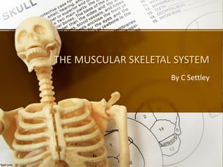
- 1. THE MUSCULAR SKELETAL SYSTEM By C Settley
- 2. At the end of the unit, the student will be able to: 1. Apply knowledge regarding: patho-physiology, disease process, clinical manifestations, specific diagnostic and therapeutic interventions (diagnostic tests and examinations) 2. Distinguish between the different health problems: medical and surgical conditions of various body systems: Congenital defects, Amputation, Osteoarthritis, Rheumatoid arthritis, Juvenile chronic, Arthritis & Osteoporosis 3. Assess, relate and apply the scientific process of nursing, provision and facilitation of nursing care 4. Evaluate, analyse and solve problems in familiar and unfamiliar context in the Comprehensive Health Care system 5. Understand the relationship between social, cultural and economic factors that may impact significantly on the health status of clients / patients and groups. (Health education) 6. Apply knowledge of emergency and trauma management principles Trauma to the muscular skeletal system 4/23/2018 Compiled by C Settley 2
- 3. What is the muscular skeletal system? • The musculoskeletal system provides form, support, stability, and movement to the body. • It is made up of the bones of the skeleton, muscles, cartilage, tendons, ligaments, joints, and other connective tissue that supports and binds tissues and organs together. 4/23/2018 Compiled by C Settley 3
- 4. 4/23/2018 Compiled by C Settley 4
- 5. 4/23/2018 Compiled by C Settley 5
- 6. The raduis and ulna 4/23/2018 Compiled by C Settley 6
- 7. Where is cartilage found in the body? 4/23/2018 Compiled by C Settley 7
- 8. 4/23/2018 Compiled by C Settley 8
- 9. 4/23/2018 Compiled by C Settley 9
- 10. 4/23/2018 Compiled by C Settley 10
- 11. 4/23/2018 Compiled by C Settley 11
- 12. Foot muscles 4/23/2018 Compiled by C Settley 12
- 13. 4/23/2018 Compiled by C Settley 13
- 14. 4/23/2018 Compiled by C Settley 14
- 15. 4/23/2018 Compiled by C Settley 15
- 16. 4/23/2018 Compiled by C Settley 16
- 17. Congenital deformities of the foot pg. 916 • Genu varum – Genu varum is a deformity marked by (outward) bowing at the knee, which means that the lower leg is angled inward (medially) in relation to the thigh's axis, giving the limb overall the appearance of an archer's bow. – Normal and resolves spontaneously 4/23/2018 Compiled by C Settley 17
- 18. Congenital deformities of the foot pg. 916 • Genu valgum – commonly called "knock-knee", is a condition in which the knees angle in and touch each other when the legs are straightened. – Individuals with severe valgus deformities are typically unable to touch their feet together while simultaneously straightening the legs. – Corrects Spontaneously 4/23/2018 Compiled by C Settley 18
- 19. Congenital deformities of the foot pg. 916 • Talipes equinovarus – A birth defect in which the foot is twisted out of shape or position. – Involves bony structures of the foot. – Adduction and supination of the forefoot, the heel, the ankle equines and includes medial deviation of the entire foot. – Soft tissue tightening might also be present. – Exact cause is unknown, but thought to be related to genetics and intrauterine positioning. – Treatment consists of gentle manipulation to the desired position and application of casts or tapes. – Treatment successful if started early. – Surgical correction might be required. 4/23/2018 Compiled by C Settley 19
- 20. Congenital deformities of the foot pg. 916 • Talipes equinovarus 4/23/2018 Compiled by C Settley 20
- 21. Congenital deformities of the foot pg. 916 • Pes cavus – Medical term for ‘claw foot’. – Abnormal arch of the foot. – Often there is clawing of the toes. – The foot is short and the bottom of the foot reaches the ground on the heel and metatarsal heads. – Increased pressure and callus formation on those areas that touches the ground. – Exercise may be advised to try and manipulate the foot into dorsi-flexion. – A brace may be ordered. – In severe cases surgery may be required. 4/23/2018 Compiled by C Settley 21
- 22. Congenital deformities of the foot pg. 916 • Pes cavus 4/23/2018 Compiled by C Settley 22
- 23. Congenital deformities of the foot pg. 916 • Pes planus – A condition in which the entire sole of the foot touches the floor when standing. – May go unnoticed. – Arch of the foot has collapsed to such a degree that the medial border of the foot reaches the ground. – May be congenital or acquired (associated with trauma of muscles and ligaments, paralysis, overweight, poor posture, poorly fitting shoes, arthritis) – In some cases it may be painful. – Management includes exercise to improve muscle strength, posture and gait. – An arch support device may be used over time. 4/23/2018 Compiled by C Settley 23
- 24. Congenital deformities of the foot pg. 916 • Pes planus 4/23/2018 Compiled by C Settley 24
- 25. Trauma to the muscular skeletal system • Fractures – pg. 919 – A complete or partial break in the continuity of a bone. 4/23/2018 Compiled by C Settley 25
- 26. Causes of fractures • Trauma – As result of direct force such as a blow or crush as in MVA’s – Indirect violence – Sports and occupational injuries 4/23/2018 Compiled by C Settley 26
- 27. Causes of fractures • Degenerative disorders – Such as those responsible for pathological fractures, which occur in elderly women as a result of osteoporosis (a condition in which bones become weak and brittle) associated with hormonal changes during menopause – Or bone metastasis 4/23/2018 Compiled by C Settley 27
- 28. Pathophysiology of fractures • Break in the continuity of a bone • Which results in the periosteum being stripped off from the bone and blood vessels supplying the bone being severed • The edges of the bone may damage surrounding tissue and blood vessels resulting in bleeding into the bone and tissues, including joint cavities if joints are involved • A haematoma may form increasing swelling in the area 4/23/2018 Compiled by C Settley 28
- 29. Pathophysiology of fractures 4/23/2018 Compiled by C Settley 29
- 30. Pathophysiology of fractures 4/23/2018 Compiled by C Settley 30
- 31. Types of fractures- pg. 919 (Table 47.1) Complete and incomplete fractures refer to the way the bone breaks. In a complete fracture, the bone snaps into two or more parts, In an incomplete fracture, the bone cracks but does not break all the way through. 4/23/2018 Compiled by C Settley 31
- 32. Types of fractures- pg. 919 (Table 47.1) Complete and Incomplete fractures (example of the radius) 4/23/2018 Compiled by C Settley 32
- 33. Types of fractures- pg. 919 (Table 47.1) Closed fractures or simple fractures A closed fracture is a broken bone that does not penetrate the skin. This is an important distinction because when a broken bone penetrates the skin (an open fracture) there is a need for immediate treatment, and an operation is often required to clean the area of the fracture. An open fracture can be defined as a broken bone that is in communication through the skin with the environment. 4/23/2018 Compiled by C Settley 33
- 34. Types of fractures- pg. 919 (Table 47.1) Closed fractures or simple fractures 4/23/2018 Compiled by C Settley 34
- 35. Types of fractures- pg. 919 (Table 47.1) Closed fractures or simple fractures 4/23/2018 Compiled by C Settley 35
- 36. Types of fractures- pg. 919 (Table 47.1) Comminuted fracture A comminuted fracture is a break or splinter of the bone into more than two fragments. 4/23/2018 Compiled by C Settley 36
- 37. Types of fractures- pg. 919 (Table 47.1) Compression fracture Fractures from osteoporosis usually occur in the front (anterior) part of the vertebral body. Osteoporosis is a disease that weakens bone. Sometimes the bones in the spine weaken to the point that even mild forces can lead to a compression fracture. These compression fractures can occur in vertebrae anywhere in the spine, but they tend to occur most commonly in the upper back (thoracic spine), particularly in the lower vertebrae of that section of the spine (e.g. T10, T11, T12). 4/23/2018 Compiled by C Settley 37
- 38. Types of fractures- pg. 919 (Table 47.1) 4/23/2018 Compiled by C Settley 38
- 39. Types of fractures- pg. 919 (Table 47.1) Depressed fracture A depressed skull fracture is a break in a cranial bone (or "crushed" portion of skull) with depression of the bone in toward the brain. 4/23/2018 Compiled by C Settley 39
- 40. Types of fractures- pg. 919 (Table 47.1) Impacted fracture An impacted fracture occurs when the broken ends of the bone are jammed together by the force of the injury or slips over the other fragment. 4/23/2018 Compiled by C Settley 40
- 41. Types of fractures- pg. 919 (Table 47.1) Incomplete fracture/ greenstick fracture An impacted fracture occurs when the broken ends of the bone are jammed together by the force of the injury or slips over the other fragment. Applies in children 4/23/2018 Compiled by C Settley 41
- 42. Types of fractures- pg. 919 (Table 47.1) Pathologic fracture A pathologic fracture (also called insufficiency fracture) is a bone fracture caused by disease that led to weakness of the bone structure. 4/23/2018 Compiled by C Settley 42
- 43. Assessment and common findings • Oedema and swelling due to soft tissue injury with bleeding into the surrounding tissue • Pain and tenderness • Loss of function • Deformity • Discoloration • Crepitation 4/23/2018 Compiled by C Settley 43
- 44. Healing of fractures 1. Haematoma formation – Bleeding around the tissues. – Clotting may also occur. 4/23/2018 Compiled by C Settley 44
- 45. Healing of fractures 2. Granulation tissue – Granulation tissue is new connective tissue and microscopic blood vessels that form on the surfaces of a wound during the healing process. This includes the formation of new cells, fibroblasts and osteoblasts. – A new bone structure, the osteoid is formed. 4/23/2018 Compiled by C Settley 45
- 46. Healing of fractures 3. Callus stage – Eventually, pain and swelling decrease and soft callus is formed. – This corresponds roughly to the time when the fragments are no longer moving freely. – A callus is an area of thickened skin in response to repeated friction, pressure, or other irritation. 4/23/2018 Compiled by C Settley 46
- 47. Healing of fractures 4. Ossification stage - The callus gradually ossified by the continuous action of osteoblasts. The soft bone is replaced by mature bone during this stage, but the fracture can still be seen on X ray. - Ossification (or osteogenesis) in bone remodeling is the process of laying down new bone material by cells called osteoblasts. It is synonymous with bone tissue formation. 4/23/2018 Compiled by C Settley 47
- 48. Healing of fractures 5. Consolidation stage - Ossification continues and more mature bone develops. The gaps between bones fills up. 6. Remodelling stage - Osteoclasts are active during this stage. Removal of unnecessary material takes place. This completes the healing process. 4/23/2018 Compiled by C Settley 48
- 49. Healing of fractures 4/23/2018 Compiled by C Settley 49
- 50. Therapeutic management of fractures • Resuscitation • Reduction of fractures • Immobilising fractures • Restoration of function • Rehabilitation 4/23/2018 Compiled by C Settley 50
- 51. Resuscitation of the patient • Blood loss and pain from the injury • Depending on extent of the fracture • Signs and symptoms of shock (Cool, clammy pale skin, rapid pulse and breathing, nausea and vomiting, enlarged pupils, weakness, dizziness or fainting). • To minimise shock: – Analgesia (Pethidine 25-50 mg IM) – Replace fluid when BP is low and the pulse is weak. – Handle fracture with care. – Temporary splinting may be required. 4/23/2018 Compiled by C Settley 51
- 52. Splinting of fracture 4/23/2018 Compiled by C Settley 52
- 53. Complications of fractures- pg. 921 • Infection – Often occurs in open fractures. – May become serious if it spreads beyond the soft tissue and infects underlying bones causing osteomyelitis. 4/23/2018 Compiled by C Settley 53
- 54. Complications of fractures- pg. 921 • Delayed union – May be due to poor blood supply or inadequate immobilization. • Malunion – Union may occur in the wrong position. • Non union – When the fractures fail to unite. Soft tissue may get interposed between bone ends. A bone graft mat be necessary to stimulate new bone growth. 4/23/2018 Compiled by C Settley 54
- 55. Complications of fractures- pg. 921 4/23/2018 Compiled by C Settley 55
- 56. Complications of fractures- pg. 921 • Fat embolism – Acute and potentially fatal condition – Fat embolism occurs when fat globules are released into the bloodstream. It’s most commonly associated with a trauma, such as a bone fracture. – Symptoms, if present, typically occur 24 to 72 hours after the trauma. They include shortness of breath, confusion and a rash. – With supportive hospital care, most people recover. – Symptoms: fast breathing or shortness of breath, fast heart rate, low oxygen in the body, mental confusion, or red spots on skin 4/23/2018 Compiled by C Settley 56
- 57. Complications of fractures- pg. 921 • Avascular necrosis – The death of bone tissue due to interruption of the blood supply; the bone structures then collapse, resulting in pain, loss of joint function and long-term joint damage. 4/23/2018 Compiled by C Settley 57
- 58. Complications of fractures- pg. 921 • Painful post traumatic osteoporosis – About two months after removal of cast – Pain, loss of function, stiff joints and swelling – Aim is to reduce pain and restore function 4/23/2018 Compiled by C Settley 58
- 59. Complications of fractures- pg. 921 • Compartment syndrome – A painful and dangerous condition caused by pressure build-up from internal bleeding or swelling of tissues. – The pressure decreases blood flow, depriving muscles and nerves of required nourishment. – Symptoms may include severe pain, sensation of pins and needles and weakness of the affected area. – For severe cases of compartment syndrome, emergency surgery is required. 4/23/2018 Compiled by C Settley 59
- 60. Complications of fractures- pg. 921 • Gas gangrene – Rapidly spreading gangrene affecting injured tissue infected by a soil bacterium and accompanied by the evolution of foul-smelling gas. 4/23/2018 Compiled by C Settley 60
- 61. Fracture of the hip- pg. 928 • A hip fracture can cause life-threatening complications. • People over the age 65 are most at risk because bones weaken and the risk of falling increases with age. • A break in the upper quarter of the thighbone, near the hip joint. 4/23/2018 Compiled by C Settley 61
- 62. Assessment and common findings • Symptoms include the inability to move after a fall and severe hip or groin pain. • Abnormality walking, bruising, swelling, or visible deformity of lower extremity • Diagnostic test: X ray 4/23/2018 Compiled by C Settley 62
- 63. Nursing management after total hip replacement- pg.928 • Monitor fluid balance and vital signs • Administer analgesics • Give prophylactic antibiotics to prevent infection • Check dressing for drainage (should not exceed 200ml per 8 hours) • Maintain bed rest for at least 4-5 days • Maintain leg abduction with pillows, and commence leg exercises on the second day post operatively 4/23/2018 Compiled by C Settley 63
- 64. Nursing management after total hip replacement- pg.928 • Ambulate early on the 4th day • Do routine neurovascular assessment • Prevent and be alert for complications of immobility, eg. Deep vein thrombosis • Provide education to enable the patient to cope at home • Provide a well balanced diet. 4/23/2018 Compiled by C Settley 64
- 65. Patient instructions • Box 47.1: – Always sit with the legs slightly apart – Avoid crossing the legs when sitting or lying down – Do not lie on the operated side for at least two months – Do not bend at the waist – Do not bend when putting on a shoe – Do not sit in a deep chair 4/23/2018 Compiled by C Settley 65
- 66. Amputation- pg. 928 • Amputation is the removal of a limb by trauma, medical illness, or surgery. • As a surgical measure, it is used to control pain or a disease process in the affected limb, such as malignancy or gangrene. • Phantom pains 4/23/2018 Compiled by C Settley 66
- 67. Amputation: Causes • There are many reasons an amputation may be necessary. • The most common is poor circulation because of damage or narrowing of the arteries, called peripheral arterial disease. • Without adequate blood flow, the body's cells cannot get oxygen and nutrients they need from the bloodstream. • As a result, the affected tissue begins to die and infection may set in. • Severe injury (from a vehicle accident or serious burn, for example) • Cancerous tumor in the bone or muscle of the limb • Serious infection that does not get better with antibiotics or other treatment • Thickening of nerve tissue, called a neuroma • Frostbite 4/23/2018 Compiled by C Settley 67
- 68. Amputation: Post operative care • Goal is to help patient to cope with the amputation • complications – Hemorrhage – Infection – Poor healing and skin breakdown – Contractures (a condition of shortening and hardening of muscles, tendons, or other tissue, often leading to deformity and rigidity of joints) – Psychological aspects with regard to limb pain 4/23/2018 Compiled by C Settley 68
- 69. Amputation: Specific Post operative nursing measures • Observe and manage bleeding • Wound monitoring • Ensure that stump rests on protected bed in extended position and in slight abduction • Elevate the foot end to prevent oedema • Control pain • Refer to psychologist • Refer to physiotherapist 4/23/2018 Compiled by C Settley 69
- 70. Amputation: Specific Post operative nursing measures 4/23/2018 Compiled by C Settley 70
- 71. Amputation: Specific Post operative nursing measures 4/23/2018 Compiled by C Settley 71
- 72. Osteoarthritis – pg. 930 • Sometimes called degenerative joint disease or degenerative arthritis, osteoarthritis (OA) is the most common chronic condition of the joints. • OA can affect any joint, but it occurs most often in knees, hips, lower back and neck, small joints of the fingers and the bases of the thumb and big toe. • Chronic non-inflammatory disease 4/23/2018 Compiled by C Settley 72
- 73. Risk factors of OA Pathophysiology- pg. 930 4/23/2018 Compiled by C Settley 73 • Congenital anatomical abnormality of the joint • Advancing age • Previous joint injury or surgery • Genetic predisposition
- 74. Causes of OA Pathophysiology- pg. 930 4/23/2018 Compiled by C Settley 74 • History of injury • Observable deformity • Wear and tear in the articular cartilage • Genetic predisposition
- 75. Clinical manifestations of OA Pathophysiology- pg. 930 4/23/2018 Compiled by C Settley 75 • Pain areas: in the joints, hands, hip, knee, lower back, or neck • Pain circumstances: can occur related to weather • Pain types: can be severe in the joints • Joints: stiffness, crackles, swelling, or tenderness • Hand: bump on the finger or bony outgrowth in fingers or toes • Also common: joint deformity or limping
- 76. OA Pathophysiology- pg. 930 4/23/2018 Compiled by C Settley 76 • In normal joints, a firm, rubbery material called cartilage covers the end of each bone. • Cartilage provides a smooth, gliding surface for joint motion and acts as a cushion between the bones. • In OA, the cartilage breaks down, causing pain, swelling and problems moving the joint. • As OA worsens over time, bones may break down and develop growths called spurs. • Bits of bone or cartilage may chip off and float around in the joint. In the body, an inflammatory process occurs and cytokines (proteins) and enzymes develop that further damage the cartilage. • In the final stages of OA, the cartilage wears away and bone rubs against bone leading to joint damage and more pain.
- 77. OA nursing management- pg. 931 4/23/2018 Compiled by C Settley 77 • Patient education • Pain management • Referrals • Encouragement to join support group
- 78. Rheumatoid arthritis – pg. 931 • A chronic inflammatory disorder affecting many joints, including those in the hands and feet. 4/23/2018 Compiled by C Settley 78
- 79. Causes of RA- pg. 931 4/23/2018 Compiled by C Settley 79 • Causes of Rheumatoid Arthritis. • Rheumatoid arthritis is an autoimmune disorder, meaning it is caused by an abnormality in the immune system.
- 80. Clinical manifestations of RA- pg. 931 4/23/2018 Compiled by C Settley 80 • Pain areas: in the joints, back, or muscles • Joints: stiffness, swelling, tenderness, or weakness • Whole body: fatigue, anaemia, or malaise • Skin: lumps or redness • Hand: bump on the finger or swelling • Also common: flare, dry mouth, physical deformity, or sensation of pins and needles
- 81. Comparison of rheumatoid arthritis and osteoarthritis- pg. 931 4/23/2018 Compiled by C Settley 81
- 82. Gout- pg. 931 • A form of arthritis characterised by severe pain, redness and tenderness in joints. 4/23/2018 Compiled by C Settley 82
- 83. Causes of gout- pg. 931 4/23/2018 Compiled by C Settley 83 • Genetic error in purine metabolism • Or following excessive consumption of a high purine diet and alcohol • More common in women and onset between 40-60
- 84. Pathophysiology of gout- pg. 932 4/23/2018 Compiled by C Settley 84 • Pain and inflammation occur when too much uric acid crystallises and deposits in the joints. • Hyperuricemia due to decreased renal excretion • Hyperuricemia is an excess of uric acid in the blood. Uric acid passes through the liver, and enters your bloodstream. Most of it is excreted (removed from your body) in your urine, or passes through your intestines to regulate "normal" levels.
- 85. Assessment and common findings of gout- pg. 932 4/23/2018 Compiled by C Settley 85 • Symptoms of gout include severe pain, redness and swelling in joints, often the big toe. Attacks can come suddenly, often at night. • Diagnostic studies may reveal elevated serum uric acid levels
- 86. Therapeutic management of gout- pg. 932 4/23/2018 Compiled by C Settley 86 • During an acute attack, anti-inflammatory medications can help relieve pain and shorten the length of the attack. • Patients with chronic gout can use behavioural modification such as diet, exercise and decreased intake of alcohol to help minimise the frequency of attacks. Allopurinol may be prescribed. • Additionally, patients with chronic gout are often put on medications such as colchicine, NSAID’s (such as aspirin).
- 87. Essential health information for patients suffering from Gout- pg. 932 4/23/2018 Compiled by C Settley 87 • Weight control • Restriction of high purine foods such as meats, fish, mushrooms • Limit alcohol intake
- 88. Osteoporosis- pg. 932 • A condition in which bones become weak and brittle. • The body constantly absorbs and replaces bone tissue. With osteoporosis, new bone creation doesn't keep up with old bone removal. • Many people have no symptoms until they have a bone fracture. 4/23/2018 Compiled by C Settley 88
- 89. Causes & risk factors of Osteoporosis- pg. 932 4/23/2018 Compiled by C Settley 89 • Female gender • Caucasian or Asian race • Thin and small body frame • Family history of osteoporosis Personal history of fracture as an adult • Cigarette smoking • Excessive alcohol consumption • Lack of exercise • Diet low in calcium
- 90. Causes of Osteoporosis- pg. 932 4/23/2018 Compiled by C Settley 90 • Poor nutrition, malabsorption of foods and poor general health, especially associated with chronic inflammation or bowel disease • Low estrogen levels in women (which may occur in menopause or with early surgical removal of both ovaries) • Low testosterone levels in men (hypogonadism) • Chemotherapy that can cause early menopause due to its toxic effects on the ovaries • Amenorrhea • Chronic inflammation, due to chronic inflammatory arthritis or diseases, such as rheumatoid arthritis or liver diseases • Immobility, such as after a stroke, or from any condition that interferes with walking
- 91. Causes of Osteoporosis- pg. 932 4/23/2018 Compiled by C Settley 91 • Hyperthyroidism, a condition wherein too much thyroid hormone is produced by the thyroid gland (as in Grave's disease) or is ingested as thyroid hormone medication • Hyperparathyroidism is a disease wherein there is excessive parathyroid hormone production by the parathyroid gland, a small gland located near or within the thyroid gland.
- 92. Causes of Osteoporosis- pg. 932 4/23/2018 Compiled by C Settley 92 • When vitamin D is lacking, the body cannot absorb adequate amounts of calcium from the diet to prevent osteoporosis. Vitamin D deficiency can result from dietary deficiency, lack of sunlight, or lack of intestinal absorption of the vitamin such as occurs in celiac sprue and primary biliary cirrhosis. • Certain medications can cause osteoporosis. These medicines include long-term use of heparin (a blood thinner), antiseizure medicine such as phenytoin (Dilantin) and phenobarbital, and long-term use of oral corticosteroids (such as prednisone).
- 93. Assessment and common findings of Osteoporosis & management therof- pg. 932 4/23/2018 Compiled by C Settley 93 • Bone x rays • Prevent further bone loss • Replacement of oestrogen in post menopausal females • Calcium supplements • Patient education
- 94. Reference list • https://www.slideshare.net/LPrakash2/biomech anics-of-fracture-healing • https://study.com/academy/lesson/periosteum- of-bone-definition-function.html • https://www.epainassist.com/sports- injuries/lower-leg-injuries/closed-tibia-or-fibula- fracture • http://safe-skipper.com/first-aid-afloat-deal- fracture/ • https://www.doereport.com/generateexhibit.ph p?ID=15292&ExhibitKeywordsRaw=&TL=&A= • http://www.pravozatelebane.com/top-five- arthritis-tips-myths/ 4/23/2018 Compiled by C Settley 94