Disease of temporal bone.ppt new copy
•Download as PPT, PDF•
15 likes•1,406 views

Diseases of the Temporal Bone INCLUDING GRANULOMAS (SYSTEMIC BONE DISEASE, INFECTIVE AND GRANULOMATOUS DISEASE, AUTOIMMUNE INNER EAR DISEASE)

Recommended
Recommended
More Related Content
What's hot
What's hot (20)
Similar to Disease of temporal bone.ppt new copy
Similar to Disease of temporal bone.ppt new copy (20)
Recently uploaded
Recently uploaded (20)
Disease of temporal bone.ppt new copy
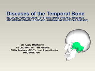
- 1. Diseases of the Temporal Bone INCLUDING GRANULOMAS (SYSTEMIC BONE DISEASE, INFECTIVE AND GRANULOMATOUS DISEASE, AUTOIMMUNE INNER EAR DISEASE) DR. RAJIV MAHASETH MS ORL- HNS, 1ST Year Resident GMSM Academy of ENT – Head & Neck Studies MMC-TUTH, IOM
- 2. Road map Disease of temporal Bone - Dr Rajeev Mahaseth 2017 •Systemic bone diseases •Infective and granulomatous disease •Autoimmune inner ear disease
- 3. Systemic bone diseaseSystemic bone disease Disease of temporal Bone - Dr Rajeev Mahaseth 2017 Paget’s disease Osteogenesis imperfecta Fibrous dysplasia Osteopetrosis Neurofibromatosis Craniofacial dysostosis Osteopathia striata
- 4. Paget’s Disease 1876 - Sir James Paget - Osteitis deformans 1888 - Sir Jonathan Hutchinson referred osteitis deformans as Paget's disease of bone Progressive,focal disorder of bone remodelling ▪ Osteoclastic - Bone resorption ▪ Osteoblastic - Bone deposition Clinical Presentation ▪ Bone pain, stiffness and fatiguability ▪ Enlargement of skull, pelvis, tibia and femur www. Google.com/image Disease of temporal bone - Dr Rajeev Mahaseth 2017 (Scott & Brown 7th edition)
- 5. 2nd most common bone disease after osteoporosis Affects -3 % of population > 40 yrs - 11 % > 80 yrs Male : Female = 3:2 Common : Britain, Australia, New Zealand, North America Uncommon: Asia (Non-white race) Disease of temporal Bone - Dr Rajeev Mahaseth 2017 Paget’s disease contd. (www.google.com/image)
- 6. Etiology : Unknown Inherited as autosomal dominant Slow viral infection Polyclonal antibodies reveal paramyxovirus antigens in osteoclasts Monoclonal antibodies - Measles, Human Parainfluenza Virus Electron microscopic study - Fingerprint pattern of osteoclast Disease of temporal Bone - Dr Rajeev Mahaseth 2017 Paget’s disease contd. (Cummings 6th edition)
- 7. Paget’s disease contd. Multinucleated osteoclast Courtesy of Pierre Delmas, MD. Disease of temporal Bone - Dr Rajeev Mahaseth 2017 Bone affected Pelvis > Femur >Skull > Tibia >Vertebrae > Clavicle > Humerus Phases: Osteolytic - Mixed - Osteoblastic - Lamellar remodeling (Temporal Bone) Bossing of temporal bone Histology Periosteum - Enchondral - Endosteal layer
- 8. Clinical Feature Bilateral progressive hearing loss: 13-40% Sensorineural , mixed or conductive Bone pain - constant deep boring Joint stiffness and fatiguability Vestibular symptoms - 20- 30% - May present with vertigo Disease of temporal Bone - Dr Rajeev Mahaseth 2017 Paget’s disease contd. (Cummings 6th edition)
- 9. Disease of temporal Bone - Dr Rajeev Mahaseth 2017 Courtesy of Pierre Delmas, MD Skull enlargement Dilated scalp veins Paget’s disease contd.
- 10. Diagnosis : Incidental finding in x - ray Early stage: Osteolytic lesion Advanced stage: Bone density Abnormal architecture Overgrowth Microfractures CT Scan: Temporal bone - washed out appearance of petrous apex Obliteration of otic capsule , narrowing of IAC Disease of temporal Bone - Dr Rajeev Mahaseth 2017 (Scott & Brown 7th edition) Paget’s disease contd.
- 11. Courtesy of Pierre Delmas, MD. Disease of temporal Bone - Dr Rajeev Mahaseth 2017 Early Stage (Osteoporosis Circumscripta) Lytic border Diffuse sclerotic changes Advanced Stage (Cotton wool skull) Paget’s disease contd.
- 12. Laboratory tests Marker of new bone formation ▪ Alkaline phosphatase Elevated markers of bone resorption: ▪ Urinary hydroxyproline/creatinine ▪ Urinary and serum deoxypyridinoline ▪ C - telopeptide ▪ N - telopeptide ) Bone scan to assess extent of disease Disease of temporal Bone - Dr Rajeev Mahaseth 2017 (Scott & Brown 7th edition) Paget’s disease contd. www. google.com/image
- 13. Pain from site of known Paget`s disease Early, potentially deforming disease Osteolytic lesions in weight bearing bones Skull disease Complications: a) Progressive neurological syndrome b) Fissure fractures c) Immobilization hypercalcaemia d) High-out put cardiac failure (Scott & Brown 7th edition) Indications forTreatment Disease of temporal Bone - Dr Rajeev Mahaseth 2017
- 14. Indications Age <55yrs Serum alkaline phosphate/urine hydroxyproline > 2 times 6 months prior to joint replacement surgery Treatment Physiotherapy Antiresorptive therapy (Bisphosphonates ) - Tab Alendronate 40mg/day PO OD for 6 mths Calcitonin was previously used, alternative therapy Analgesics Disease of temporal Bone - Dr Rajeev Mahaseth 2017 (Scott & Brown 7th edition) Paget’s disease contd.
- 15. Surgery : Fractures Bone deformities Osteoarthritis Complication Development of osteosarcoma : < 0.1 % Cranial nerve involvement Disease of temporal Bone - Dr Rajeev Mahaseth 2017 (Scott & Brown 7th edition) Paget’s disease contd.
- 16. Hereditary disorder of collagen synthesis Incidence : 2 - 15/100000 births Type 1 AD,Mild Blue sclera, non deforming fracture Normal stature, hearing loss Type 2 Severe form, multiple fractures in utero Still birth Disease of temporal Bone - Dr Rajeev Mahaseth 2017 (www.google.com/ima ge) (Cummings 6th edition) OSTEOGENESIS IMPERFECTA
- 17. Type 3 Multiple fracture, progressive bone deformity Hearing loss Type 4 Similar to type 1 but there is white sclera 95% spontaneous fracture 50-60% hearing loss Hypermobility/laxity of joints Thin skin/subcutaneous bruising Disease of temporal Bone - Dr Rajeev Mahaseth 2017 (Cummings 6th edition) Osteogenesis imperfecta contd.
- 18. Clinical Feature ▪ 95% spontaneous fractures ▪ 85% blue sclera ▪ 50-60% hearing loss ▪ Hypermobility/laxity of joints ▪ Thin skin/subcutaneous bruising CHL accompanies blue sclera appear in - 20-25 yrs of age SNHL seen in 40 % of patients - grey/white sclera Disease of temporal Bone - Dr Rajeev Mahaseth 2017 (Cummings 6th edition) (www.google.com/ima ge) Osteogenesis imperfecta contd.
- 19. Investigations Level of FGF 23 is raised PCR - mutations of the gnas gene Low serum phosphate Radiology: Enlarged temporal bone Sclerosis(23%) Ground glass appearance (56%) Cystic lesion(21%) Disease of temporal Bone - Dr Rajeev Mahaseth 2017 (www.google.com/ima ge) Osteogenesis imperfecta contd.
- 20. History of fracture after minor trauma Positive family history Blue sclerae Early onset of hearing loss Stapes footplate - Thick, soft, chalk-like/granular, fixed Stapedectomy - Similar result otosclerosis Treatment - Biphosphonate Disease of temporal Bone - Dr Rajeev Mahaseth 2017 (Cummings 6th edition) D/Dwith otosclerosis
- 21. FIBROUS DYSPLASIA ▪ 1900 - Von Recklingshausen - Ostitis Fibrosa ▪ Genetic noninherited disease ▪ Somatic activating missense mutations of GNAS1 ▪ Age – 2 – 5th decade ▪ Tumour like lesion - Replacement of normal bone with cellular connective tissue Types: Monostotic : Skull base in 15 % ,maxilla . mandible Polyostotic : More than one bone lower limbs,skull Disease of temporal Bone - Dr Rajeev Mahaseth 2017 (Scott Brown 6th edition) (www.google.com/image)
- 22. McCune-Albright syndrome Polyostotic fibrous dysplasia Skin hyperpigmentation Endocrine dysfunction Presentation Skull base involved in 15 % cases 1/3rd cases are located in maxilla or mandible Palpable visible swelling in 75 % cases Disease of temporal Bone - Dr Rajeev Mahaseth 2017 Fibrous dysplasia contd.
- 23. Fibrous dysplasia contd. Disease of temporal Bone - Dr Rajeev Mahaseth 2017 (www.google.com/image)
- 24. Presentation Pain Progressive hearing impairment - 57% Localized temporal bone swelling - 51% Bony occlusion of EAC - 42% SNHL (erosion of otic capsule) ,pain, vertigo VII Nerve paralysis Decreased vision – compression of optic nerve Disease of temporal Bone - Dr Rajeev Mahaseth 2017 Fibrous dysplasia contd.
- 25. Differential Diagnosis Ossifying fibroma Histiocytosis X Paget’s disease Aneurysmal bone cyst Giant cell tumour Brown tumour of hyperthyroidism Disease of temporal Bone - Dr Rajeev Mahaseth 2017 Fibrous dysplasia contd.
- 26. Treatment No known conservative treatment Surgery Curettage External auditory meatus stenosis Decompression of optic nerve Radiotherapy : Predispose to malignant degeneration (Scott Brown 6th edition) Disease of temporal Bone - Dr Rajeev Mahaseth 2017 Fibrous dysplasia contd.
- 27. OSTEOPETROSIS Rare genetic disorder Defective osteoclast - Failure of normal bone resorption Normal bone formation by osteoblast Thick, dense, brittle bones Malignant AR, rapidly progressive, Infancy Encroachment of bone marrow - Anemia, thrombocytopenia, susceptibility to infection, hepatospleenomegaly Disease of temporal Bone - Dr Rajeev Mahaseth 2017 (www.google.com/imag e) (Cummings 6th edition)
- 28. Osteopetrosis contd. Optic atrophy, facial paralysis, SNHL, hydrocephalus, mental retardation AD type1 Rare disorder , asymptomatic Some have pain and hearing loss No fracture AD type 2 (Marble bone disease) Frequent type with normal life expectancy May be asymptomatic Increased fracture of bones Disease of temporal Bone - Dr Rajeev Mahaseth 2017
- 29. Otological manifestation Dense calcified cartilage Non-pneumatized mastoid Fetal form stapes Normal inner ear Dehiscence of tympanic segment of VII Nerve Facial nerve paralysis : - Unilateral/Bilateral Recurrent AOM, CHL, SNHL Disease of temporal Bone - Dr Rajeev Mahaseth 2017 Osteopetrosis contd. (Cummings 6th edition)
- 30. Radiological: Increase density all bones Increase serum acid phosphatase Treatment Not definitive Steroid Interferon BMT Disease of temporal Bone - Dr Rajeev Mahaseth 2017 Osteopetrosis contd. (Cummings 6th edition)
- 31. Infective and granulomatous Disease Tuberculosis Syphilis Sarcoidosis Histiocytosis X Wegener’s granulomatosis Disease of temporal Bone - Dr Rajeev Mahaseth 2017
- 32. TUBERCULOSIS Chronic granulomatous infection Age: 1 - 5 yrs Incidence : 0.05 – 0.9 % of COM cases Etiology: M. tuberculosis, M.Bovis, M.avium, M.fortuitum Risk factors: Active pulmonary tuberculosis, HIV infection Mode of transmission: Hematogenous/Lymphatic Eustachian tube Intracranial, extracranial infection Maternal systemic tuberculosisDisease of temporal Bone - Dr Rajeev Mahaseth 2017 (Cummings 6th edition)
- 33. Diagnosis Chronic otorrhoea resistant to usual antibiotic treatment, surgery Non tender high jugular chain lymphadenopathy, cold abscess Otoscopic examination TM thickened ,landmark obliterated Multiple perforation in TM with seropurulent discharge Middle ear mucosa - Pale Conductive hearing loss Effusion in middle ear & ossicles destructionDisease of temporal Bone - Dr Rajeev Mahaseth 2017 (Cummings 6th edition) Tuberculosis contd.
- 34. Otic capsule – Loss of auditory and vestibular function Histopathology Granuloma with Langerhans cells – 1/3rd case Acid fast organism Culture – Gold standard Skin test , biopsy & PCR - TB CT Scan – Soft tissue in middle ear, intracranial/extracranial foci ANCA (Antineutrophil cytoplasmic antibody) - WG Tuberculosis contd. (Cummings 6th edition) Disease of temporal Bone - Dr Rajeev Mahaseth 2017
- 35. Management Antituberculous therapy - Dry ear in 1-3 months Ear surgery minimal role Complications Profound SNHL Facial paralysis Petrous apicitis Labyrinthitis, meningitis Post auricular abscesses Disease of temporal Bone - Dr Rajeev Mahaseth 2017 (Scott Brown 7th edition) Tuberculosis contd.
- 36. SYPHILIS Congenital and acquired syphilis affect middle ear Less frequently seen in clinical practice Male : Female – 4:1 Acquired form 25 times more frequent than congenital SNHL (sudden/fluctuating ) tinnitus & vertigo Systemic feature - Cardiac and aortic involvement Disease of temporal Bone - Dr Rajeev Mahaseth 2017 (Scott Brown 6th edition)
- 37. Syphilis contd. Early syphilis: Congenital bilateral hearing loss – 50 % Secondary syphilis - Transient vestibular symptoms (Positional with tinnitus) - Ocular palsy ,facial paralysis SNHL – High frequency involved Speech descrimination worse Caloric response reduced Disease of temporal Bone - Dr Rajeev Mahaseth 2017
- 38. Late syphilis: 10 - 50 yrs of primary infection Otological features : SNHL – B/L in congenital , U/L in acquired Onset of aural symptoms : - Sudden, fluctuant , episodic vertigo Severe vestibular damage - Imbalance, ataxia Disease of temporal Bone - Dr Rajeev Mahaseth 2017 (Scott Brown 6th edition) Syphilis contd.
- 39. Hennebert’s sign Ocular deviation with positive or negative pressure in external auditory canal Fistula between middle and inner ear due to rarefying osteitis Tullio’s sign Transient vertigo and nystagmus due to exposue to loud sound Diagnosis: VDRL, TPHA, TPI, FTA-ABS CSF examination - Raised globulin, IgG level & lymphocytosisDisease of temporal Bone - Dr Rajeev Mahaseth 2017 Syphilis contd.
- 40. Treatment: Penicillin - 600000units I/M OD x 21 days Probenecid - 500mg x 6hrly Ampicillin - 1.5 gm x 6hrly x 4wks. Prednisolone - 30mg x 8hrly x 4wks (Jarish –Herxeimer rxn) Disease of temporal Bone - Dr Rajeev Mahaseth 2017 Syphilis contd.
- 41. Sarcoidosis Rare systemic granulomatous disease of unknown etiology Non-caseating granuloma Female predominance Onset - 3rd - 4th decade Bilateral hilar lymphadenopathy, cough and granulomatous skin rash Organs: Parotid gland, facial nerve, nasal cavity and larynx Uveitis 80 %, lymphadenopathy 55 %, parotid swelling 20 %, facial nerve palsy 43% Disease of temporal Bone - Dr Rajeev Mahaseth 2017 (Scott Brown 6th edition)
- 42. Otological Feature SNHL – sudden fluctuating or progressive vestibular dysfunction Granulomatous disease external , middle ear & mastoid PTA - Low or high frequency hearing loss Heerfordt syndrome Parotitis, uveitis, facial nerve paralysis and pyrexia Histopathology Perivascular lymphocytic infiltration and granulomatous Disease of temporal Bone - Dr Rajeev Mahaseth 2017 (Scott Brown 6th edition) Sarcoidosis contd.
- 43. Laboratory Tests Serum ACE ( Angiotensin converting enzyme) elevated Hypercalcemia & hypercalciuria Raised ESR Treatment: Steroids Methotrexate, Cyclophosphamide Hydroxychloroquine Infliximab Disease of temporal Bone - Dr Rajeev Mahaseth 2017 Sarcoidosis contd.
- 44. Histiocytosis X Proliferation of cytologically benign histiocytes, lymphocytes, eosinophlis in the affected tissue Immunologic dysfunction Organs: Reticuloendothelial system 3 types: Eosinophilic granuloma Hand-Schuller-Christian disease Letterer-Siwe disease Disease of temporal Bone - Dr Rajeev Mahaseth 2017
- 45. Eosinophilic granuloma Children , young adults Male : Female - 2 : 1 Osteolytic lesion - Femur , pelvis , scapula, vertebrae ,ribs, mandible Maxilla of skull Clinical features: Asymptomatic Local Pain Local swelling Pathologic fracture Disease of temporal Bone - Dr Rajeev Mahaseth 2017 Histiocytosis x contd.
- 46. Histopathology : • Sheets of histiocytes • Scattered eosinophils • Plasma cells • Areas of haemorrhage • Necrosis with giant cell Radiological appearance • Punched out / lytic appearance Disease of temporal Bone - Dr Rajeev Mahaseth 2017 www.google.com/image Histiocytosis x contd.
- 47. Hand-Schuller Christian disease Multiple focal lesions Early onset (<5yrs) Common site – Skull bone Destruction of temporal bone - Secondary infection & otorrhoea Systemic manifestation Fever / Recurrent URTI Otitis media Cervical Lymphadenopathy, hepatosplenomegaly Disease of temporal Bone - Dr Rajeev Mahaseth 2017 www.google.com/image Histiocytosis x contd.
- 48. Letterer- Siwe Disease Disseminated disease Early onset (< 3 yrs) Multiple organ involvement (Skin, Lymph node, viscera) Systemic manifestation: Fever Eczema like rash ,oral lesions Lymphadenopathy Multiple bony lesion Respiratory failure- pulmonary infiltration Disease of temporal Bone - Dr Rajeev Mahaseth 2017 (Cummings 6th Edition) Histiocytosis x contd. (www.google.com/image)
- 49. Disease of temporal Bone - Dr Rajeev Mahaseth 2017 Letterer- Siwe Disease contd. Otological manifestation (15-61%) Otorrhoea Post auricular swelling, hearing loss, vertigo Granulation /Polyp in EAC Diagnosis Inflammatory disorder not responding to antibiotic Bilateral destructive ear disease High ESR Immunohistochemistry Treatment Aural polypectomy +/- steroid Low dose chemotherapy Histiocytosis x contd.
- 50. Autoimmune inner ear disease 1. Vasculitis ▪ Wegener granulomatosis ▪ Polyarteritis nodosa ▪ Temporal arteritis ▪ Cogan syndrome ▪ Behcet’s disease 2. Relapsing polychondritis 3. Systemic lupus erythmatosis Disease of temporal Bone - Dr Rajeev Mahaseth 2017 4. Immunodeficiency disease T cell deficiency B cell deficiency Disorder of phagocytosis Complement system disorder
- 51. Wegener’s Granulomatosis Friedrich Wegener – German pathologist Also called as Granulomatosis with polyangiitis Age : 15 - 73 yrs Necrotizing vasculitis small to medium sized vessels Triad : Airway, lung & renal disease Clinical features: Epistaxis, nasal obstruction, crusting Saddle nose deformity (perforated septum) Disease of temporal Bone - Dr Rajeev Mahaseth 2017 (Scott- Brown 7th edition) (www.google.com/ima ge)
- 52. Wegener’s Granulomatosis contd. Otological features: Ear involved in 15 - 35% Seromucinous otitis media, frank otorrhoea Necrotizing granulation tissue - Tympanic cavity TM - Multiple perforations (granulation) Rapidly progressive SNHL Loss of vestibular function Disease of temporal Bone - Dr Rajeev Mahaseth 2017 (Scott- Brown 7th edition)
- 53. Wegener’s Granulomatosis contd. Diagnosis: C-ANCA - Positive in 95% patient Diagnosis & activity of disease Anaemia, thrombocytosis, hyperglobulinemia [IgA] Raised ESR & CRP level Treatments: Steroids - Prednisolone 60-80 mg/day Cyclophosphamide 2mg/kg, azathioprine 200mg/day Complete remission - 90% Duration : 3 – 6 months Disease of temporal Bone - Dr Rajeev Mahaseth 2017 (Scott-Brown 6th edition)
- 54. Polyarteritis nodosa Systemic necrotizing vasculitis of small and medium sized arteries Male : Female - 3:1 Renal, coronary, hepatic and visceral circulation involved Otological feature: SNHL - Sudden, bilateral symmetrical CHL - Middle ear granulation Disease of temporal Bone - Dr Rajeev Mahaseth 2017 (Scott-Brown 6th edition)
- 55. Polyarteritis nodosa contd. Lab tests : Anaemia, raised ESR Leukocytosis Treatment Systemic steroid Immunosuppressive drug - Chlorambucil Disease of temporal Bone - Dr Rajeev Mahaseth 2017
- 56. Temporal arteritis Older age group Vasculitis of extracranial arteries Aneurysm formation Stenosis Occlusion Features: Pyrexia Bitemporal headache Tender palpable thickening of temporal arteries Blindness: 30% (Ophthalmic artery)Disease of temporal Bone - Dr Rajeev Mahaseth 2017 (www.google.com/image)
- 57. Temporal arteritis contd. Otological features: Hearing loss (rapid, progressive, vertigo) Lab.tests: ESR Serum globulin raised Biopsy Ultrasound/ Doppler help to identify involved area Treatments: Systemic steroidsDisease of temporal Bone - Dr Rajeev Mahaseth 2017 (www.google.com/image)
- 58. Cogan Syndrome Dr. David Cogan in 1945 Rare, autoimmune, young adults Non syphilitic interstitial keratitis Fluctuant, aggressive cochleovestibular damage Acute episode - Nausea, vomiting, tinnitus followed by SNHL Otological features: - B/L SNHL (Progressive , fluctuating ,episodic ) Disease of temporal Bone - Dr Rajeev Mahaseth 2017 (www.google.com/image) (Ballenger’s 17th edition)
- 59. Cogan`s Syndrome contd. Ocular symptoms: ▪ Excessive lacrimation, ▪ Photophobia ▪ Blurred vision Treatment: ▪ Steroid ▪ Immunosuppresive drugs Disease of temporal Bone - Dr Rajeev Mahaseth 2017 (www.google.com/image)
- 60. Behcet’s disease Prof. Hulusi Behcet named in 1973 Uncommmon Chronic relapsing inflammatory disorder Recurrent mouth ulcers (89%) Genital ulceration Inflammed eyes Disease of temporal Bone - Dr Rajeev Mahaseth 2017 (www.google.com/image)
- 61. Behcet’s disease contd. Otological: Slow progressive, bilateral SNHL Vestibular symtoms Treatment: Immunosuppresive Steroids Disease of temporal Bone - Dr Rajeev Mahaseth 2017
- 62. Relapsing polychondritis Rare disease of episodic inflammation Degeneration of multiple cartilagenous structure Pearson coined the term in 1960 Pathology Cartilage and proteoglycan rich structures (eyes and cardiovascular system) Neutrophil infilteration Loss of basophilia of cartilage Etiology: Autoimmune Disease of temporal Bone - Dr Rajeev Mahaseth 2017 (www.google.com/image)
- 63. Disease of temporal Bone - Dr Rajeev Mahaseth 2017 Relapsing polychondritis contd.
- 64. Relapsing polychondritis contd. Disease of temporal Bone - Dr Rajeev Mahaseth 2017 (Wallace et al.)
- 65. Clinical presentation Otological Pinna involved in 89 % Eustachian tube - Otitis media with effusion Inner ear - Hearing loss and dysequilibrium Nasal chondritis: Pain, feeling of fullness , sudden painless saddle deformity Larynx and trachea Hoarseness , tenderness, airway obstruction Occular inflammationDisease of temporal Bone - Dr Rajeev Mahaseth 2017 Relapsing polychondritis contd.
- 66. Outcomes Follows a course of acute exacerbation and remission Treatments I. Medical Steroids – Prednisolone 30-60mg/daily Immunosupressive ( Azathioprine,Methotrexate) Anti-CD4monoclonal antibody, Minocycline. II. Surgical TracheostomyDisease of temporal Bone - Dr Rajeev Mahaseth 2017 Relapsing polychondritis contd.
- 67. Systemic lupus erythematosus Multisystem connective tissue disorder Etiology: Autoimmune Immune complex deposition Otological features Bilateral SSNHL - 8 % Loss of spiral ganglion cell and hair cell and atrophy of the stria vascularis Circulating immune complex sludge in stria vascularis Disease of temporal Bone - Dr Rajeev Mahaseth 2017
- 68. Systemic lupus erythematosus contd. Lab Tests: Antinuclear antibody Anti dsDNA Anaemia Raised ESR Treatment: Steroids - Prednisolone 40 mg daily for 2 wks Plasma exchange at 6 months interval Disease of temporal Bone - Dr Rajeev Mahaseth 2017
- 69. References 1. Scott-Brown 6th & 7th edition 2. Cumming’s otorhinolaryngology, 6th edition 3. Ballenger’s otorhinolaryngology 17th edition 4. https://scholar.google.com 5. Current Diagnosis and treatment 3rd edition 6. Glasscock and sambough surgery of ear 6th edition Disease of temporal Bone - Dr Rajeev Mahaseth 2017
Editor's Notes
- History This drawing of Paget&apos;s first patient, published in his original paper, demonstrates the characteristic appearance that may occur with severe disease. In his original description, Paget wrote, &quot;The disease begins in middle age or later (and) affects most frequently the long bones of the lower extremities and the skull in shape, size.... The bones enlarge and soften, and those bearing weight yield and become unnaturally curved and misshapen.“1 At the bottom of the slide, a marked increase in hat size is apparent. Recent evidence supports genetic and environmental etiologic factors.2 References The Paget Foundation. Available at: www.paget.org/Information/slide/paget_diag.ppt. Accessed August 10, 2005. Altman RD. Paget’s disease of bone. In: Coe FL, Favus MJ, eds. Disorders of Bone and Mineral Metabolism. 2nd ed. Philadelphia, Pa: Lippincott Williams & Wilkins; 2002:985-1020.
- It is caused by the excessive breakdown and formation of bone, followed by disorganised bone remodelling. This causes affected bone to weaken, resulting in pain, fractures and arthritis in the joints near the affected bones.
- Paramyxovirus – Measles and RSV
- Paget’s Disease: An Osteoclast-Mediated Disorder Osteoclasts play a crucial role in certain metabolic bone diseases, such as Paget’s disease and osteoporosis.1 The constant renewal of the skeleton entails a process called bone remodeling. In this process, osteoclasts remove old bone and osteoblasts form new bone. The osteoclasts are responsible for bone resorption, while the osteoblasts lay down new bone in the resorbed areas. This new bone later mineralizes.2 In affected areas there is an increase in both the size and number of osteoclasts (as shown on the left). The image on the right shows that pagetic osteoclasts are also multinucleated and can contain up to 100 nuclei per cell.1 References Siris ES, Roodman GD. Paget’s disease of bone. In: Favus MJ, ed. Primer on the Metabolic Bone Diseases and Disorders of Mineral Metabolism. 5th ed. Washington, DC: American Society for Bone and Mineral Research; 2003:495-506. Roodman GD, Windle JJ. Paget disease of bone. J Clin Invest. 2005;115:200-208.
- Paget’s Disease: Clinical Presentation (growing periosteal blood flow increase intraosseous pressure—stimulate bone pain fibres) Paget’s disease is usually mild or symptomatic,1,2 and is most commonly found incidentally during examination for another complaint.1 Hearing loss high frequency attributed to loss in bone density narrow EAC and nerve compression In one typical diagnostic scenario, an elderly person suffers a broken bone in an accident and a radiologist recognizes pagetic lesions in the x-rays. Another example is a patient who presents with abdominal pain and is found to have Paget’s disease on radiography. Symptoms and complications of Paget’s relate to the site and extent of the disease and include bone, muscle, or joint pain; fractures; bone deformity; osteoarthritis; hearing loss; and dental complaints such as tooth loosening, all of which can contribute to a significantly decreased quality of life.1-4 Pain related to osteoarthritis in a hip or knee adjacent to a pagetic bone is a common complaint among patients with Paget’s disease.1 Bone pain, when present, is often mild to moderate; it often persists during the night, a feature that generally differentiates it from osteoarthritis.1 Pain in the tibia or femur may be aggravated by weight bearing.1 Dental problems such as loosening of the teeth or swollen, infected gums can occur in patients with Paget’s disease in the skull.4 References Siris ES, Roodman GD. Paget’s disease of bone. In: Favus MJ, ed. Primer on the Metabolic Bone Diseases and Disorders of Mineral Metabolism. 5th ed. Washington, DC: American Society for Bone and Mineral Research; 2003:495-506. Altman RD. Paget’s disease of bone. In: Coe FL, Favus MJ (eds). Disorders of Bone and Mineral Metabolism. 2nd ed. Philadelphia, Pa: Lippincott Williams & Wilkins; 2002:985-1020. Gold DT, Boisture J, Shipp KM, et al. Paget’s disease of bone and quality of life. J Bone Miner Res. 1996;11:1897-1904. Ankrom MA, Shapiro JR. Paget’s disease of bone (osteitis deformans). J Am Geriatr Soc. 1998;46:1025-1033.
- Paget’s Disease in the Skull Typical changes in the skull associated with Paget’s disease include diffuse enlargement, and dilated scalp veins. Some patients may show frontal bossing or knobby deformity, or there may be enlargement of the maxilla or mandible.
- Diagnosing Paget’s Disease: Tests Diagnosis of Paget’s disease includes clinical, radiological, biochemical, and, in some rare cases, histological investigations.1,2 Diagnosis usually involves measurement of biochemical markers associated with increased bone turnover, especially serum alkaline phosphatase. In rare cases, bone biopsies may be required to establish a diagnosis.1,2 Biomarkers associated with increased bone measures provide a more immediate indication of response to therapy than do markers of bone formation.1 It is important to note that alkaline phosphatase can be elevated in other disorders, notably vitamin D deficiency, osteomalacia, and gall bladder and liver disease, which must be ruled out. Radiographic findings of Paget’s disease are characteristic and rarely confused with x-ray findings suggestive of other diseases. Osteolytic lesions in long bones advance upward or downward at about 1 cm/y.1 As Paget’s disease progresses, there is cortical thickening, loss of corticomedullary distinction, and accentuated trabecular markings. The last phase of Paget’s disease is primarily sclerotic, with enlargement and thickening of long bones.3 Plain radiographs of Paget’s lesions are usually confirmatory and rarely confused with x-ray findings of other conditions. Scintigraphic bone scans may be used to assess the extent of skeletal involvement.1,2 References Lyles KW, Siris ES, Singer FR, et al. A clinical approach to diagnosis and management of Paget’s disease of bone. J Bone Miner Res. 2001;16:1379-1387. Selby PL, Davie MW, Ralston SH, Stone MD, Bone and Tooth Society of Great Britain, National Association for the Relief of Paget&apos;s Disease. Guidelines on the management of Paget&apos;s disease of bone. Bone. 2002;31:366-373. Siris ES, Roodman GD. Paget’s disease of bone. In: Favus MJ, ed. Primer on the Metabolic Bone Diseases and Disorders of Mineral Metabolism. 5th ed. Washington, DC: American Society for Bone and Mineral Research; 2003:495-506.
- Early-Stage (Lytic) Paget’s Disease in the Skull: Known as “Osteoporosis Circumscripta” A typical lesion of early-stage Paget’s disease is the resorptive border in the skull known as osteoporosis circumscripta. Reference Lyles KW, Siris ES, Singer FR, et al. A clinical approach to diagnosis and management of Paget’s disease of bone. J Bone Miner Res. 2001;16:1379-1387.
- The primary objectives of treatment for Paget&apos;s disease should be to relieve current symptoms, prevent progression of the disease and thereby help avoid complications. &gt; 2 times upper limit than normal 6mths prior – reduce hypervascularity in active pagetic disease
- The primary objectives of treatment for Paget&apos;s disease should be to relieve current symptoms, prevent progression of the disease and thereby help avoid complications. &gt; 2 times upper limit than normal 6mths prior – reduce hypervascularity in active pagetic disease
- The primary objectives of treatment for Paget&apos;s disease should be to relieve current symptoms, prevent progression of the disease and thereby help avoid complications. &gt; 2 times upper limit than normal 6mths prior – reduce hypervascularity in active pagetic disease
- H.L in child and adolescence Platinum ribbon used for prosthesis
- Guanine nucleotide binding proteins (G proteins) are membrane-associated, heterotrimeric proteins composed of three subunits: alpha beta ( and gamma G proteins and their receptors (GPCRs) form one of the most prevalent signalling systems in mammalian cells, regulating systems as diverse as sensory perception, cell growth and hormonal regulation Mono – temporal bone GNAS (guanine nucleotide-binding protein/α-subunit Polyostotic fibrous dysplasia Endocrine dysfunction Skin hyperpigmentation
- Better demarcated expansile lesion with Smooth margin more aggressive
- Albers schonberg
- Autosomal dominant
- Middle ear involved
- Middle ear involved
- Pt may have systemic symptoms of TB , pale middle ear mucosa
- Middle ear involved
- Middle ear involved
- Caloric test – Reduced /No response
- Anti TNF @ infliximab
- Electron micro Bierbeck granule
- Middle ear involved
- Capillary,venule,arteriole arteriesis Kidney: rapidly progressive glomerulonephritis (75%), leading to chronic kidney failure Upper airway, eye and ear disease Nose: pain, stuffiness, nosebleeds, rhinitis, crusting, saddle-nose deformity due to a perforated septum Ears: conductive hearing loss due to auditory tube dysfunction, sensorineural hearing loss (unclear mechanism) Oral cavity: strawberry gingivitis,[6] underlying bone destruction with loosening of teeth, non-specific ulcerations throughout oral mucosa Eyes: pseudotumours, scleritis, conjunctivitis, uveitis, episcleritis
- Capillary,venule,arteriole arteriesis Kidney: rapidly progressive glomerulonephritis (75%), leading to chronic kidney failure Upper airway, eye and ear disease Nose: pain, stuffiness, nosebleeds, rhinitis, crusting, saddle-nose deformity due to a perforated septum Ears: conductive hearing loss due to auditory tube dysfunction, sensorineural hearing loss (unclear mechanism) Oral cavity: strawberry gingivitis,[6] underlying bone destruction with loosening of teeth, non-specific ulcerations throughout oral mucosa Eyes: pseudotumours, scleritis, conjunctivitis, uveitis, episcleritis
- Middle ear involved
- Middle ear involved
- Exact etiology unknown
- 89% involves pinna. cartilagenous portion-Flopp pinna ET involve: OME