
Recommended
More Related Content
What's hot
What's hot (20)
Similar to 9-Indirect Ophthalmoscope.ppt
Similar to 9-Indirect Ophthalmoscope.ppt (20)
More from bakanangemmahpholoan
More from bakanangemmahpholoan (20)
Recently uploaded
Recently uploaded (20)
9-Indirect Ophthalmoscope.ppt
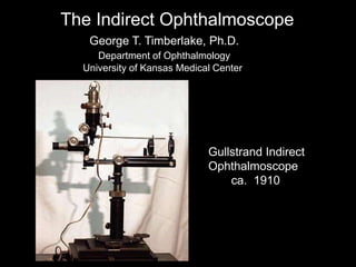
- 1. The Indirect Ophthalmoscope Gullstrand Indirect Ophthalmoscope ca. 1910 George T. Timberlake, Ph.D. Department of Ophthalmology University of Kansas Medical Center
- 2. Name Trial 1 Trial 2 Trial 3 Comments FAMILIARITY WITH DEVICE 3. The resident should be able to identify the components of the BIO. Instructions to Faculty: Please observe the resident perform the following tasks, and grade each step, "P" (successful completion of step) or "X" (needs additional training or practice). The resident must successfully perform each step in order to pass t THEORY 1. The resident should be able to explain and draw a simplified diagram of the optics of the binocular indirect ophthalmoscope. 2. The resident should be able to explain the relative image orientation, field, and magnification of the image using the binocular indirect ophthalmoscope and various condensing lenses (at least the 20 and 28 diopter lenses). What You Need to Know
- 3. If the retina could light up…. Emmetropic eye Image of retina on distant surface GTT 04 Fundamental Principle of the Indirect Ophthalmoscope
- 4. Ophthalmoscopic lens Aerial image of retina Fundamental Principle of Indirect Ophthalmoscope GTT 04
- 5. Viewing the aerial image with a magnifier GTT 04
- 6. Allvar Gullstrand Swedish Ophthalmologist 1862 - 1930 Professor of Physical & Physiological Optics, University of Uppsala Nobel Prize 1911 for work on optics of eye First “reflex free” ophthamoscope GTT 04
- 7. Light entering eye Light leaving eye Pupil Gullstrand Principle for Reflex-free Ophthalmoscopy Light entrance and exit separated in pupil plane GTT 04
- 8. 20 D Nikon Aspheric Lens Optical Conjugates 70 44* 152 168 Retina Pupil Cornea (Distances in mm) Emmetropic Gullstrand Eye * back focal plane GTT 04
- 10. 1.75 mm 2.0 mm entrance pupil 7 mm diameter pupil Gullstrand Principle—Entrance Pupil GTT 04
- 11. 74.1 153.6 20 D Pupil Conjugate Plane Gullstrand Retinal Illumination Light introduced in pupil conjugate plane In focus in pupil plane Mpupil conjugate = 153.6 74.1 = 2.1 M pupil conjugate = 1 2.1 = 0.48 GTT 04
- 12. Radius of Distance to next Index of Curvature (mm) surface (mm) Material Refraction, n retina 11.198 17.051 vitreous 1.336 post lens 6.000 0.636 lens surface 1.386 post lens core 5.760 2.419 lens core 1.406 ant lens core -7.911 0.546 lens surface 1.386 ant lens -10.000 3.100 aqueous 1.336 post cornea -6.800 0.500 cornea 1.376 ant cornea -7.700 air 1.000 cornea crystalline lens retina aqueous vitreous optic axis pupil iris lens core Gullstrand Model Eye GTT 04
- 13. pupil conjugate plane pupil plane A point in the pupil conjugate plane is Imaged in the eye’s pupil plane SIDE VIEW GTT 04
- 14. pupil pupil conjugate Light leaving the eye from a disk-shaped area in the pupil is imaged in the pupil conjugate plane SIDE VIEW GTT 04
- 15. 1 mm 2.1 mm Real example of entrance pupil continued SIDE VIEW GTT 04
- 16. Pupil conjugate plane Lamp Aperture 45 mirror Practical Retinal Illumination System GTT 04
- 18. 2.0 mm entrance pupil 7 mm diameter pupil 4.0 mm 1.0 mm exit pupil Gullstrand Principle--Exit Pupils GTT 04
- 19. Pupil Conjugate Plane Gullstrand Retinal Imaging Light from retina passes through area in pupil plane Pupil-plane area is in focus in pupil conjugate plane Mpupil conjugate = 153.6 74.1 = 2.1 M pupil conjugate = 1 2.1 = 0.48 20 D GTT 04
- 20. Light from retina passing through pupil plane conjugate….. …also passes through an aperture in the conjugate pupil plane. PP RIP TOP VIEW GTT 05 RAYS FROM THE EXIT PUPIL PASS THROUGH THE CONJUGATE PUPIL
- 21. Light from retina not passing through pupil plane conjugate….. ….doesn’t get past stop in the conjugate pupil plane. RAYS FROM RETINA MUST GO THROUGH PUPIL CONJUGATE TO BE SEEN GTT 04
- 22. stop in pupil plane conjugate circular aperture in stop optic axis GTT 04
- 23. PP OL RIP PP conjugate Observer’s pupil at PP conjugate VIEWING THE RETINAL IMAGE out of focus rays—poor image
- 24. optic axis observer’s pupil in conjugate pupil plane SUBJECT OBSERVER
- 25. VIEWING THE RETINAL IMAGE Weak + lens brings rays to focus on retina
- 26. FIRST ATTEMPT AT BINOCULAR VIEW Obs. L eye Obs. R eye S’s eye Combine L and R eye views Observer’s eyes have to be too close
- 32. GTT 05
- 34. Subject’s eye Observer R Eye L exit pupil R exit pupil Observer L Eye aerial image left-to-right reversed subject’s retina appears reversed L to R SUBJECT’S RETINAAPPEARS REVERSED LEFT TO RIGHT TOP VIEW
- 35. Subject’s eye SIDE VIEW sup inf RIP Observer’s eye aerial image top-to-bottom reversed subject’s retina appears reversed top-to-bottom SUBJECT’S RETINA APPEARS REVERSED TOP-TO-BOTTOM
- 36. 20 D lens RI 60 D eye OPHTHALMOSCOPE MAGNIFICATION Mag of RI Peye Plens = 60 D 20 D = 3.0 M =
- 37. 42 40 mm 50 mm 20 D 1 mm dia exit pupil 2.0 mm MONOCULAR FIELD OF VIEW GTT 04
- 38. 20 D 40 Area of binocular view BINOCULAR FIELD OF VIEW GTT 04
- 39. Clear Aperture: CLAP Working Distance: WD CLAP 2 WD 54.72 mm 51.04 mm = 24 mm 47 mm CLAP WD = tan -1 ½ ( ( = 23.7 FOV = 47.4 = 25.2 FOV = 50.3 = 25.2 FOV = 50.3 Example: OI Maxlight 20 D CLAP = 48 mm WD = 47 mm FOV = 50 ESTIMATING FIELD OF VIEW
- 40. SUMMARY Draw a simplified diagram of the optics of the binocular indirect ophthalmoscope. Illumination planes Pupil planes Retinal image planes Be able to explain: Image orientation Field of view Magnification
- 41. BIO RETINAL ILLUMINATION RIP’ Subject O.L. Observer PP light source Mirror O.L.: Ophthalmoscopic lens RIP: Retinal Image Plane PP: Pupil Plane entry pupil
- 42. O.L.: Ophthalmoscopic lens RIP: Retinal Image Plane BIO PUPIL & RETINAL IMAGE PLANES & IMAGE ORIENTATION M: Mirror GTT 04 RIP’ Subject O.L. Observer M PP: Pupil Plane PP PP
- 44. Clear Aperture: CLAP Working Distance: WD CLAP WD = tan -1 ½ ( ( BIO APPROXIMATE FIELD OF VIEW CLAP 2 WD FOV = 2
- 46. prism Retinal Image Plane Pupil Plane RETINAL IMAGING SYSTEM fixing diverging rays from retina Pupil Plane PP RIP Rays diverging from optic axis Rays parallel to optic axis GTT 04
- 47. optic axis axis parallel to optic axis prism GTT 04
- 48. lens retinal image retinal image Formation of Final Aerial Retinal Image GTT 04
- 51. fobj fep objective eyepiece Example: fobj fep M = fobj fep = 50.0 mm = - 5.0 mm M = 50.0 - 5.0 = 10.0 MAGNIFICATION OF GALILEAN TELESCOPE GTT 04
- 52. Viewing Through a Galilean Telescope parallel rays emmetropic eye object iImage UPRIGHT OBJECT APPEARS UPRIGHT GTT 04
- 53. Subject Observer Retinal image plane--begin new raytrace Galilean telescope Complete (monocular) System GTT 04
- 55. GTT 04
- 57. Retinal Image Orientation Subject Observer Subject’s superior retina appears upward Subject’s temporal retina appears rightward sup. inf. GTT 04
- 58. 7.77 mm RIP PP 70 mm 35.5 mm 2.547 mm 56.4 D emmetropic Gullstrand Eye Asphere f = 53 mm = 68 mm 18.86 D M = Peye Plens = 56.4 18.86 = 2.99 M = Himage Hobject = 7.77 2.55 = 3.05 2.99 3.05 (2% diff) RETINAL IMAGE MAGNIFICATION
- 59. 20 D lens RI1 RI2 25D lens 12.5X telescope p 230 mm q 60 D eye OPHTHALMOSCOPE MAGNIFICATION Mag of RI1 Mag of RI2 1 1 1 p f + q = 1 1 0.230 + q = 25 D q = 48 mm M= q p = 230 48 = 0.21 Peye Plens = 60 D 20 D = 3.0 M = GTT 04
- 60. Mtotal = M1 M2 Mtelescope OPHTHALMOSCOPE MAGNIFICATION (CONTINUED) = 3.0 X 0.21 X 12.5 Mtotal = 7.9 Indirec opthalmoscope magnification depends on: Power of ophthalmoscopic lens Power of intemediate field lens Power of Galilean telescope GTT 04
- 61. OBSERVER AND SUBJECT PUPILS ARE CONJUGATE Observer Subject field lens Ophthalmoscopic lens telescope THE INDIRECT OPHTHALMOSCOPE IS A CO-PUPILLARY OPTICAL SYSTEM
- 62. GTT/98
- 63. GTT/98 Confocal Scanning Laser Ophthalmoscope