Corneal Cross-Linking
Ayienwi FRUBIH WILSON
COA,SRN,BSN
OBJECTIVES
• WHAT IS CORNEAL CROSS-LINKING?
• TYPES OF CROSS-LINKING
• Procedure and process
• WHAT IS KERATOCONUS AND ITS CAUSE?
• HOW IS KERATOCONUS DIAGNOSED?
• TREATMENT?
CORNEAL CROSS-LINKING
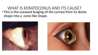
• Corneal cross-linking is a treatment for an EYE problem
called KERATOCONUS
TYPES OF CROSS-LINKING
• Epithelium-off cross-linking. In this procedure, the thin outer layer
(epithelium) of the cornea is removed to allow the liquid riboflavin to
more easily penetrate the deeper corneal tissue.
• Epithelium-on cross-linking. In this procedure (also called
transepithelial cross-linking), the protective corneal epithelium is left
intact.
Procedure and process
• Eyedrop medication and ultraviolet (UV) light are used to make the tissues
in your cornea stronger. The goal is to keep the cornea from bulging more.
• It’s called “cross-linking” because it adds bonds between the collagen
fibers in your eye. They work like support beams to help the cornea stay
stable.
• specially formulated riboflavin (vitamin B2) eyedrops, which allow your
cornea to better absorb light. It takes about 30 minutes for the drops to
soak into your cornea.
Who Should Get Corneal Cross-Linking?
Every victim of keratoconus.
Best at the early stages when cornea not severely irregular
WHO CAN’T GET IT
• PREGNANT WOMEN
• CORNEA KERATITIS
• CORNEA THICKNESS LESS THAN 400MICRO METERS
• ACTIVE OCCULAR DISEASE
• DIABETES
• IMMUNOCOMPROMISED PATIENT
WHAT IS KERATOCONUS AND ITS CAUSE?
•This is the outward bulging of the cornea from its dome
shape into a cone-like shape.
Causes
•Keratoconus is caused by a decrease in
protective antioxidants in the cornea which leads to weakness
in collagen fibers.
•Commonly starts at the age of 10 years and NOT GENETIC
BUT RUNS IN FAMILIES.
DIAGNOSING KERATOCONUS
• As the cornea changes from a ball shape to a cone shape.
•A sudden change of vision in one eye double when looking
with just one eye
• Objects both near and far looking distorted
• Being uncomfortable driving due to blurry vision, especially at
night
•Performing a cornea topography
TREATMENT
•Eye Glasses
•Contacts
•Collagen cross-linking
•Intacts
•Cornea transplant
MECHANISM OF ACTION
THANKS FOR YOUR ATTENTION

Corneal Cross-Linking .pptx

  • 1.
  • 2.
    OBJECTIVES • WHAT ISCORNEAL CROSS-LINKING? • TYPES OF CROSS-LINKING • Procedure and process • WHAT IS KERATOCONUS AND ITS CAUSE? • HOW IS KERATOCONUS DIAGNOSED? • TREATMENT?
  • 3.
    CORNEAL CROSS-LINKING • Cornealcross-linking is a treatment for an EYE problem called KERATOCONUS
  • 4.
    TYPES OF CROSS-LINKING •Epithelium-off cross-linking. In this procedure, the thin outer layer (epithelium) of the cornea is removed to allow the liquid riboflavin to more easily penetrate the deeper corneal tissue. • Epithelium-on cross-linking. In this procedure (also called transepithelial cross-linking), the protective corneal epithelium is left intact.
  • 5.
    Procedure and process •Eyedrop medication and ultraviolet (UV) light are used to make the tissues in your cornea stronger. The goal is to keep the cornea from bulging more. • It’s called “cross-linking” because it adds bonds between the collagen fibers in your eye. They work like support beams to help the cornea stay stable. • specially formulated riboflavin (vitamin B2) eyedrops, which allow your cornea to better absorb light. It takes about 30 minutes for the drops to soak into your cornea.
  • 6.
    Who Should GetCorneal Cross-Linking? Every victim of keratoconus. Best at the early stages when cornea not severely irregular
  • 7.
    WHO CAN’T GETIT • PREGNANT WOMEN • CORNEA KERATITIS • CORNEA THICKNESS LESS THAN 400MICRO METERS • ACTIVE OCCULAR DISEASE • DIABETES • IMMUNOCOMPROMISED PATIENT
  • 8.
    WHAT IS KERATOCONUSAND ITS CAUSE? •This is the outward bulging of the cornea from its dome shape into a cone-like shape.
  • 9.
    Causes •Keratoconus is causedby a decrease in protective antioxidants in the cornea which leads to weakness in collagen fibers. •Commonly starts at the age of 10 years and NOT GENETIC BUT RUNS IN FAMILIES.
  • 10.
    DIAGNOSING KERATOCONUS • Asthe cornea changes from a ball shape to a cone shape. •A sudden change of vision in one eye double when looking with just one eye • Objects both near and far looking distorted • Being uncomfortable driving due to blurry vision, especially at night •Performing a cornea topography
  • 11.
  • 12.
  • 13.
    THANKS FOR YOURATTENTION

Editor's Notes

  • #4 In this condition, the front part of your eye, called the cornea, thins out and gets weaker over time. This makes it bulge into a cone shape, which can distort your vision and make it hard to see. If the symptoms of keratoconus get severe, you will need a corneal transplant.