SlideShare a Scribd company logo
1 of 36
Download to read offline
liposomes
Dr. Atish S. Mundada
Associate Professor,
SNJB’s SSDJ College of Pharmacy,
Chandwad, Dist. Nashik
Introduction:Introduction:
• The name liposome is derived from two Greek words: 'Lipos'
meaning fat and 'Soma' meaning body.
• Liposomes were first described by British haematologist Dr Alec D
Bangham in 1961 at the Babraham Institute, in Cambridge.
• They were discovered when Bangham and R. W. Horne were
testing the institute's new electron microscope by adding
negative stain to dry phospholipids. The microscope pictures
served as the first real evidence for the liposomes resemblance to
the cell membrane like a bilayer lipid structure.
• Liposomes are small artificial vesicles of spherical shape that can
be created from cholesterol and natural nontoxic phospholipids.
• Generally, liposomes are definite as spherical vesicles with
particle sizes ranging from 30 nm to several micrometers.
• Due to their size and hydrophobic and hydrophilic character
(besides biocompatibility), liposomes are promising systems for
drug delivery.
• Liposome properties differ considerably with lipid composition,
surface charge, size, and the method of preparation.
• Furthermore, the choice of bilayer components determines the
‘rigidity’ or ‘fluidity’ and the charge of the bilayer.
• For instance, unsaturated phosphatidylcholine species from
natural sources (egg or soybean phosphatidylcholine) give much
more permeable and less stable bilayers, whereas the saturated
phospholipids with long acyl chains (for example, dipalmitoylphos
phatidylcholine) form a rigid, rather impermeable bilayer
structure.
• Because of their biocompatibility, biodegradability, low toxicity,
and aptitude to trap both hydrophilic and lipophilic drugs and
simplify site-specific drug delivery to tumor tissues, liposomes
have increased rate both as an investigational system and
commercially as a drug delivery system.
Advantages of liposome:-
• Liposomes increased efficacy and therapeutic index of drug
(actinomycin-D)
• Liposome increased stability via encapsulation
• Liposomes are non-toxic, flexible, biocompatible, completely
biodegradable, and nonimmunogenic for systemic and non-
systemic administrations
• Liposomes reduce the toxicity of the encapsulated agent
(amphotericin B, Taxol)
• Liposomes help reduce the exposure of sensitive tissues to toxic
drugs
• Flexibility to couple with site-specific ligands to achieve active
targeting
Disadvantages of liposome:-
• Low solubility
• Sterilization
• Short half-life
• Sometimes phospholipid undergoes oxidation and hydrolysis-like
reaction
• Leakage and fusion of encapsulated drug/ molecules
• Production cost is high
• Fewer stables
Classification of liposomes:Classification of liposomes:
• Based on the size and number of bilayers:
• Multilamellar vesicles (MLV),
• Large unilamellar vesicles (LUV) and
• Small unilamellar vesicles (SUV).
• Based on composition:
• Conventional liposomes (CL),
• pH-sensitive liposomes,
• Cationic liposomes,
• Long circulating liposomes (LCL) and
• Immuno-liposomes.
• Based on the method of preparation:
• Reverse phase evaporation vesicles (REV),
• French press vesicles (FPV) and
• Ether injection vesicles (EIV)
Methods of liposome preparation:Methods of liposome preparation:
• The correct choice of liposome preparation method depends on
the following parameters:
1) The physicochemical characteristics of the material to be
entrapped and those of the liposomal ingredients;
2) The nature of the medium in which the lipid vesicles are
dispersed;
3) The effective concentration of the entrapped substance and its
potential toxicity;
4) Additional processes involved during application/ delivery of the
vesicles;
5) Optimum size, polydispersity and shelf-life of the vesicles for the
intended application and
6) Batch-to-batch reproducibility and possibility of large-scale
production of safe and efficient liposomal products.
• All the methods of preparing the liposomes involve four basic
stages:
– Drying down lipids from organic solvent.
– Dispersing the lipid in aqueous media.
– Purifying the resultant liposome.
– Analyzing the final product.
• Handling of Liposomes:
• The lipids used in the preparation of liposomes are unsaturated
and hence susceptible to oxidation.
• Volatile solvents such as chloroform which are used will tend to
evaporate from the container.
• Thus liposomes must be stored in an inert atmosphere of
nitrogen and in the dark, in glass vessels with a securely fastened
cap.
Preparation of liposomes by lipid film hydration:
• When preparing liposomes with mixed lipid composition, the
lipids must first be dissolved and mixed in an organic solvent to
assure a homogeneous mixture of lipids.
• Usually this process is carried out using chloroform or chloroform:
methanol mixtures. The intent is to obtain a clear lipid solution for
complete mixing of lipids.
• Typically lipid solutions are prepared at 10-20mg lipid/ml of
organic solvent, although higher concentrations may be used if the
lipid solubility and mixing are acceptable.
• Once the lipids are thoroughly mixed in the organic solvent, the
solvent is removed to yield a lipid film.
• For small volumes of organic solvent (<1mL), the solvent may be
evaporated using a dry nitrogen or argon stream in a fume hood.
For larger volumes, the organic solvent should be removed by
rotary evaporation yielding a thin lipid film on the sides of a round
bottom flask.
Mechanical Dispersion Method:Mechanical Dispersion Method:
• The lipid film is thoroughly dried to remove residual organic
solvent by placing the vial or flask on a vacuum pump overnight.
• Hydration of the dry lipid film/cake is accomplished simply by
adding an aqueous medium to the container of dry lipid and
agitating.
• The temperature of the hydrating medium should be above the
gel liquid crystal transition temperature (Tc or Tm) of the lipid.
• After addition of the hydrating medium, the lipid suspension
should be maintained above the Tc during the hydration period.
• Spinning the round bottom flask in the warm water bath
maintained at a temperature above the Tc of the lipid suspension
allows the lipid to hydrate in its fluid phase with adequate
agitation.
• Hydration time may differ slightly among lipid species and
structure, however, a hydration time of 1 hour with vigorous
shaking, mixing, or stirring is highly recommended.
• The hydration medium is generally determined by the application
of the lipid vesicles.
• Suitable hydration media include distilled water, buffer solutions,
saline, and non-electrolytes such as sugar solutions. Generally
accepted solutions which meet these conditions are 0.9% saline,
5% dextrose and 10% sucrose.
• During hydration some lipids form complexes unique to their
structure. Highly charged lipids have been observed to form a
viscous gel when hydrated with low ionic strength solutions. The
problem can be alleviated by addition of salt or by downsizing the
lipid suspension.
• Poorly hydrating lipids such as phosphatidyl ethanolamine have a
tendency to self aggregate upon hydration.
Sonication:
• Sonication is perhaps the most extensively used method for the
preparation of SUV. Here, MLVs are sonicated either with a bath
type sonicator or a probe sonicator under a passive atmosphere.
• The main disadvantages of this method are very low internal
volume/ encapsulation efficacy, possible degradation of
phospholipids and compounds to be encapsulated, elimination of
large molecules, metal pollution from probe tip, and presence of
MLV along with SUV.
There are two sonication techniques:
• a) Probe sonication: The tip of a sonicator is directly engrossed
into the liposome dispersion.
• b) Bath sonication: The liposome dispersion in a cylinder is placed
into a bath sonicator.
French pressure cell:
• Extrusion French pressure cell involves the extrusion of MLV
through a small orifice at 20,000 psi at 4°C.
• An interesting comment is that French press vesicle appears to
recall entrapped solutes significantly longer than SUVs do,
produced by sonication or detergent removal.
• The method has several advantages over sonication method.
– The method is simple, rapid and reproducible and it involves
gentle handling of unstable materials.
– The resulting liposomes are rather larger than sonicated SUVs.
• The drawbacks of the method are that
– the high temperature is difficult to attain, and
– the working volumes are comparatively small (about 50 mL as
the maximum).
Freeze-thawed liposomes:
• SUVs are rapidly frozen and thawed slowly. The short-lived
sonication disperses aggregated materials to LUV. The creation of
unilamellar vesicles is as a result of the fusion of SUV throughout
the processes of freezing and thawing.
• This type of synthesis is strongly inhibited by increasing the
phospholipid concentration and by increasing the ionic strength of
the medium.
• The encapsulation efficacies from 20% to 30% were obtained.
Solvent dispersion method:Solvent dispersion method:
Ether injection (solvent vaporization):
• A solution of lipids dissolved in diethyl ether or ether-methanol
mixture is gradually injected to an aqueous solution of the
material to be encapsulated at 55°C to 65°C or under reduced
pressure.
• The consequent removal of ether under vacuum leads to the
creation of liposomes.
• The main disadvantages of the technique are that the population
is heterogeneous (70 to 200 nm) and the exposure of compounds
to be encapsulated to organic solvents at high temperature.
Ethanol injection:
• A lipid solution of ethanol is rapidly injected to a huge excess of
buffer. The MLVs are at once formed.
• The disadvantages of the method are
– that the population is heterogeneous (30 to 110 nm),
– liposomes are very dilute,
– the removal all ethanol is difficult because it forms into
azeotrope with water, and
– the probability of the various biologically active
macromolecules to inactivate in the presence of even low
amounts of ethanol is high.
Reverse phase evaporation method:-
• This method provided a progress in liposome technology, since it
allowed for the first time the preparation of liposomes with a high
aqueous space-to-lipid ratio and a capability to entrap a large
percentage of the aqueous material presented.
• Reverse-phase evaporation is based on the creation of inverted
micelles. These inverted micelles are shaped upon sonication of a
mixture of a buffered aqueous phase, which contains the water-
soluble molecules to be encapsulated into the liposomes and an
organic phase in which the amphiphilic molecules are solubilized.
• The slow elimination of the organic solvent leads to the
conversion of these inverted micelles into viscous state and gel
form.
• At a critical point in this process, the gel state collapses, and some
of the inverted micelles were disturbed. The excess of
phospholipids in the environment donates to the formation of a
complete bilayer around the residual micelles, which results in
the creation of liposomes.
Detergent removal method:Detergent removal method:
Dialysis:
• The detergents at their critical micelle concentrations (CMC)
have been used to solubilize lipids.
• As the detergent is detached, the micelles become increasingly
better-off in phospholipid and lastly combine to form LUVs.
• The detergents were removed by dialysis.
• A commercial device called LipoPrep (Diachema AG,
Switzerland), which is a version of dialysis system, is obtainable
for the elimination of detergents.
• The dialysis can be performed in dialysis bags engrossed in large
detergent free buffers (equilibrium dialysis)
Detergent (cholate, alkyl glycoside, Triton X-100) removal of
mixed micelles (absorption):
• Detergent absorption is attained by shaking mixed micelle
solution with beaded organic polystyrene adsorbers such as
XAD-2 beads (SERVA Electrophoresis GmbH, Heidelberg,
Germany) and Bio-beads SM2 (Bio-Rad Laboratories, Inc.,
Hercules, USA).
• The great benefit of using detergent adsorbers is that they can
eliminate detergents with a very low CMC, which are not
entirely depleted.
Gel-permeation chromatography:-
• In this method, the detergent is depleted by size special
chromatography.
• Sephadex G-50, Sephadex G-l 00 (Sigma-Aldrich, MO, USA),
Sepharose 2B-6B, and Sephacryl S200-S1000 (General Electric
Company, Tehran, Iran) can be used for gel filtration.
• The liposomes do not penetrate into the pores of the beads
packed in a column. They percolate through the inter-bead
spaces. At slow flow rates, the separation of liposomes from
detergent monomers is very good.
• The swollen polysaccharide beads adsorb substantial amounts of
amphiphilic lipids; therefore, pretreatment is necessary.
• The pre-treatment is done by pre-saturation of the gel filtration
column by lipids using empty liposome suspensions.
Dilution:-
• Upon dilution of aqueous mixed micellar solution of detergent
and phospholipids with buffer, the micellar size and the
polydispersity increase fundamentally, and as the system is
diluted beyond the mixed micellar phase boundary, a
spontaneous transition from poly-dispersed micelles to
vesicles occurs.
Stealth liposomes and conventional liposomes:-
• Although liposomes are like biomembranes, they are still foreign
objects of the body. Therefore, liposomes are known by the
mononuclear phagocytic system (MPS) after contact with plasma
proteins. Accordingly, liposomes are cleared from the blood
stream.
• These stability difficulties are solved through the use of synthetic
phospholipids, particle coated with amphipathic polyethylene
glycol, coating liposomes with chitin derivatives, freeze drying,
polymerization, micro-encapsulation of gangliosides.
• Coating liposomes with PEG reduces the percentage of uptake by
macrophages and leads to a prolonged presence of liposomes in
the circulation and, therefore, make available abundant time for
these liposomes to leak from the circulation through leaky
endothelium.
• A stealth liposome is a sphere-shaped vesicle with a membrane
composed of phospholipid bilayer used to deliver drugs or genetic
material into a cell.
• A liposome can be composed of naturally derived phospholipids
with mixed lipid chains coated or steadied by polymers of PEG and
colloidal in nature.
• Stealth liposomes are attained and grown in new drug delivery
and in controlled release.
• This stealth principle has been used to develop the successful
doxorubicin-loaded liposome product that is presently marketed
as Doxil (Janssen Biotech, Inc., Horsham, USA) or Caelyx (Schering-
Plough Corporation, Kenilworth, USA) for the treatment of solid
tumors.
• Recently impressive therapeutic improvements were described
with the useof corticosteroidloaded liposome in experimental
arthritic models.
Mechanism of Liposome Formation:Mechanism of Liposome Formation:
• The basic part of liposome is formed by phospholipids, which are
amphiphilic molecules (having a hydrophilic head and
hydrophobic tail).
• The hydrophilic part is mainly phosphoric acid bound to a water
soluble molecule, whereas, the hydrophobic part consists of two
fatty acid chains with 10 – 24 carbon atoms and 0 – 6 double
bonds in each chain.
• When these phospholipids are dispersed in aqueous medium,
they form lamellar sheets by organizing in such a way that, the
polar head group faces outwards to the aqueous region while the
fatty acid groups face each other and finally form spherical/
vesicle like structures called as liposomes.
• The polar portion remains in contact with aqueous region along
with shielding of the non-polar part (which is oriented at an angle
to the membrane surface).
• When phospholipids are hydrated in water, along with the input
of energy like sonication, shaking, heating, homogenization, etc.
• It is the hydrophilic/ hydrophobic interactions between lipid –
lipid, lipid – water molecules that lead to the formation of
bilayered vesicles in order to achieve a thermodynamic
equilibrium in the aqueous phase.
• The reasons for bilayered formation include:
– The unfavorable interactions created between hydrophilic and
hydrophobic phase can be minimized by folding into closed
concentric vesicles.
– Large bilayered vesicle formation promotes the reduction of
large free energy difference present between the hydrophilic
and hydrophobic environment.
– Maximum stability to supramolecular self assembled structure
can be attained by forming into vesicles.
Evaluation of Liposomes:Evaluation of Liposomes:
• The characterization parameters for purpose of evaluation could
be classified into three broad Categories, which include physical,
chemical and biological parameters.
• Physical characterization evaluates various parameters including
size, shape, surface features, lamellarity phase-behaviour and drug
release profile.
• Chemical characterization includes those studies which establish
the purity and potency of various lipophillic constituents.
• Biological characterization parameters are helpful in establishing
the safety and suitability of formulation for therapeutic
application.
Size and size distribution:-
• When liposomes are intended for inhalation or parenteral
administration, the size distribution is of primary consideration,
since it influences the in vivo fate of liposomes along with the
encapsulated drug molecules.
• Various techniques of determining the size of the vesicles include
– microscopy (optical microscopy, negative stain transmission
electron microscopy, cryo-transmission electron microscopy,
freeze fracture electron microscopy and scanning electron
microscopy
– diffraction and scattering techniques (laser light scattering and
photon correlation spectroscopy) and
– hydrodynamic techniques (field flow fractionation, gel
permeation and ultracentrifugation).
Percent drug encapsulation:-
• The amount of drug encapsulated/ entrapped in liposome vesicle
is given by percent drug encapsulation.
• Column chromatography can be used to estimate the percent
drug encapsulation of liposomes.
• The formulation consists of both free (un-encapsulated) and
encapsulated drug. So as to know the exact amount of drug
encapsulated, the free drug is separated from the encapsulated
one.
• Then the fraction of liposomes containing the encapsulated drug
is treated with a detergent, so as to attain lysis, which leads to the
discharge of the drug from the vesicles into the surrounding
medium.
• This exposed drug is assayed by a suitable technique which gives
the percent drug encapsulated from which encapsulation
efficiency can be calculated.
• Trapped volume per lipid weight can also give the percent drug
encapsulated in a liposome vesicle. It is generally expressed as
aqueous volume entrapped per unit quantity of lipid, Âľl/Âľmol or
Âľg/mg of total lipid.
• In order to determine the trapped volume, various materials like
radioactive markers, fluorescent markers and spectroscopically
inert fluid can be used.
• Radioactive method is mostly used for determining trapped
volume. It is determined by dispersing lipid in an aqueous
medium containing a non-permeable radioactive solute like
[22Na] or [14C] inulin.
• Alternatively, water soluble markers like 6-carboxyfluorescein,
14C or 3H-glucose or sucrose can be used to determine the
trapped volume
Surface charge:-
• Since the charge on the liposome surface plays a key role in the in vivo
disposition, it is essential to know the surface charge on the vesicle
surface.
• Two methods namely, free-flow electrophoresis and zeta potential
measurement can be used to estimate the surface charge of the vesicle.
• The surface charge can be calculated by estimating the mobility of the
liposomal dispersion in a suitable buffer (determined using Helmholtz–
Smolochowski equation).
Vesicle shape and lamellarity:-
• Various electron microscopic techniques can be used to assess the shape
of the vesicles. The number of bilayers present in the liposome, i.e.,
lamellarity can be determined using freeze fracture electron microscopy
and 31P-Nuclear magnetic resonance analysis.
• Apart from knowing the shape and lamellarity, the surface morphology
of liposomes can be assessed using freeze-fracture and freeze-etch
electron microscopy.
Phospholipid identification and assay:-
• The chemical components of liposomes must be analyzed prior to
and after the preparation.
• Barlett assay, Stewart assay and thin layer chromatography can be
used to estimate the phospholipid concentration in the liposomal
formulation.
• A spectrophotometric method to quantify total phosphorous in a
sample was given in literature, which measure the intensity of
blue color developed at 825 nm against water.
• Cholesterol oxidase assay or ferric perchlorate method and Gas
liquid chromatography techniques can be used to determine the
cholesterol concentration.
Stability of Liposomes:-
• During the development of liposomal drug products, the stability
of the developed formulation is of major consideration.
• The therapeutic activity of the drug is governed by the stability of
the liposomes right from the manufacturing steps to storage to
delivery.
• A stable dosage forms is the one which maintains the physical
stability and chemical integrity of the active molecule during its
developmental procedure and storage.
• A well designed stability study includes the evaluation of its
physical, chemical and microbial parameters along with the
assurance of product’s integrity throughout its storage period.
• Hence a stability protocol is essential to study the physical and
chemical integrity of the drug product in its storage.
Liposomes dr. asm

More Related Content

What's hot

Microcapsules: types, preparation and evaluation
Microcapsules: types, preparation and evaluationMicrocapsules: types, preparation and evaluation
Microcapsules: types, preparation and evaluationMOHAMMAD ASIM
 
Formulation and invitro evaluation of microspheres
Formulation and invitro evaluation of  microspheresFormulation and invitro evaluation of  microspheres
Formulation and invitro evaluation of microspheresTejaswi Kurma
 
Microencapsulation
MicroencapsulationMicroencapsulation
MicroencapsulationChirag Goda
 
Microencapsulation
MicroencapsulationMicroencapsulation
MicroencapsulationArpitSuralkar
 
Microencapsulation by Mali vv
Microencapsulation by Mali vv Microencapsulation by Mali vv
Microencapsulation by Mali vv VidhyaMali1
 
MANUFACTURING EQUIPMENTS,EVALUATION & STABILITY ASPECTS OF MICROCAPSULE
MANUFACTURING EQUIPMENTS,EVALUATION &STABILITY ASPECTS OF MICROCAPSULEMANUFACTURING EQUIPMENTS,EVALUATION &STABILITY ASPECTS OF MICROCAPSULE
MANUFACTURING EQUIPMENTS,EVALUATION & STABILITY ASPECTS OF MICROCAPSULESagar Savale
 
Nanosuspension
NanosuspensionNanosuspension
NanosuspensionAnil Pethe
 
Microencapsulation
MicroencapsulationMicroencapsulation
MicroencapsulationPooja Sadgir
 
Ppt microencapsulation
Ppt microencapsulationPpt microencapsulation
Ppt microencapsulationSANA TABASSUM
 
Microencapsulation
MicroencapsulationMicroencapsulation
MicroencapsulationSamiksha Sawant
 
Microencapsulation
Microencapsulation Microencapsulation
Microencapsulation Gauri Kaphare
 
Coacervation Phase Separation Techniques
Coacervation Phase Separation TechniquesCoacervation Phase Separation Techniques
Coacervation Phase Separation TechniquesGargi Nanda
 
microsphere and microencapsulation
microsphere and microencapsulationmicrosphere and microencapsulation
microsphere and microencapsulationDebash8
 
Solubility enhancement by using various techniques
Solubility enhancement by using various techniques Solubility enhancement by using various techniques
Solubility enhancement by using various techniques Prajakta Chavan
 
Microencapsulation (2)
Microencapsulation (2)Microencapsulation (2)
Microencapsulation (2)Shivaram
 
Cubosomes a drug delivery system by Shree
Cubosomes a drug delivery system by Shree Cubosomes a drug delivery system by Shree
Cubosomes a drug delivery system by Shree ShrikantDivekar
 

What's hot (20)

Microcapsules: types, preparation and evaluation
Microcapsules: types, preparation and evaluationMicrocapsules: types, preparation and evaluation
Microcapsules: types, preparation and evaluation
 
Formulation and invitro evaluation of microspheres
Formulation and invitro evaluation of  microspheresFormulation and invitro evaluation of  microspheres
Formulation and invitro evaluation of microspheres
 
Microencapsulation
MicroencapsulationMicroencapsulation
Microencapsulation
 
Microencapsulation
MicroencapsulationMicroencapsulation
Microencapsulation
 
Microencapsulation by Mali vv
Microencapsulation by Mali vv Microencapsulation by Mali vv
Microencapsulation by Mali vv
 
Niosomes
NiosomesNiosomes
Niosomes
 
MANUFACTURING EQUIPMENTS,EVALUATION & STABILITY ASPECTS OF MICROCAPSULE
MANUFACTURING EQUIPMENTS,EVALUATION &STABILITY ASPECTS OF MICROCAPSULEMANUFACTURING EQUIPMENTS,EVALUATION &STABILITY ASPECTS OF MICROCAPSULE
MANUFACTURING EQUIPMENTS,EVALUATION & STABILITY ASPECTS OF MICROCAPSULE
 
Nanosuspension
NanosuspensionNanosuspension
Nanosuspension
 
Microencapsulation
MicroencapsulationMicroencapsulation
Microencapsulation
 
Ppt microencapsulation
Ppt microencapsulationPpt microencapsulation
Ppt microencapsulation
 
Microencapsulation
MicroencapsulationMicroencapsulation
Microencapsulation
 
Microencapsulation
MicroencapsulationMicroencapsulation
Microencapsulation
 
Microencapsulation
Microencapsulation Microencapsulation
Microencapsulation
 
Coacervation Phase Separation Techniques
Coacervation Phase Separation TechniquesCoacervation Phase Separation Techniques
Coacervation Phase Separation Techniques
 
microsphere and microencapsulation
microsphere and microencapsulationmicrosphere and microencapsulation
microsphere and microencapsulation
 
Solubility enhancement by using various techniques
Solubility enhancement by using various techniques Solubility enhancement by using various techniques
Solubility enhancement by using various techniques
 
Microencapsulation
MicroencapsulationMicroencapsulation
Microencapsulation
 
Microencapsulation
Microencapsulation Microencapsulation
Microencapsulation
 
Microencapsulation (2)
Microencapsulation (2)Microencapsulation (2)
Microencapsulation (2)
 
Cubosomes a drug delivery system by Shree
Cubosomes a drug delivery system by Shree Cubosomes a drug delivery system by Shree
Cubosomes a drug delivery system by Shree
 

Similar to Liposomes dr. asm

Liposomes-Classification, methods of preparation and application
Liposomes-Classification, methods of preparation and application Liposomes-Classification, methods of preparation and application
Liposomes-Classification, methods of preparation and application Vijay Hemmadi
 
Liposomal drug delivery systems an overview
Liposomal drug delivery systems an overviewLiposomal drug delivery systems an overview
Liposomal drug delivery systems an overviewsana916816
 
Liposomes- A Novel Drug Delivery System
Liposomes- A Novel Drug Delivery SystemLiposomes- A Novel Drug Delivery System
Liposomes- A Novel Drug Delivery SystemSnehal Dhobale
 
Liposomes- A Novel Drug Delivery System
Liposomes- A Novel Drug Delivery SystemLiposomes- A Novel Drug Delivery System
Liposomes- A Novel Drug Delivery SystemSnehal Dhobale
 
Snehal liposomes
Snehal liposomesSnehal liposomes
Snehal liposomessnehal dhobale
 
Preparation of liposomes.methods
Preparation of liposomes.methodsPreparation of liposomes.methods
Preparation of liposomes.methodsSushma Rajuri
 
LIPOSOMES USED AS AN DRUG DELIVERY SYSTEMS
LIPOSOMES USED AS AN DRUG DELIVERY SYSTEMSLIPOSOMES USED AS AN DRUG DELIVERY SYSTEMS
LIPOSOMES USED AS AN DRUG DELIVERY SYSTEMSROHIT
 
Liposomes - Targeted drug delivery system
Liposomes - Targeted drug delivery systemLiposomes - Targeted drug delivery system
Liposomes - Targeted drug delivery systemJyoti Nautiyal
 
liposomes.pptx
liposomes.pptxliposomes.pptx
liposomes.pptxTenzinPema20
 

Similar to Liposomes dr. asm (20)

Liposomes-Classification, methods of preparation and application
Liposomes-Classification, methods of preparation and application Liposomes-Classification, methods of preparation and application
Liposomes-Classification, methods of preparation and application
 
Liposomes- overview
Liposomes- overview Liposomes- overview
Liposomes- overview
 
Liposomal drug delivery systems an overview
Liposomal drug delivery systems an overviewLiposomal drug delivery systems an overview
Liposomal drug delivery systems an overview
 
Liposomes
LiposomesLiposomes
Liposomes
 
Liposomes- A Novel Drug Delivery System
Liposomes- A Novel Drug Delivery SystemLiposomes- A Novel Drug Delivery System
Liposomes- A Novel Drug Delivery System
 
Liposomes- A Novel Drug Delivery System
Liposomes- A Novel Drug Delivery SystemLiposomes- A Novel Drug Delivery System
Liposomes- A Novel Drug Delivery System
 
Snehal liposomes
Snehal liposomesSnehal liposomes
Snehal liposomes
 
Preparation of liposomes.methods
Preparation of liposomes.methodsPreparation of liposomes.methods
Preparation of liposomes.methods
 
Liposomes
LiposomesLiposomes
Liposomes
 
LIPOSOMES USED AS AN DRUG DELIVERY SYSTEMS
LIPOSOMES USED AS AN DRUG DELIVERY SYSTEMSLIPOSOMES USED AS AN DRUG DELIVERY SYSTEMS
LIPOSOMES USED AS AN DRUG DELIVERY SYSTEMS
 
Liposomes
LiposomesLiposomes
Liposomes
 
Liposome
LiposomeLiposome
Liposome
 
LIPOSOME
LIPOSOMELIPOSOME
LIPOSOME
 
Liposomes - Targeted drug delivery system
Liposomes - Targeted drug delivery systemLiposomes - Targeted drug delivery system
Liposomes - Targeted drug delivery system
 
LIPOSOMES@.pptx
LIPOSOMES@.pptxLIPOSOMES@.pptx
LIPOSOMES@.pptx
 
Liposomes
LiposomesLiposomes
Liposomes
 
liposome
liposomeliposome
liposome
 
liposomes.pptx
liposomes.pptxliposomes.pptx
liposomes.pptx
 
Niosomes
NiosomesNiosomes
Niosomes
 
Liposome
LiposomeLiposome
Liposome
 

More from SNJBs SSDJ College of Pharmacy, Chandwad (7)

Gene therapy dr. asm
Gene therapy dr. asmGene therapy dr. asm
Gene therapy dr. asm
 
Nucleic acid based therapeutics dr. asm
Nucleic acid based therapeutics dr. asmNucleic acid based therapeutics dr. asm
Nucleic acid based therapeutics dr. asm
 
Monoclonal antibodies dr. asm
Monoclonal antibodies dr. asmMonoclonal antibodies dr. asm
Monoclonal antibodies dr. asm
 
Niosomes
NiosomesNiosomes
Niosomes
 
Targeting methods nanoparticles dr. asm
Targeting methods nanoparticles dr. asmTargeting methods nanoparticles dr. asm
Targeting methods nanoparticles dr. asm
 
Chemical kinetics dr. asm
Chemical kinetics dr. asmChemical kinetics dr. asm
Chemical kinetics dr. asm
 
Solubility of drugs by Dr. Atish Mundada
Solubility of drugs by Dr. Atish MundadaSolubility of drugs by Dr. Atish Mundada
Solubility of drugs by Dr. Atish Mundada
 

Recently uploaded

Class 11 Legal Studies Ch-1 Concept of State .pdf
Class 11 Legal Studies Ch-1 Concept of State .pdfClass 11 Legal Studies Ch-1 Concept of State .pdf
Class 11 Legal Studies Ch-1 Concept of State .pdfakmcokerachita
 
The Most Excellent Way | 1 Corinthians 13
The Most Excellent Way | 1 Corinthians 13The Most Excellent Way | 1 Corinthians 13
The Most Excellent Way | 1 Corinthians 13Steve Thomason
 
The basics of sentences session 2pptx copy.pptx
The basics of sentences session 2pptx copy.pptxThe basics of sentences session 2pptx copy.pptx
The basics of sentences session 2pptx copy.pptxheathfieldcps1
 
Enzyme, Pharmaceutical Aids, Miscellaneous Last Part of Chapter no 5th.pdf
Enzyme, Pharmaceutical Aids, Miscellaneous Last Part of Chapter no 5th.pdfEnzyme, Pharmaceutical Aids, Miscellaneous Last Part of Chapter no 5th.pdf
Enzyme, Pharmaceutical Aids, Miscellaneous Last Part of Chapter no 5th.pdfSumit Tiwari
 
Call Girls in Dwarka Mor Delhi Contact Us 9654467111
Call Girls in Dwarka Mor Delhi Contact Us 9654467111Call Girls in Dwarka Mor Delhi Contact Us 9654467111
Call Girls in Dwarka Mor Delhi Contact Us 9654467111Sapana Sha
 
call girls in Kamla Market (DELHI) 🔝 >༒9953330565🔝 genuine Escort Service 🔝✔️✔️
call girls in Kamla Market (DELHI) 🔝 >༒9953330565🔝 genuine Escort Service 🔝✔️✔️call girls in Kamla Market (DELHI) 🔝 >༒9953330565🔝 genuine Escort Service 🔝✔️✔️
call girls in Kamla Market (DELHI) 🔝 >༒9953330565🔝 genuine Escort Service 🔝✔️✔️9953056974 Low Rate Call Girls In Saket, Delhi NCR
 
ECONOMIC CONTEXT - LONG FORM TV DRAMA - PPT
ECONOMIC CONTEXT - LONG FORM TV DRAMA - PPTECONOMIC CONTEXT - LONG FORM TV DRAMA - PPT
ECONOMIC CONTEXT - LONG FORM TV DRAMA - PPTiammrhaywood
 
Science 7 - LAND and SEA BREEZE and its Characteristics
Science 7 - LAND and SEA BREEZE and its CharacteristicsScience 7 - LAND and SEA BREEZE and its Characteristics
Science 7 - LAND and SEA BREEZE and its CharacteristicsKarinaGenton
 
KSHARA STURA .pptx---KSHARA KARMA THERAPY (CAUSTIC THERAPY)————IMP.OF KSHARA ...
KSHARA STURA .pptx---KSHARA KARMA THERAPY (CAUSTIC THERAPY)————IMP.OF KSHARA ...KSHARA STURA .pptx---KSHARA KARMA THERAPY (CAUSTIC THERAPY)————IMP.OF KSHARA ...
KSHARA STURA .pptx---KSHARA KARMA THERAPY (CAUSTIC THERAPY)————IMP.OF KSHARA ...M56BOOKSTORE PRODUCT/SERVICE
 
Hybridoma Technology ( Production , Purification , and Application )
Hybridoma Technology  ( Production , Purification , and Application  ) Hybridoma Technology  ( Production , Purification , and Application  )
Hybridoma Technology ( Production , Purification , and Application ) Sakshi Ghasle
 
_Math 4-Q4 Week 5.pptx Steps in Collecting Data
_Math 4-Q4 Week 5.pptx Steps in Collecting Data_Math 4-Q4 Week 5.pptx Steps in Collecting Data
_Math 4-Q4 Week 5.pptx Steps in Collecting DataJhengPantaleon
 
How to Configure Email Server in Odoo 17
How to Configure Email Server in Odoo 17How to Configure Email Server in Odoo 17
How to Configure Email Server in Odoo 17Celine George
 
18-04-UA_REPORT_MEDIALITERAĐĄY_INDEX-DM_23-1-final-eng.pdf
18-04-UA_REPORT_MEDIALITERAĐĄY_INDEX-DM_23-1-final-eng.pdf18-04-UA_REPORT_MEDIALITERAĐĄY_INDEX-DM_23-1-final-eng.pdf
18-04-UA_REPORT_MEDIALITERAĐĄY_INDEX-DM_23-1-final-eng.pdfssuser54595a
 
Accessible design: Minimum effort, maximum impact
Accessible design: Minimum effort, maximum impactAccessible design: Minimum effort, maximum impact
Accessible design: Minimum effort, maximum impactdawncurless
 
microwave assisted reaction. General introduction
microwave assisted reaction. General introductionmicrowave assisted reaction. General introduction
microwave assisted reaction. General introductionMaksud Ahmed
 
Sanyam Choudhary Chemistry practical.pdf
Sanyam Choudhary Chemistry practical.pdfSanyam Choudhary Chemistry practical.pdf
Sanyam Choudhary Chemistry practical.pdfsanyamsingh5019
 
Incoming and Outgoing Shipments in 1 STEP Using Odoo 17
Incoming and Outgoing Shipments in 1 STEP Using Odoo 17Incoming and Outgoing Shipments in 1 STEP Using Odoo 17
Incoming and Outgoing Shipments in 1 STEP Using Odoo 17Celine George
 
Solving Puzzles Benefits Everyone (English).pptx
Solving Puzzles Benefits Everyone (English).pptxSolving Puzzles Benefits Everyone (English).pptx
Solving Puzzles Benefits Everyone (English).pptxOH TEIK BIN
 
A Critique of the Proposed National Education Policy Reform
A Critique of the Proposed National Education Policy ReformA Critique of the Proposed National Education Policy Reform
A Critique of the Proposed National Education Policy ReformChameera Dedduwage
 

Recently uploaded (20)

Class 11 Legal Studies Ch-1 Concept of State .pdf
Class 11 Legal Studies Ch-1 Concept of State .pdfClass 11 Legal Studies Ch-1 Concept of State .pdf
Class 11 Legal Studies Ch-1 Concept of State .pdf
 
The Most Excellent Way | 1 Corinthians 13
The Most Excellent Way | 1 Corinthians 13The Most Excellent Way | 1 Corinthians 13
The Most Excellent Way | 1 Corinthians 13
 
TataKelola dan KamSiber Kecerdasan Buatan v022.pdf
TataKelola dan KamSiber Kecerdasan Buatan v022.pdfTataKelola dan KamSiber Kecerdasan Buatan v022.pdf
TataKelola dan KamSiber Kecerdasan Buatan v022.pdf
 
The basics of sentences session 2pptx copy.pptx
The basics of sentences session 2pptx copy.pptxThe basics of sentences session 2pptx copy.pptx
The basics of sentences session 2pptx copy.pptx
 
Enzyme, Pharmaceutical Aids, Miscellaneous Last Part of Chapter no 5th.pdf
Enzyme, Pharmaceutical Aids, Miscellaneous Last Part of Chapter no 5th.pdfEnzyme, Pharmaceutical Aids, Miscellaneous Last Part of Chapter no 5th.pdf
Enzyme, Pharmaceutical Aids, Miscellaneous Last Part of Chapter no 5th.pdf
 
Call Girls in Dwarka Mor Delhi Contact Us 9654467111
Call Girls in Dwarka Mor Delhi Contact Us 9654467111Call Girls in Dwarka Mor Delhi Contact Us 9654467111
Call Girls in Dwarka Mor Delhi Contact Us 9654467111
 
call girls in Kamla Market (DELHI) 🔝 >༒9953330565🔝 genuine Escort Service 🔝✔️✔️
call girls in Kamla Market (DELHI) 🔝 >༒9953330565🔝 genuine Escort Service 🔝✔️✔️call girls in Kamla Market (DELHI) 🔝 >༒9953330565🔝 genuine Escort Service 🔝✔️✔️
call girls in Kamla Market (DELHI) 🔝 >༒9953330565🔝 genuine Escort Service 🔝✔️✔️
 
ECONOMIC CONTEXT - LONG FORM TV DRAMA - PPT
ECONOMIC CONTEXT - LONG FORM TV DRAMA - PPTECONOMIC CONTEXT - LONG FORM TV DRAMA - PPT
ECONOMIC CONTEXT - LONG FORM TV DRAMA - PPT
 
Science 7 - LAND and SEA BREEZE and its Characteristics
Science 7 - LAND and SEA BREEZE and its CharacteristicsScience 7 - LAND and SEA BREEZE and its Characteristics
Science 7 - LAND and SEA BREEZE and its Characteristics
 
KSHARA STURA .pptx---KSHARA KARMA THERAPY (CAUSTIC THERAPY)————IMP.OF KSHARA ...
KSHARA STURA .pptx---KSHARA KARMA THERAPY (CAUSTIC THERAPY)————IMP.OF KSHARA ...KSHARA STURA .pptx---KSHARA KARMA THERAPY (CAUSTIC THERAPY)————IMP.OF KSHARA ...
KSHARA STURA .pptx---KSHARA KARMA THERAPY (CAUSTIC THERAPY)————IMP.OF KSHARA ...
 
Hybridoma Technology ( Production , Purification , and Application )
Hybridoma Technology  ( Production , Purification , and Application  ) Hybridoma Technology  ( Production , Purification , and Application  )
Hybridoma Technology ( Production , Purification , and Application )
 
_Math 4-Q4 Week 5.pptx Steps in Collecting Data
_Math 4-Q4 Week 5.pptx Steps in Collecting Data_Math 4-Q4 Week 5.pptx Steps in Collecting Data
_Math 4-Q4 Week 5.pptx Steps in Collecting Data
 
How to Configure Email Server in Odoo 17
How to Configure Email Server in Odoo 17How to Configure Email Server in Odoo 17
How to Configure Email Server in Odoo 17
 
18-04-UA_REPORT_MEDIALITERAĐĄY_INDEX-DM_23-1-final-eng.pdf
18-04-UA_REPORT_MEDIALITERAĐĄY_INDEX-DM_23-1-final-eng.pdf18-04-UA_REPORT_MEDIALITERAĐĄY_INDEX-DM_23-1-final-eng.pdf
18-04-UA_REPORT_MEDIALITERAĐĄY_INDEX-DM_23-1-final-eng.pdf
 
Accessible design: Minimum effort, maximum impact
Accessible design: Minimum effort, maximum impactAccessible design: Minimum effort, maximum impact
Accessible design: Minimum effort, maximum impact
 
microwave assisted reaction. General introduction
microwave assisted reaction. General introductionmicrowave assisted reaction. General introduction
microwave assisted reaction. General introduction
 
Sanyam Choudhary Chemistry practical.pdf
Sanyam Choudhary Chemistry practical.pdfSanyam Choudhary Chemistry practical.pdf
Sanyam Choudhary Chemistry practical.pdf
 
Incoming and Outgoing Shipments in 1 STEP Using Odoo 17
Incoming and Outgoing Shipments in 1 STEP Using Odoo 17Incoming and Outgoing Shipments in 1 STEP Using Odoo 17
Incoming and Outgoing Shipments in 1 STEP Using Odoo 17
 
Solving Puzzles Benefits Everyone (English).pptx
Solving Puzzles Benefits Everyone (English).pptxSolving Puzzles Benefits Everyone (English).pptx
Solving Puzzles Benefits Everyone (English).pptx
 
A Critique of the Proposed National Education Policy Reform
A Critique of the Proposed National Education Policy ReformA Critique of the Proposed National Education Policy Reform
A Critique of the Proposed National Education Policy Reform
 

Liposomes dr. asm

  • 1. liposomes Dr. Atish S. Mundada Associate Professor, SNJB’s SSDJ College of Pharmacy, Chandwad, Dist. Nashik
  • 2. Introduction:Introduction: • The name liposome is derived from two Greek words: 'Lipos' meaning fat and 'Soma' meaning body. • Liposomes were first described by British haematologist Dr Alec D Bangham in 1961 at the Babraham Institute, in Cambridge. • They were discovered when Bangham and R. W. Horne were testing the institute's new electron microscope by adding negative stain to dry phospholipids. The microscope pictures served as the first real evidence for the liposomes resemblance to the cell membrane like a bilayer lipid structure. • Liposomes are small artificial vesicles of spherical shape that can be created from cholesterol and natural nontoxic phospholipids. • Generally, liposomes are definite as spherical vesicles with particle sizes ranging from 30 nm to several micrometers. • Due to their size and hydrophobic and hydrophilic character (besides biocompatibility), liposomes are promising systems for drug delivery.
  • 3. • Liposome properties differ considerably with lipid composition, surface charge, size, and the method of preparation. • Furthermore, the choice of bilayer components determines the ‘rigidity’ or ‘fluidity’ and the charge of the bilayer. • For instance, unsaturated phosphatidylcholine species from natural sources (egg or soybean phosphatidylcholine) give much more permeable and less stable bilayers, whereas the saturated phospholipids with long acyl chains (for example, dipalmitoylphos phatidylcholine) form a rigid, rather impermeable bilayer structure. • Because of their biocompatibility, biodegradability, low toxicity, and aptitude to trap both hydrophilic and lipophilic drugs and simplify site-specific drug delivery to tumor tissues, liposomes have increased rate both as an investigational system and commercially as a drug delivery system.
  • 4. Advantages of liposome:- • Liposomes increased efficacy and therapeutic index of drug (actinomycin-D) • Liposome increased stability via encapsulation • Liposomes are non-toxic, flexible, biocompatible, completely biodegradable, and nonimmunogenic for systemic and non- systemic administrations • Liposomes reduce the toxicity of the encapsulated agent (amphotericin B, Taxol) • Liposomes help reduce the exposure of sensitive tissues to toxic drugs • Flexibility to couple with site-specific ligands to achieve active targeting
  • 5. Disadvantages of liposome:- • Low solubility • Sterilization • Short half-life • Sometimes phospholipid undergoes oxidation and hydrolysis-like reaction • Leakage and fusion of encapsulated drug/ molecules • Production cost is high • Fewer stables
  • 6. Classification of liposomes:Classification of liposomes: • Based on the size and number of bilayers: • Multilamellar vesicles (MLV), • Large unilamellar vesicles (LUV) and • Small unilamellar vesicles (SUV). • Based on composition: • Conventional liposomes (CL), • pH-sensitive liposomes, • Cationic liposomes, • Long circulating liposomes (LCL) and • Immuno-liposomes. • Based on the method of preparation: • Reverse phase evaporation vesicles (REV), • French press vesicles (FPV) and • Ether injection vesicles (EIV)
  • 7.
  • 8.
  • 9. Methods of liposome preparation:Methods of liposome preparation: • The correct choice of liposome preparation method depends on the following parameters: 1) The physicochemical characteristics of the material to be entrapped and those of the liposomal ingredients; 2) The nature of the medium in which the lipid vesicles are dispersed; 3) The effective concentration of the entrapped substance and its potential toxicity; 4) Additional processes involved during application/ delivery of the vesicles; 5) Optimum size, polydispersity and shelf-life of the vesicles for the intended application and 6) Batch-to-batch reproducibility and possibility of large-scale production of safe and efficient liposomal products.
  • 10.
  • 11. • All the methods of preparing the liposomes involve four basic stages: – Drying down lipids from organic solvent. – Dispersing the lipid in aqueous media. – Purifying the resultant liposome. – Analyzing the final product. • Handling of Liposomes: • The lipids used in the preparation of liposomes are unsaturated and hence susceptible to oxidation. • Volatile solvents such as chloroform which are used will tend to evaporate from the container. • Thus liposomes must be stored in an inert atmosphere of nitrogen and in the dark, in glass vessels with a securely fastened cap.
  • 12. Preparation of liposomes by lipid film hydration: • When preparing liposomes with mixed lipid composition, the lipids must first be dissolved and mixed in an organic solvent to assure a homogeneous mixture of lipids. • Usually this process is carried out using chloroform or chloroform: methanol mixtures. The intent is to obtain a clear lipid solution for complete mixing of lipids. • Typically lipid solutions are prepared at 10-20mg lipid/ml of organic solvent, although higher concentrations may be used if the lipid solubility and mixing are acceptable. • Once the lipids are thoroughly mixed in the organic solvent, the solvent is removed to yield a lipid film. • For small volumes of organic solvent (<1mL), the solvent may be evaporated using a dry nitrogen or argon stream in a fume hood. For larger volumes, the organic solvent should be removed by rotary evaporation yielding a thin lipid film on the sides of a round bottom flask. Mechanical Dispersion Method:Mechanical Dispersion Method:
  • 13. • The lipid film is thoroughly dried to remove residual organic solvent by placing the vial or flask on a vacuum pump overnight. • Hydration of the dry lipid film/cake is accomplished simply by adding an aqueous medium to the container of dry lipid and agitating. • The temperature of the hydrating medium should be above the gel liquid crystal transition temperature (Tc or Tm) of the lipid. • After addition of the hydrating medium, the lipid suspension should be maintained above the Tc during the hydration period. • Spinning the round bottom flask in the warm water bath maintained at a temperature above the Tc of the lipid suspension allows the lipid to hydrate in its fluid phase with adequate agitation. • Hydration time may differ slightly among lipid species and structure, however, a hydration time of 1 hour with vigorous shaking, mixing, or stirring is highly recommended.
  • 14. • The hydration medium is generally determined by the application of the lipid vesicles. • Suitable hydration media include distilled water, buffer solutions, saline, and non-electrolytes such as sugar solutions. Generally accepted solutions which meet these conditions are 0.9% saline, 5% dextrose and 10% sucrose. • During hydration some lipids form complexes unique to their structure. Highly charged lipids have been observed to form a viscous gel when hydrated with low ionic strength solutions. The problem can be alleviated by addition of salt or by downsizing the lipid suspension. • Poorly hydrating lipids such as phosphatidyl ethanolamine have a tendency to self aggregate upon hydration.
  • 15. Sonication: • Sonication is perhaps the most extensively used method for the preparation of SUV. Here, MLVs are sonicated either with a bath type sonicator or a probe sonicator under a passive atmosphere. • The main disadvantages of this method are very low internal volume/ encapsulation efficacy, possible degradation of phospholipids and compounds to be encapsulated, elimination of large molecules, metal pollution from probe tip, and presence of MLV along with SUV. There are two sonication techniques: • a) Probe sonication: The tip of a sonicator is directly engrossed into the liposome dispersion. • b) Bath sonication: The liposome dispersion in a cylinder is placed into a bath sonicator.
  • 16. French pressure cell: • Extrusion French pressure cell involves the extrusion of MLV through a small orifice at 20,000 psi at 4°C. • An interesting comment is that French press vesicle appears to recall entrapped solutes significantly longer than SUVs do, produced by sonication or detergent removal. • The method has several advantages over sonication method. – The method is simple, rapid and reproducible and it involves gentle handling of unstable materials. – The resulting liposomes are rather larger than sonicated SUVs. • The drawbacks of the method are that – the high temperature is difficult to attain, and – the working volumes are comparatively small (about 50 mL as the maximum).
  • 17. Freeze-thawed liposomes: • SUVs are rapidly frozen and thawed slowly. The short-lived sonication disperses aggregated materials to LUV. The creation of unilamellar vesicles is as a result of the fusion of SUV throughout the processes of freezing and thawing. • This type of synthesis is strongly inhibited by increasing the phospholipid concentration and by increasing the ionic strength of the medium. • The encapsulation efficacies from 20% to 30% were obtained.
  • 18. Solvent dispersion method:Solvent dispersion method: Ether injection (solvent vaporization): • A solution of lipids dissolved in diethyl ether or ether-methanol mixture is gradually injected to an aqueous solution of the material to be encapsulated at 55°C to 65°C or under reduced pressure. • The consequent removal of ether under vacuum leads to the creation of liposomes. • The main disadvantages of the technique are that the population is heterogeneous (70 to 200 nm) and the exposure of compounds to be encapsulated to organic solvents at high temperature.
  • 19. Ethanol injection: • A lipid solution of ethanol is rapidly injected to a huge excess of buffer. The MLVs are at once formed. • The disadvantages of the method are – that the population is heterogeneous (30 to 110 nm), – liposomes are very dilute, – the removal all ethanol is difficult because it forms into azeotrope with water, and – the probability of the various biologically active macromolecules to inactivate in the presence of even low amounts of ethanol is high.
  • 20. Reverse phase evaporation method:- • This method provided a progress in liposome technology, since it allowed for the first time the preparation of liposomes with a high aqueous space-to-lipid ratio and a capability to entrap a large percentage of the aqueous material presented. • Reverse-phase evaporation is based on the creation of inverted micelles. These inverted micelles are shaped upon sonication of a mixture of a buffered aqueous phase, which contains the water- soluble molecules to be encapsulated into the liposomes and an organic phase in which the amphiphilic molecules are solubilized. • The slow elimination of the organic solvent leads to the conversion of these inverted micelles into viscous state and gel form. • At a critical point in this process, the gel state collapses, and some of the inverted micelles were disturbed. The excess of phospholipids in the environment donates to the formation of a complete bilayer around the residual micelles, which results in the creation of liposomes.
  • 21. Detergent removal method:Detergent removal method: Dialysis: • The detergents at their critical micelle concentrations (CMC) have been used to solubilize lipids. • As the detergent is detached, the micelles become increasingly better-off in phospholipid and lastly combine to form LUVs. • The detergents were removed by dialysis. • A commercial device called LipoPrep (Diachema AG, Switzerland), which is a version of dialysis system, is obtainable for the elimination of detergents. • The dialysis can be performed in dialysis bags engrossed in large detergent free buffers (equilibrium dialysis)
  • 22. Detergent (cholate, alkyl glycoside, Triton X-100) removal of mixed micelles (absorption): • Detergent absorption is attained by shaking mixed micelle solution with beaded organic polystyrene adsorbers such as XAD-2 beads (SERVA Electrophoresis GmbH, Heidelberg, Germany) and Bio-beads SM2 (Bio-Rad Laboratories, Inc., Hercules, USA). • The great benefit of using detergent adsorbers is that they can eliminate detergents with a very low CMC, which are not entirely depleted.
  • 23. Gel-permeation chromatography:- • In this method, the detergent is depleted by size special chromatography. • Sephadex G-50, Sephadex G-l 00 (Sigma-Aldrich, MO, USA), Sepharose 2B-6B, and Sephacryl S200-S1000 (General Electric Company, Tehran, Iran) can be used for gel filtration. • The liposomes do not penetrate into the pores of the beads packed in a column. They percolate through the inter-bead spaces. At slow flow rates, the separation of liposomes from detergent monomers is very good. • The swollen polysaccharide beads adsorb substantial amounts of amphiphilic lipids; therefore, pretreatment is necessary. • The pre-treatment is done by pre-saturation of the gel filtration column by lipids using empty liposome suspensions.
  • 24. Dilution:- • Upon dilution of aqueous mixed micellar solution of detergent and phospholipids with buffer, the micellar size and the polydispersity increase fundamentally, and as the system is diluted beyond the mixed micellar phase boundary, a spontaneous transition from poly-dispersed micelles to vesicles occurs.
  • 25. Stealth liposomes and conventional liposomes:- • Although liposomes are like biomembranes, they are still foreign objects of the body. Therefore, liposomes are known by the mononuclear phagocytic system (MPS) after contact with plasma proteins. Accordingly, liposomes are cleared from the blood stream. • These stability difficulties are solved through the use of synthetic phospholipids, particle coated with amphipathic polyethylene glycol, coating liposomes with chitin derivatives, freeze drying, polymerization, micro-encapsulation of gangliosides. • Coating liposomes with PEG reduces the percentage of uptake by macrophages and leads to a prolonged presence of liposomes in the circulation and, therefore, make available abundant time for these liposomes to leak from the circulation through leaky endothelium.
  • 26. • A stealth liposome is a sphere-shaped vesicle with a membrane composed of phospholipid bilayer used to deliver drugs or genetic material into a cell. • A liposome can be composed of naturally derived phospholipids with mixed lipid chains coated or steadied by polymers of PEG and colloidal in nature. • Stealth liposomes are attained and grown in new drug delivery and in controlled release. • This stealth principle has been used to develop the successful doxorubicin-loaded liposome product that is presently marketed as Doxil (Janssen Biotech, Inc., Horsham, USA) or Caelyx (Schering- Plough Corporation, Kenilworth, USA) for the treatment of solid tumors. • Recently impressive therapeutic improvements were described with the useof corticosteroidloaded liposome in experimental arthritic models.
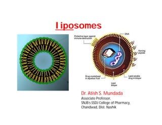
  • 27. Mechanism of Liposome Formation:Mechanism of Liposome Formation: • The basic part of liposome is formed by phospholipids, which are amphiphilic molecules (having a hydrophilic head and hydrophobic tail). • The hydrophilic part is mainly phosphoric acid bound to a water soluble molecule, whereas, the hydrophobic part consists of two fatty acid chains with 10 – 24 carbon atoms and 0 – 6 double bonds in each chain. • When these phospholipids are dispersed in aqueous medium, they form lamellar sheets by organizing in such a way that, the polar head group faces outwards to the aqueous region while the fatty acid groups face each other and finally form spherical/ vesicle like structures called as liposomes. • The polar portion remains in contact with aqueous region along with shielding of the non-polar part (which is oriented at an angle to the membrane surface).
  • 28. • When phospholipids are hydrated in water, along with the input of energy like sonication, shaking, heating, homogenization, etc. • It is the hydrophilic/ hydrophobic interactions between lipid – lipid, lipid – water molecules that lead to the formation of bilayered vesicles in order to achieve a thermodynamic equilibrium in the aqueous phase. • The reasons for bilayered formation include: – The unfavorable interactions created between hydrophilic and hydrophobic phase can be minimized by folding into closed concentric vesicles. – Large bilayered vesicle formation promotes the reduction of large free energy difference present between the hydrophilic and hydrophobic environment. – Maximum stability to supramolecular self assembled structure can be attained by forming into vesicles.
  • 29. Evaluation of Liposomes:Evaluation of Liposomes: • The characterization parameters for purpose of evaluation could be classified into three broad Categories, which include physical, chemical and biological parameters. • Physical characterization evaluates various parameters including size, shape, surface features, lamellarity phase-behaviour and drug release profile. • Chemical characterization includes those studies which establish the purity and potency of various lipophillic constituents. • Biological characterization parameters are helpful in establishing the safety and suitability of formulation for therapeutic application.
  • 30. Size and size distribution:- • When liposomes are intended for inhalation or parenteral administration, the size distribution is of primary consideration, since it influences the in vivo fate of liposomes along with the encapsulated drug molecules. • Various techniques of determining the size of the vesicles include – microscopy (optical microscopy, negative stain transmission electron microscopy, cryo-transmission electron microscopy, freeze fracture electron microscopy and scanning electron microscopy – diffraction and scattering techniques (laser light scattering and photon correlation spectroscopy) and – hydrodynamic techniques (field flow fractionation, gel permeation and ultracentrifugation).
  • 31. Percent drug encapsulation:- • The amount of drug encapsulated/ entrapped in liposome vesicle is given by percent drug encapsulation. • Column chromatography can be used to estimate the percent drug encapsulation of liposomes. • The formulation consists of both free (un-encapsulated) and encapsulated drug. So as to know the exact amount of drug encapsulated, the free drug is separated from the encapsulated one. • Then the fraction of liposomes containing the encapsulated drug is treated with a detergent, so as to attain lysis, which leads to the discharge of the drug from the vesicles into the surrounding medium. • This exposed drug is assayed by a suitable technique which gives the percent drug encapsulated from which encapsulation efficiency can be calculated.
  • 32. • Trapped volume per lipid weight can also give the percent drug encapsulated in a liposome vesicle. It is generally expressed as aqueous volume entrapped per unit quantity of lipid, Âľl/Âľmol or Âľg/mg of total lipid. • In order to determine the trapped volume, various materials like radioactive markers, fluorescent markers and spectroscopically inert fluid can be used. • Radioactive method is mostly used for determining trapped volume. It is determined by dispersing lipid in an aqueous medium containing a non-permeable radioactive solute like [22Na] or [14C] inulin. • Alternatively, water soluble markers like 6-carboxyfluorescein, 14C or 3H-glucose or sucrose can be used to determine the trapped volume
  • 33. Surface charge:- • Since the charge on the liposome surface plays a key role in the in vivo disposition, it is essential to know the surface charge on the vesicle surface. • Two methods namely, free-flow electrophoresis and zeta potential measurement can be used to estimate the surface charge of the vesicle. • The surface charge can be calculated by estimating the mobility of the liposomal dispersion in a suitable buffer (determined using Helmholtz– Smolochowski equation). Vesicle shape and lamellarity:- • Various electron microscopic techniques can be used to assess the shape of the vesicles. The number of bilayers present in the liposome, i.e., lamellarity can be determined using freeze fracture electron microscopy and 31P-Nuclear magnetic resonance analysis. • Apart from knowing the shape and lamellarity, the surface morphology of liposomes can be assessed using freeze-fracture and freeze-etch electron microscopy.
  • 34. Phospholipid identification and assay:- • The chemical components of liposomes must be analyzed prior to and after the preparation. • Barlett assay, Stewart assay and thin layer chromatography can be used to estimate the phospholipid concentration in the liposomal formulation. • A spectrophotometric method to quantify total phosphorous in a sample was given in literature, which measure the intensity of blue color developed at 825 nm against water. • Cholesterol oxidase assay or ferric perchlorate method and Gas liquid chromatography techniques can be used to determine the cholesterol concentration.
  • 35. Stability of Liposomes:- • During the development of liposomal drug products, the stability of the developed formulation is of major consideration. • The therapeutic activity of the drug is governed by the stability of the liposomes right from the manufacturing steps to storage to delivery. • A stable dosage forms is the one which maintains the physical stability and chemical integrity of the active molecule during its developmental procedure and storage. • A well designed stability study includes the evaluation of its physical, chemical and microbial parameters along with the assurance of product’s integrity throughout its storage period. • Hence a stability protocol is essential to study the physical and chemical integrity of the drug product in its storage.