27. OVARIAN TUMORS
• Solid vs Cystic
• Functional vs. NON-functional
• Benign vs. Malignant
• First clinical presentation may be ascites
• Malignant ascites in a woman is ovarian
cancer until proven otherwise
• CA-125 is THE important tumor marker in
ovarian cancer, especially as a follow up.
36. “GERM CELL” Tumors
• Teratomas (usually benign in ovary), i.e.,
“mature” cystic teratoma or dermoid cyst
• “Immature” teratomas are regarded as
malignant
• Dysgerminoma (look exactly like the
testicular seminoma), malignant
• Endodermal Sinus (Yolk Sac),
malignant, Just like testicular
• Choriocarcinoma, malignant, just like
testicular
51. Ectopic Pregnancy
• Chiefly TUBAL, but ovarian or
abdominal rare
•1% OF NORMAL WOMEN
•35%-50% OF WOMEN with
previous SALPINGITIS/PID
• + HCG, Abdominal pain, 1st
trimester, ultrasound
64. Placental Infections
• Villitis vs. chorionamnionitis vs. funisitis
• ASCENDING vs. hematogenous
• ASCENDING are usually bacterial,
and chorionamnionitis
• HEMATOGENOUS
are often TORCH,
and villitis
65. Placental Neoplasms,
i.e. gestational trophoblastic disease
• Benign: MOLES (Hydatidiform moles)
• Malignant: CHORIOCARCINOMA
• BOTH are associated with increased or
persistent levels of the placental
hormone HCG
66.
67. Hydatidiform Mole
• 1/1000 in USA
• 1% in Indonesia
• Also called NON-invasive mole in
its most common benign variant,
but can also be “invasive”
• Complete (2% chorioCA incidence)
or partial (0% incidence)
• Grapelike clusters, i.e., swollen villi
68.
69. The MAIN thing
differentiating benign
from malignant from
worrisome trophoblastic
neoplasms is
INVASIVENESS
of the trophoblast
Editor's Notes
Primary germ cells, male or female, first arise in the yolk sac and migrate to the genital ridge, which is in close proximity to the mesonephros.
Eventually, retroperitoneal testes migrate through the inguinal canal to the scrotum, covered by peritoneum.
Ovaries stay in the pelvis, and are covered by serosa, and are therefore intraperitoneal, but POSTERIOR to the fallopian tubes.
The CORTEX is the site of developing follicles.
The MEDULLA is relatively free of developing follicles, and rich in connective tissue (stroma) and blood vessels.
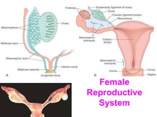
Major internal female genitalia structures, landmarks, and interrelationships.
In which ligament does the ovarian artery lie?
Through which structure does the round ligament travel.
Normally the uterus is a bit ANTE-VERTED and ANTE-FLEXED
Major internal female genitalia structures, landmarks, and interrelationships.
GREAT whole mount to demonstrate overall cortex vs. medullary differentiation.
Zona pellucida, arrow, becomes “atretic” follicle.
Is this a primary follicle? Ans: YES Why?
Secondary = Graffian = Antral follicle
Where is the antrum?
Find the cumulus oophorus, liquor folliculi, and corona radiata
Granulosa and theca INTERNA cells make estrogen.
LUTEAL cells, under LUTEINIZING hormone and FSH too, make progesterone.
LUTEUM means YELLOW. Why? Why is ANYTHING bright yellow?
A corpus luteum of pregnancy is considerably larger than a regular, NON-pregnancy, corpus luteum, often, perhaps about a half or third the size of the ovary.
Corpus albicans.
ALBA means WHITE.
Why is it white?
Most common PRE-menopausal cyst
Any EXTREMELY yellow cyst of a premenopausal ovary, is regarded as luteal in origin.
Very common PRE-menopausal cyst
Although the cortical area of the normal ovary contains cysts, i.e., various stages of follicular development, true PCOD (PolyCystic Ovarian Disease, or Stein-Leventhall) ovaries are BIGGER (2x) than normal premenopaosal ovaries and have “true” cysts, NON-ovulatory, NOT just stages of follicular development.
Is a “cyst” a “tumor” (i.e. swelling) in the classical sense of the word, like a bump on the head. Is a cyst usually a true neoplasm? Ans: Of course not!
Always think of true ovarian tumors as following the normal anatomy/histology in these FOUR groups---mullerian, germ, sex-cord, metastatic.
In contrast to the testicle, the ovary DOES occasionally get metastases.
Gross, microscopic, physiologic, behavioral classification factors for ovarian tumors.
The HUGEST tumors ever reported in human beings (50-100 lbs.?) are frequently benign mucinous ovarian tumors.
Q: What other adjective can we give to this tumor besides serous? Ans: Papillary
Close up of papillae
Why is this serous and NOT mucinous?
PSAMMOMA bodies
Less common Müllerian carcinomas
I TOLD you this looks the same as TESTICULAR germ cell tumors.
Dermoid “cyst” = BENIGN CYSTIC TERATOMA, BY FAR the most common ovarian NEOPLASM of younger women, usually BENIGN
Whether the teratomatous elements are “mature” or “immature” determine, greatly, the behavior of the teratoma, i.e., benign or malignant.
IMMATURE looking neural tissue.
This is much more likely to behave badly (i.e., malignant) than a mature one.
Often, you might see retinal tissue, like you see here.
Female dysgerminomas are IDENTICAL in appearance to male seminomas, i.e., germ cells + lymphocytes. You’d have to tell the pathologist whether this was a male or female in order for him to diagnose seminoma vs. dysgerminoma.
Schiller-Duvall Body, just like in the testis yolk sac tumor!
EXACTLY the same as a malignant HCG producing testicular choriocarcinoma or a malignant HCG producing placental choriocarcinoma
“Sex cord” = “stroma”
MANY are functional, i.e., associated with hyper estrogenism (or androgenism)
Call-Exner bodies are virtually diagnostic of granulosa cell tumors.
Q: Do they remind you of “rosettes”? Ans: YES
Q: Would a “thecoma” derived from theca INTERNA be more likely to be functional than a thecoma derived from theca EXTERNA? Ans: YES
Why?
Note the “theca” has both a vesicular and spindle cell appearance. The juicy vesicular cells, theca interna, and tumors derived from them, can secrete estrogen. The spindly theca externa cells, usually do not, and may look simply like fibromas.
Many thecomas look white and fibrous, That is why the term fibrothecoma is often used?
Is a fibrothecoma or fibroma less likely to be functional than a thecoma? Ans: YES
Why? Ans: It is derived from NON-estrogen producing cells.
Accessory placental lobe.
An extreme lobe might be called a BI-partite placenta.
In a circumvallate placenta the amnionic, i.e., amniotic, membranes “thicken” or “double back”
You might guess this kind of placenta would be VERY difficult to remove, and remnants (retained POC) might result in endometritis
And don’t forget placenta “abruptio” or premature separation of placenta with hemorrhage (i.e., hematoma)
Twin zygosity (mon- or di-) is related to the number of CHORIONS, NOT amnions or umbilical cords!
Toxemia of pregnancy occurs in an amazing 6% of all pregnancies. Toxemia is also called PRE-eclampsia.
When PRE-eclampsia is particularly severe and associated with more serious systemic effects such as DIC or convulsions, it is called ECLAMPSIA.
What does TORCH stand for?
T-oxo
O-ther
R-ubella
C-MV (Do you see the BASOPHILIC intranuclear inclusion in the above villitis pic?)
H-erpes
Syncytial cells are FUSED, CYTO-trophoblastic cells are deeper stem cells.
Is this chorionic villus mature or IM-mature? Ans: mature
Why? Ans: It has blood vessels in its core. If it was IMMATURE, it would NOT need secondary blood vessels and can diffuse oxygen and nutrients WITHOUT secondary blood vessel formation.
In COMPLETE moles, ALL the villi are swollen. They turn into choriocarcinomas 2% of the time.
In PARTIAL moles, only some are. They NEVER turn into choriocarcinomas.
NOTE trophoblast looks NORMAL, i.e., NON-invasive and NON-proliferative, and NON atypical.
Choriocarcinoma. Note invasive trophoblast.
Choriocarcinoma. Note extreme pleomorphism of trophoblastic cells.