Ahmedabad Call Girls Book Now 9630942363 Top Class Ahmedabad Escort Service A...
Mycetoma
1. 1
Amjad Khan Afridi Date: 12th
May, 2017
MYCETOMA
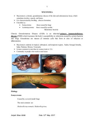
Mycetoma is a chronic, granulomatous disease of the skin and subcutaneous tissue, which
sometimes involves muscle, and bones.
It is characterized by Swelling , abscess formation,
Classified as :
Eumycetoma: those caused by fungi
Actinomycetoma: those caused by aerobic
filamentous bacteria
Chronic Granulomatous Disease (CGD) is an inherited primary immunodeficiency
disease (PIDD) which increases the body’s susceptibility to infections caused by certain bacteria
and fungi. Granulomas are masses of immune cells that form at sites of infection or
inflammation.
Mycetoma is endemic in tropical, subtropical, and temperate regions. Sudan, Senegal, Somalia,
India, Pakistan, Mexico, Venezuela
Is more common in men than in women (ratio is 3:1).
Commonly in people who work in rural areas
Etiology
Eumycetomas
Caused by a several mould fungi
The most common are
Madurella mycetomatis, Madurella grisea,
2. 2
Amjad Khan Afridi Date: 12th
May, 2017
Actinomycetomas
Caused by aerobic filamentous bacteria , gram positive
Streptomycessomaliensis
Nocardia brasiliensis
SIGN AND SYMPTOMS
The initial lesion may be a papule
Subcutaneous nodule,
Subcutaneous abscess
Fibrosis is common in and around early lesions.
Infection progresses slowly over months or years,gradually extending to and destroying
muscles, tendons, and bones
Subcutaneous nodule:A subcutaneous nodule refers to a firm lump under a person’s skin.
Subcutaneous nodules are lumps under the skin, often caused due to an infection or inflammation.
Papule:A papule is an area of abnormal skin tissue. A papule has distinct borders, and it can
appear in a variety of shapes. Papules are often called skin lesions, which are essentially
changes in the color or texture of your skin. Sometimes, papules cluster together to form a rash.
Fibrosis:the thickening and scarring of connective tissue, usually as a result of injury.
3. 3
Amjad Khan Afridi Date: 12th
May, 2017
PATHOGENSIS
The causative organism enters through sites of local trauma (eg, cut on the hand, foot splinter)
A neutrophilic response initially occurs, which may be followed by a granulomatous
reaction.
Spread occurs through
• skin .
• Hematogenous
• lymphatic spread (uncommon).
The body parts affected most commonly in persons with mycetoma include
• foot or lower leg,
• The hand
The chest and back (frequently caused by Nocardia species,) whereas lesions on the head and
neck are usually caused by Streptomyces somaliensis
Diagnosis:
Clinical samples:
• Biopsy tissue
• Pus
• Blood (for serology only)
1. Direct microscopic examination
Microscopic examination of tissue
a. Histological sections: Hematoxylin-Eosin,
b. Smears: Stain with Giemsa , methenamine silver
c. Stain by: Gram, ZN (Actinomycetes)
2. Culture
a. Media such as Sabouraud dextrose agar (SDA) to isolate fungi
b. Blood agar to isolate bacteria.
3. Serology:
Antibodies can be determined by immunodiffusion, , enzyme-linked immunosorbent assay
Treatment
A. Eumycetoma : Ketoconazole
Itraconazole
Also Voriconazole and Amphotericin B
B. Actinomycetoma: Trimethoprim-sulfamethoxazole
Dapsone
Streptomycin
Combination of 2 drugs is used
Therapy is suggested for several months or years (1-2 years or more)