SlideShare a Scribd company logo
1 of 42
Multifocal sallmonellosis
Invasion of the ileum of a calf by Salmonella
typhimurium
Multifocal ulceration ( cecum )
Multifocal, ulceration (colon)
Etiology: Clostridium
perfringens type D epsilon
toxin Disease: Clostridial
enterotoxemia
Blood injected intestine typical of
Type C enterotoxemia in a young
lamb.
Clostridium perfringens Type C
enterotoxemia typically presents
as what appear to be blood-
filled small intestines. Intestinal
walls are dark red to black ...
Gross lesions include fluid cecal
contents with serosal hemorrhages
and edema on the cecum, and
occasionally the ileum and proximal
colon (A)
Microscopically, there is necrotic erosive
or ulcerative typhlitis with swollen or
vacuolated enterocytes, pseudomembrane
formation, heterophilic infiltration, as
well as submucosal edema and
hemorrhage (B). Large Gram-positive
bacilli are present on the mucosal
surface.
Accumulation and puch nucleus to
periphral
Yellow color due to
Fat cell from liver
Enlargement, edges
and filled with
fluid like albumin
Diffusely the hepatocytes are markedly
enlarged by a discrete clear cytoplasmic
vacuole (lipid) that flattens and displaces
the nucleus to the periphery
Zone 3 around central vein
Congestive Hepatopathy
nutmeg liver
Periportal necrosis
Ex: Both Carbon Tetrachloride
and Blue-Green Algae cause
periportal necrosis of the liver.
centrilobular necrosis means
necrosis around the central vein
(called zone 3) ● Causes of
Centrilobular Necrosis In Liver:
1= Acetaminophen toxicity
2= Chronic passive congestion as in
Cardiac failure (pt:Right sided)
n cases of anemia, centrilobular
area is the area that will the
most hypoxic and therefore
undergo the most significant
necrosis.
This is because the has blood
that is the least well oxygenated
massive necrosis liver, pig.
● Acute centrilobular necrosis is the
principal lesion of this disorder.
Midzonal necrosis
Cocklebur plant is an
example of a plant that
causes mid-zonal necrosis.
Other forms of hepatic necrosis
are paracentral (a wedge-
shaped band of necrosis
extending from the
centrilobular to the portal area)
and midzonal necrosis (affects
hepatocytes halfway between
the centrilobular area and the
periportal area).
liver looks lobated
like a kidney. This is
due to scaring or
fibrosis from a
chronic disease.
The word
"cirrhosis" comes
from the greek
word "kirrhos"
that means orange
colored and that
is the way many
cirrhotic livers
appear grossly.
To have cirrhosis, there must
be three features:
● necrosis of hepatocytes
● hyperplasia of nodules, and
● fibrosis
dog had severe
hepatic cirrhosis
or "end-stage"
liver disease.
Intranuclear inclusion bodies
in the hepatocytes is
diagnostic for Infectious
Canine Hepatitis.
Replecate in lymphocyte
and cause necrosis in
centrilobar area
The enlarged, swollen liver,
icteric mucous membranes,
and swollen, reddened tonsils
are all suggestive of this
disease.In the microscopic
section, esinophilic to
amphiophilic inclusion bodies
are present in many
hepatocytes clinching the
diagnosis.
Infectious Canine Hepatitis
Rift Valley Fever
(ruminant)
● Section reveals
that the liver is
pale, swollen and
contains multiple
foci of hemorrhage.
Sheep, liver.
Liver is pale and
swollen and contains
many areas of severe
congestion.
Hemorrhagic fever
viruses such as
dengue virus, yellow
fever virus, and Rift
Valley virus.
Campylobacter fetus
and Campylobacter
jejuni
there is a
GRANULOMATOUS
REACTION with
MULTINUCLEATED
GIANT CELL
Tuberculosis
FASCIOLA HEPATICA causes
necrosis and secondary fibrosis
of the biliary tree in cattle.
Corn can be contaminated
with Aspergillus flavus, a
mold that produces
AFLATOXIN
Liver of a dog that died of
leptospirosis.
Multifocal hepatic necrosis
is associated with a mottled
appearance of this organ
there is loss of hepatic
plate architecture.
Heptaocytes are
individualized, rounded
up, and exhibit single cell
necrosis
leptospora
macrophages and hepatocytes contain
abundant red globular pigment (copper).
Kideny-tubules necrosos
without nuclus
(pyknotic,karyolysis)
The cause : ischemia / toxemia
Heamorrhagic nephrosis
Morphological diagnosis :
Congestion, glomerullitis,
nephrosis, nephritis.
Congestion ( RBC's ‫م‬
‫ا‬
‫خ‬
‫ذ‬
‫ه‬
‫ش‬
‫ك‬
‫ل‬
‫ا‬
‫ل‬
‫ع‬
‫ض‬
‫و‬ )
Hemorrhage
RBC's located between the cell
Proximal tubules
Glomerulus
Morphological diagnosis:
Nephrosis, heamorrhage, glomerulitis
Edema
Proximal tubules : the nuclus
disappear
MDx: nephrosis, heamorrhage,
MDx : glomerulitis
Glomerulitis,
nephrosis
Unilateral renal
aplasia, abdominal
cavity, gross,
rabbit. Single
kidney only
present.
Renal aplasia, piglets.
No renal tissue is
present at the tip of
the two black arrow
marked piglets.
Unilateral
hypoplastic kidney,
young cat. The left
kidney (ventro-
dorsal view) is
normal in shape
and structure but
reduced in overall
size.
Unilateral hypoplastic kidney,
dorsal sections, young dog.
Grossly, affected kidney is
nearly identical structurally to
left kidney but smaller in size.
Congenital Horseshoe Kidney,
calf. Both kidneys are fused
together at one pole.
Polycystic disease, dorsal section,
cat. Numerous variably sized
tubular cysts are present in the
cortex and medulla and affect
approximately 60% of the kidney.
The cyst contain clear colorless
fluid.This condition is hereditary,
and Persia cats are predisposed.
Embolic nephritis, kidney, horse.
Multiple, small white necrotic foci
and abscesses are present
subcapsulary.
Embolic nephritis, kidney,
horse.
Dorsal section.Variably sized
abscesses are scattered
throughout the cortex
(arrows).
Cause : actinobacillas
Proliferative
glomerulonephritis, kidney,
dorsal section, dog. The
small, white, round foci in
cortex are enlarged
glomeruli.
Haemorrhage, kideny,↑permiability, necrosis, leakage blood cell.
Cause : actinobasillas
Embolic nephritis, kidney,
horse. Causative bacteria
(arrow) enter kidney via
the vasculature
(bactermia) and lodge in
capillaries of glomeruli,
where they replicate and
induce necrosis and
inflammation.
Pyelonephritis, kidney. Dorsal
section, dog. Extensive pelvic
inflammation has destroyed
areas of the inner medulla
and extends focally
into the outer medulla.
Pyelonephritis, kidney.
Dorsal section, cow. Renal
calyces in the cow
contain suppurative exudate.
Pyelonephritis, kidney. Dog.
There is both intratubular
and interstitial
inflammation,
characterized by infiltrates
of principally neutrophils
(arrow).
Inset: Higher magnification
of intratubular neutrophils.
Inflamatory cell
Hydronephrosis, kidney, dorsal section. Sheep. The pelvis of each kidney is
markedly dilated.
Bacterial-induced septicemic renal
cortical hemorrhage, erysipelas,
kidney, pig. Petechial hemorrhages
caused by septic emboli of
erysipelothrix
are randomly scattered over capsular
surface of kidney.
Extreme left and right ventricle
with visible areas of myocardial
fibrisis.
Epicardial hemorrhage, heart, left atrium
Epicardial hemorrhage, Petechiae and ecchymoses
Briskit edema, high altuatid disease, due
to heart congestive heart faiuler
Hydrothoraxs
Patent ductus arteriosus, vascular channel between
the pulmonary artery and aorta that allows blood to
bypass the lungs.
Atrial septal defect, heart, opened right heart.
The prominent opening (arrow) low in the atrial
septum (AS) and just above the atrioventricular
valve is the septal defect, (VS) ventricular
septum
Ventricular septal defect (high defect),
heart, opened left side, calf. Note the large
opening in the basal portion of the
ventricular septum (arrow) immediately
below the aortic valve through which the
tube has been passed. A, Aorta; LV, Left
Ventricle.
Tetralogy of Fallot, heart, dissected, dog.
Cause :
1. ventricular septal defect is an
overlying ,
2. straddling aorta (A),
3. sever pulmonic stenosis (arrow)
4. massive right ventricular
hypertrophy.
Subaortic stenosis,
Valvular hematocyst, heart, Opened
left side, mitral valve
Valvular lymphocyst, A lymph-
filled cyst on the atrioventricular
valve
CHRONIC SEPTIC PERICARDITIS
SECONDARY TO HARDWARE
DISEASE
Hydropericardium, Pericardial sac, pig. The thin-walled
pericardial sac contains serous fluid that has accumulated
secondary to alteration in hydrostatic pressure between the
pericardial cavity, circlatory system and lymphatic system
Hemopericardium, heart,
dog. The pericadial sac is
filled with clotted blood
Cardiac tamponade from
hemopericardium, heart, dog. The
pericardium is distended and dark
blue it contains whole blood
Hemopericardium, heart, dog. The
pericadial sac is filled with clotted
blood
Fibrinous pericarditis, heart, epicardium,
horse.
The epicardium is covered dorsally by a
thick, yellow layer of fibrin (arrows) and
ventrally by granulation tissue (finely
granular surface), thus indicating the
chronicity of the inflammatory process.
This lesion occurs in horses with
streptococcus zooepidemicus.
Fibrinous pericarditis, heart,
epicardium. Note eosinophilic fibrin
depositis (left), on the epicardial
surface (E). This lesion occurs with
bacterial septicemia.
Chronic Suppurative pericarditis,
traumatic reticuloperitonitis (hard
ware disease), heart, opened
pericardial sac , cow.
The exposed epicardial and parietal
surfaces are thickened by fibrous
connective tissue and covered with
fibrinopurulent exudate. On clinical
examination the heart sounds are
muffled. P, reflected parietal
pericardium.
Dark pale greenish thickened valves verrucous endocardiosis
Vegetative valvualr
endocarditis, bacterial
infection, heart, tricuspid
valve, cow.
Note abundant masses of
fibrin and bacterial colonies
(arrow).
Hypertrophic cardiomyopathy,
myocyte hypertrophy, heart,
myocardium, cat.
Cardiac myocytes are
hypertrophied and there is
increase in interstitial fibroblasts.
Dirofilariasis, heart, dog.
The adult hypertrophy of right (RV) and
adult Dirofilaria immitis in the pulmonary
artery and it’s branch (PA). LV, Left
ventricle.
The adult parasite
Dirofilariasis, heart, Opened right
ventricle, right atrium, and pulmonary
artery, dog.
Numerous adult
Dirofilaria immitis
are present in the right Ventricle (RV),
right Atrium, and pulmonary Artery
(PA).
Myocardial Necrosis, (heart-brain
syndrome), heart, transverse section
of ventricles, dog. Necrotic areas are
pale and are concentered in the inner
half of the wall of the left ventricle
(LV) and in the ventricular septum.
Myocardial necrosis, acute
monensin toxicosis, heart, cross
section, left ventricular
myocardium, calf.
Note the pale, mottled, necrotic,
areas (arrows) distributed
throughout the ventricular
myocardium.
The prominent white chalky areas of necrosis with minerlization
(arrows) of the myocardium.
Myocardial necrosis, selenium-vitamin E deficiency, heart, left
ventricular myocardium, calf.
Subepicardium and subendocardium necrosis.
Acute myocardial necrosis with
mineralization, heart, ventricular
myocardium, pig, toxicity.
The darker red myocytes are
necrotic, and some mineralization
occurred (purplish area).
Healing, postmyocardial
necrosis, heart, ventricle, dog.
The necrotic myocytes have
been removed by phagocytosis
by macrophages, and the area
is now undergoing fibrosis.
Cutaneous infarcts, diamond skin disease,
Erysipelothrix septicemia, skin, pig.
Emboli of have lodge in cutaneous vessels
and cause localized vasculitis, which
resulted in thrombosis followed by
ischemia and cutaneous infarction.
Necrohemorrhagic myocarditis,
heart, steer.
Not the area of hemorrhgic
myocarditis (arrows) in the wall
of the ventricular myocardium.
This disease is caused by
Clostridium chauvoei, and the
lesions are most common in
skeletal muscle.
Parvovirus myocarditis,
heart, dog.
A, note the multifocal pale
areas (arrows) in the
ventricular myocardium. B,
Parovirus infection, section
of myocardium. An
intracellular basophilic
inclusion body is in a
myocyte (arrow).
Dissecting aneurysm, copper
deficiency, heart, pulmonary
artery, right ventricle, pig. The
dark blood –filled, bulging
segment of the wall of the
pulmonary artery (arrows) has
resulted from disruption of
elastic fibers.
Dissecting aneurysm, aorta,
turkey. Blood has dissected
through the tunica media and
has to come to lie in the outer
layers of tunica media and
adventia
Medial hypertrophy, periarteritis,
dirofilariasis, lung, small pulmonary
arteries, cat.
Note the massive thickened tunica
media of the small branches of
pulmonary arteries and their
pericardial cuff of chronic
inflammatory cells and some
eosinophils.
Coronary atherosclerosis,
hypothyroidism, heart, left
ventricle, dog.
The affected coronary arteries
are prominent and cord-like
(arrows) with thickened walls.
The diffuse and focal yellow
areas in the wals of the
arteries are sites of
atheromatous depositis.
Atherosclerosis, meningeal
artery, horse.
Note extensive accumulation of
lipid-laden (clear vacuoles)
(foamy cells) throughout the
thickened media.
Medial calcification, aorta, cow.
Note the layer of minerlization
in the middle of tunica media.
Aortic thrombosis, aorta and
external iliac arteries, dog.
The tan thrombus occluding the
caudal abdominal aorta is a
cranial extension of the red
saddle thrombus at the aortic
biforcation and external iliac
arteries. (arrows).
Verminous arteritis and mural
thrombosis, strongylosis, abdominla
aorta (A), and cranial mesentric
artery.
A pale friable thrmobotic mass in
which several strongylus larvae
(arrows) are embedded.
Arteial thrombus, pulmonary artery,
dog.
Arterial thrombi are composed of
platelets and fibrin because of the
rapid flow of blood tends to exclude
erythrocytes from the thrombus, and
thus arterial thrombi are usually pale
to gray (arrow).
Myocardial infarction, heart,
left and right ventricles, dog.
Pale, necrotic, circumscribed
areas (arrows) are present in
the ventricular walls and are
most prominent at the apex.
Inset: the cardiac myocytes
are eosinophilic (ischemic
necrosis) and have lost their
nuclei (karyolysis).
Distal tubule
( necrosis
due to
toxins )
Tounge, cotagious
ecthyma
Congeestion
Liver, first stage
inflammation, necrosis,

More Related Content

Similar to pathology photo

Diagnosis of abdominal tuberculosis
Diagnosis of abdominal tuberculosisDiagnosis of abdominal tuberculosis
Diagnosis of abdominal tuberculosis
drranjithmp
 
Liver Cirrhosis
Liver CirrhosisLiver Cirrhosis
Liver Cirrhosis
dorai
 
liver disease presentation .pptxmmmmmmmmm
liver disease presentation .pptxmmmmmmmmmliver disease presentation .pptxmmmmmmmmm
liver disease presentation .pptxmmmmmmmmm
IbrahemIssacGaied
 

Similar to pathology photo (20)

Ascites by dr. p.b.sadiq_ulla
Ascites by dr. p.b.sadiq_ullaAscites by dr. p.b.sadiq_ulla
Ascites by dr. p.b.sadiq_ulla
 
Lecture 25 diseases of liver and pancreas
Lecture 25 diseases of liver and pancreasLecture 25 diseases of liver and pancreas
Lecture 25 diseases of liver and pancreas
 
Diagnosis of abdominal tuberculosis
Diagnosis of abdominal tuberculosisDiagnosis of abdominal tuberculosis
Diagnosis of abdominal tuberculosis
 
The biliary tract
The biliary tractThe biliary tract
The biliary tract
 
Theileriosis
TheileriosisTheileriosis
Theileriosis
 
ACUTE ABDOMEN SONOGRAPHIC FINDINGS ppt
ACUTE ABDOMEN SONOGRAPHIC FINDINGS pptACUTE ABDOMEN SONOGRAPHIC FINDINGS ppt
ACUTE ABDOMEN SONOGRAPHIC FINDINGS ppt
 
Liver Cirrhosis
Liver CirrhosisLiver Cirrhosis
Liver Cirrhosis
 
Approach to ascites
Approach to ascitesApproach to ascites
Approach to ascites
 
Hydatid Cyst Of Liver
Hydatid Cyst Of LiverHydatid Cyst Of Liver
Hydatid Cyst Of Liver
 
Liver Disease in General Surgery
Liver Disease in General SurgeryLiver Disease in General Surgery
Liver Disease in General Surgery
 
AMOEBIASIS
AMOEBIASISAMOEBIASIS
AMOEBIASIS
 
liver disease presentation .pptxmmmmmmmmm
liver disease presentation .pptxmmmmmmmmmliver disease presentation .pptxmmmmmmmmm
liver disease presentation .pptxmmmmmmmmm
 
Spleen
SpleenSpleen
Spleen
 
Liver abscess
Liver abscessLiver abscess
Liver abscess
 
6 bovine leukemia (leucosis)
6 bovine leukemia (leucosis)6 bovine leukemia (leucosis)
6 bovine leukemia (leucosis)
 
Hydatid cyst
Hydatid cystHydatid cyst
Hydatid cyst
 
Hemodynamic disturbances
Hemodynamic disturbancesHemodynamic disturbances
Hemodynamic disturbances
 
benign lesions of the Liver.pptx
benign lesions of the Liver.pptxbenign lesions of the Liver.pptx
benign lesions of the Liver.pptx
 
Haemodynamic disorders , thromboembolism and shock by Dr Nadeem (RMC)
Haemodynamic disorders , thromboembolism and shock by Dr Nadeem (RMC)Haemodynamic disorders , thromboembolism and shock by Dr Nadeem (RMC)
Haemodynamic disorders , thromboembolism and shock by Dr Nadeem (RMC)
 
LIVER & GALLBLADDER PATHOLOGY
LIVER & GALLBLADDER PATHOLOGYLIVER & GALLBLADDER PATHOLOGY
LIVER & GALLBLADDER PATHOLOGY
 

Recently uploaded

Bacterial Identification and Classifications
Bacterial Identification and ClassificationsBacterial Identification and Classifications
Bacterial Identification and Classifications
Areesha Ahmad
 
Module for Grade 9 for Asynchronous/Distance learning
Module for Grade 9 for Asynchronous/Distance learningModule for Grade 9 for Asynchronous/Distance learning
Module for Grade 9 for Asynchronous/Distance learning
levieagacer
 
Formation of low mass protostars and their circumstellar disks
Formation of low mass protostars and their circumstellar disksFormation of low mass protostars and their circumstellar disks
Formation of low mass protostars and their circumstellar disks
Sérgio Sacani
 
biology HL practice questions IB BIOLOGY
biology HL practice questions IB BIOLOGYbiology HL practice questions IB BIOLOGY
biology HL practice questions IB BIOLOGY
1301aanya
 
Asymmetry in the atmosphere of the ultra-hot Jupiter WASP-76 b
Asymmetry in the atmosphere of the ultra-hot Jupiter WASP-76 bAsymmetry in the atmosphere of the ultra-hot Jupiter WASP-76 b
Asymmetry in the atmosphere of the ultra-hot Jupiter WASP-76 b
Sérgio Sacani
 
Pests of cotton_Borer_Pests_Binomics_Dr.UPR.pdf
Pests of cotton_Borer_Pests_Binomics_Dr.UPR.pdfPests of cotton_Borer_Pests_Binomics_Dr.UPR.pdf
Pests of cotton_Borer_Pests_Binomics_Dr.UPR.pdf
PirithiRaju
 

Recently uploaded (20)

Bacterial Identification and Classifications
Bacterial Identification and ClassificationsBacterial Identification and Classifications
Bacterial Identification and Classifications
 
COST ESTIMATION FOR A RESEARCH PROJECT.pptx
COST ESTIMATION FOR A RESEARCH PROJECT.pptxCOST ESTIMATION FOR A RESEARCH PROJECT.pptx
COST ESTIMATION FOR A RESEARCH PROJECT.pptx
 
Site Acceptance Test .
Site Acceptance Test                    .Site Acceptance Test                    .
Site Acceptance Test .
 
FAIRSpectra - Enabling the FAIRification of Spectroscopy and Spectrometry
FAIRSpectra - Enabling the FAIRification of Spectroscopy and SpectrometryFAIRSpectra - Enabling the FAIRification of Spectroscopy and Spectrometry
FAIRSpectra - Enabling the FAIRification of Spectroscopy and Spectrometry
 
Module for Grade 9 for Asynchronous/Distance learning
Module for Grade 9 for Asynchronous/Distance learningModule for Grade 9 for Asynchronous/Distance learning
Module for Grade 9 for Asynchronous/Distance learning
 
CELL -Structural and Functional unit of life.pdf
CELL -Structural and Functional unit of life.pdfCELL -Structural and Functional unit of life.pdf
CELL -Structural and Functional unit of life.pdf
 
Formation of low mass protostars and their circumstellar disks
Formation of low mass protostars and their circumstellar disksFormation of low mass protostars and their circumstellar disks
Formation of low mass protostars and their circumstellar disks
 
pumpkin fruit fly, water melon fruit fly, cucumber fruit fly
pumpkin fruit fly, water melon fruit fly, cucumber fruit flypumpkin fruit fly, water melon fruit fly, cucumber fruit fly
pumpkin fruit fly, water melon fruit fly, cucumber fruit fly
 
9999266834 Call Girls In Noida Sector 22 (Delhi) Call Girl Service
9999266834 Call Girls In Noida Sector 22 (Delhi) Call Girl Service9999266834 Call Girls In Noida Sector 22 (Delhi) Call Girl Service
9999266834 Call Girls In Noida Sector 22 (Delhi) Call Girl Service
 
Clean In Place(CIP).pptx .
Clean In Place(CIP).pptx                 .Clean In Place(CIP).pptx                 .
Clean In Place(CIP).pptx .
 
IDENTIFICATION OF THE LIVING- forensic medicine
IDENTIFICATION OF THE LIVING- forensic medicineIDENTIFICATION OF THE LIVING- forensic medicine
IDENTIFICATION OF THE LIVING- forensic medicine
 
biology HL practice questions IB BIOLOGY
biology HL practice questions IB BIOLOGYbiology HL practice questions IB BIOLOGY
biology HL practice questions IB BIOLOGY
 
GBSN - Microbiology (Unit 3)
GBSN - Microbiology (Unit 3)GBSN - Microbiology (Unit 3)
GBSN - Microbiology (Unit 3)
 
Asymmetry in the atmosphere of the ultra-hot Jupiter WASP-76 b
Asymmetry in the atmosphere of the ultra-hot Jupiter WASP-76 bAsymmetry in the atmosphere of the ultra-hot Jupiter WASP-76 b
Asymmetry in the atmosphere of the ultra-hot Jupiter WASP-76 b
 
Vip profile Call Girls In Lonavala 9748763073 For Genuine Sex Service At Just...
Vip profile Call Girls In Lonavala 9748763073 For Genuine Sex Service At Just...Vip profile Call Girls In Lonavala 9748763073 For Genuine Sex Service At Just...
Vip profile Call Girls In Lonavala 9748763073 For Genuine Sex Service At Just...
 
Unit5-Cloud.pptx for lpu course cse121 o
Unit5-Cloud.pptx for lpu course cse121 oUnit5-Cloud.pptx for lpu course cse121 o
Unit5-Cloud.pptx for lpu course cse121 o
 
module for grade 9 for distance learning
module for grade 9 for distance learningmodule for grade 9 for distance learning
module for grade 9 for distance learning
 
Kochi ❤CALL GIRL 84099*07087 ❤CALL GIRLS IN Kochi ESCORT SERVICE❤CALL GIRL
Kochi ❤CALL GIRL 84099*07087 ❤CALL GIRLS IN Kochi ESCORT SERVICE❤CALL GIRLKochi ❤CALL GIRL 84099*07087 ❤CALL GIRLS IN Kochi ESCORT SERVICE❤CALL GIRL
Kochi ❤CALL GIRL 84099*07087 ❤CALL GIRLS IN Kochi ESCORT SERVICE❤CALL GIRL
 
Pests of cotton_Borer_Pests_Binomics_Dr.UPR.pdf
Pests of cotton_Borer_Pests_Binomics_Dr.UPR.pdfPests of cotton_Borer_Pests_Binomics_Dr.UPR.pdf
Pests of cotton_Borer_Pests_Binomics_Dr.UPR.pdf
 
9654467111 Call Girls In Raj Nagar Delhi Short 1500 Night 6000
9654467111 Call Girls In Raj Nagar Delhi Short 1500 Night 60009654467111 Call Girls In Raj Nagar Delhi Short 1500 Night 6000
9654467111 Call Girls In Raj Nagar Delhi Short 1500 Night 6000
 

pathology photo

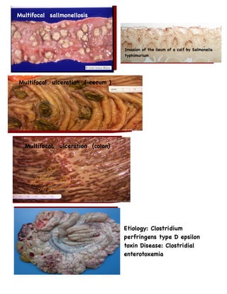
  • 1. Multifocal sallmonellosis Invasion of the ileum of a calf by Salmonella typhimurium Multifocal ulceration ( cecum ) Multifocal, ulceration (colon) Etiology: Clostridium perfringens type D epsilon toxin Disease: Clostridial enterotoxemia
  • 2. Blood injected intestine typical of Type C enterotoxemia in a young lamb. Clostridium perfringens Type C enterotoxemia typically presents as what appear to be blood- filled small intestines. Intestinal walls are dark red to black ... Gross lesions include fluid cecal contents with serosal hemorrhages and edema on the cecum, and occasionally the ileum and proximal colon (A) Microscopically, there is necrotic erosive or ulcerative typhlitis with swollen or vacuolated enterocytes, pseudomembrane formation, heterophilic infiltration, as well as submucosal edema and hemorrhage (B). Large Gram-positive bacilli are present on the mucosal surface.
  • 3. Accumulation and puch nucleus to periphral Yellow color due to Fat cell from liver Enlargement, edges and filled with fluid like albumin Diffusely the hepatocytes are markedly enlarged by a discrete clear cytoplasmic vacuole (lipid) that flattens and displaces the nucleus to the periphery
  • 4. Zone 3 around central vein Congestive Hepatopathy nutmeg liver
  • 5. Periportal necrosis Ex: Both Carbon Tetrachloride and Blue-Green Algae cause periportal necrosis of the liver. centrilobular necrosis means necrosis around the central vein (called zone 3) ● Causes of Centrilobular Necrosis In Liver: 1= Acetaminophen toxicity 2= Chronic passive congestion as in Cardiac failure (pt:Right sided) n cases of anemia, centrilobular area is the area that will the most hypoxic and therefore undergo the most significant necrosis. This is because the has blood that is the least well oxygenated massive necrosis liver, pig. ● Acute centrilobular necrosis is the principal lesion of this disorder.
  • 6. Midzonal necrosis Cocklebur plant is an example of a plant that causes mid-zonal necrosis. Other forms of hepatic necrosis are paracentral (a wedge- shaped band of necrosis extending from the centrilobular to the portal area) and midzonal necrosis (affects hepatocytes halfway between the centrilobular area and the periportal area).
  • 7. liver looks lobated like a kidney. This is due to scaring or fibrosis from a chronic disease. The word "cirrhosis" comes from the greek word "kirrhos" that means orange colored and that is the way many cirrhotic livers appear grossly.
  • 8. To have cirrhosis, there must be three features: ● necrosis of hepatocytes ● hyperplasia of nodules, and ● fibrosis dog had severe hepatic cirrhosis or "end-stage" liver disease.
  • 9. Intranuclear inclusion bodies in the hepatocytes is diagnostic for Infectious Canine Hepatitis. Replecate in lymphocyte and cause necrosis in centrilobar area The enlarged, swollen liver, icteric mucous membranes, and swollen, reddened tonsils are all suggestive of this disease.In the microscopic section, esinophilic to amphiophilic inclusion bodies are present in many hepatocytes clinching the diagnosis. Infectious Canine Hepatitis
  • 10. Rift Valley Fever (ruminant) ● Section reveals that the liver is pale, swollen and contains multiple foci of hemorrhage. Sheep, liver. Liver is pale and swollen and contains many areas of severe congestion. Hemorrhagic fever viruses such as dengue virus, yellow fever virus, and Rift Valley virus.
  • 11. Campylobacter fetus and Campylobacter jejuni there is a GRANULOMATOUS REACTION with MULTINUCLEATED GIANT CELL Tuberculosis
  • 12. FASCIOLA HEPATICA causes necrosis and secondary fibrosis of the biliary tree in cattle. Corn can be contaminated with Aspergillus flavus, a mold that produces AFLATOXIN Liver of a dog that died of leptospirosis. Multifocal hepatic necrosis is associated with a mottled appearance of this organ
  • 13. there is loss of hepatic plate architecture. Heptaocytes are individualized, rounded up, and exhibit single cell necrosis leptospora macrophages and hepatocytes contain abundant red globular pigment (copper).
  • 14. Kideny-tubules necrosos without nuclus (pyknotic,karyolysis) The cause : ischemia / toxemia Heamorrhagic nephrosis Morphological diagnosis : Congestion, glomerullitis, nephrosis, nephritis. Congestion ( RBC's ‫م‬ ‫ا‬ ‫خ‬ ‫ذ‬ ‫ه‬ ‫ش‬ ‫ك‬ ‫ل‬ ‫ا‬ ‫ل‬ ‫ع‬ ‫ض‬ ‫و‬ )
  • 15. Hemorrhage RBC's located between the cell Proximal tubules Glomerulus Morphological diagnosis: Nephrosis, heamorrhage, glomerulitis Edema Proximal tubules : the nuclus disappear MDx: nephrosis, heamorrhage,
  • 17. Unilateral renal aplasia, abdominal cavity, gross, rabbit. Single kidney only present. Renal aplasia, piglets. No renal tissue is present at the tip of the two black arrow marked piglets. Unilateral hypoplastic kidney, young cat. The left kidney (ventro- dorsal view) is normal in shape and structure but reduced in overall size.
  • 18. Unilateral hypoplastic kidney, dorsal sections, young dog. Grossly, affected kidney is nearly identical structurally to left kidney but smaller in size. Congenital Horseshoe Kidney, calf. Both kidneys are fused together at one pole. Polycystic disease, dorsal section, cat. Numerous variably sized tubular cysts are present in the cortex and medulla and affect approximately 60% of the kidney. The cyst contain clear colorless fluid.This condition is hereditary, and Persia cats are predisposed.
  • 19. Embolic nephritis, kidney, horse. Multiple, small white necrotic foci and abscesses are present subcapsulary. Embolic nephritis, kidney, horse. Dorsal section.Variably sized abscesses are scattered throughout the cortex (arrows). Cause : actinobacillas Proliferative glomerulonephritis, kidney, dorsal section, dog. The small, white, round foci in cortex are enlarged glomeruli.
  • 20. Haemorrhage, kideny,↑permiability, necrosis, leakage blood cell. Cause : actinobasillas Embolic nephritis, kidney, horse. Causative bacteria (arrow) enter kidney via the vasculature (bactermia) and lodge in capillaries of glomeruli, where they replicate and induce necrosis and inflammation. Pyelonephritis, kidney. Dorsal section, dog. Extensive pelvic inflammation has destroyed areas of the inner medulla and extends focally into the outer medulla. Pyelonephritis, kidney. Dorsal section, cow. Renal calyces in the cow contain suppurative exudate.
  • 21. Pyelonephritis, kidney. Dog. There is both intratubular and interstitial inflammation, characterized by infiltrates of principally neutrophils (arrow). Inset: Higher magnification of intratubular neutrophils. Inflamatory cell Hydronephrosis, kidney, dorsal section. Sheep. The pelvis of each kidney is markedly dilated. Bacterial-induced septicemic renal cortical hemorrhage, erysipelas, kidney, pig. Petechial hemorrhages caused by septic emboli of erysipelothrix are randomly scattered over capsular surface of kidney.
  • 22. Extreme left and right ventricle with visible areas of myocardial fibrisis. Epicardial hemorrhage, heart, left atrium Epicardial hemorrhage, Petechiae and ecchymoses Briskit edema, high altuatid disease, due to heart congestive heart faiuler
  • 23. Hydrothoraxs Patent ductus arteriosus, vascular channel between the pulmonary artery and aorta that allows blood to bypass the lungs. Atrial septal defect, heart, opened right heart. The prominent opening (arrow) low in the atrial septum (AS) and just above the atrioventricular valve is the septal defect, (VS) ventricular septum Ventricular septal defect (high defect), heart, opened left side, calf. Note the large opening in the basal portion of the ventricular septum (arrow) immediately below the aortic valve through which the tube has been passed. A, Aorta; LV, Left Ventricle.
  • 24. Tetralogy of Fallot, heart, dissected, dog. Cause : 1. ventricular septal defect is an overlying , 2. straddling aorta (A), 3. sever pulmonic stenosis (arrow) 4. massive right ventricular hypertrophy. Subaortic stenosis, Valvular hematocyst, heart, Opened left side, mitral valve Valvular lymphocyst, A lymph- filled cyst on the atrioventricular valve CHRONIC SEPTIC PERICARDITIS SECONDARY TO HARDWARE DISEASE
  • 25. Hydropericardium, Pericardial sac, pig. The thin-walled pericardial sac contains serous fluid that has accumulated secondary to alteration in hydrostatic pressure between the pericardial cavity, circlatory system and lymphatic system Hemopericardium, heart, dog. The pericadial sac is filled with clotted blood Cardiac tamponade from hemopericardium, heart, dog. The pericardium is distended and dark blue it contains whole blood Hemopericardium, heart, dog. The pericadial sac is filled with clotted blood
  • 26. Fibrinous pericarditis, heart, epicardium, horse. The epicardium is covered dorsally by a thick, yellow layer of fibrin (arrows) and ventrally by granulation tissue (finely granular surface), thus indicating the chronicity of the inflammatory process. This lesion occurs in horses with streptococcus zooepidemicus. Fibrinous pericarditis, heart, epicardium. Note eosinophilic fibrin depositis (left), on the epicardial surface (E). This lesion occurs with bacterial septicemia. Chronic Suppurative pericarditis, traumatic reticuloperitonitis (hard ware disease), heart, opened pericardial sac , cow. The exposed epicardial and parietal surfaces are thickened by fibrous connective tissue and covered with fibrinopurulent exudate. On clinical examination the heart sounds are muffled. P, reflected parietal pericardium.
  • 27. Dark pale greenish thickened valves verrucous endocardiosis Vegetative valvualr endocarditis, bacterial infection, heart, tricuspid valve, cow. Note abundant masses of fibrin and bacterial colonies (arrow). Hypertrophic cardiomyopathy, myocyte hypertrophy, heart, myocardium, cat. Cardiac myocytes are hypertrophied and there is increase in interstitial fibroblasts.
  • 28. Dirofilariasis, heart, dog. The adult hypertrophy of right (RV) and adult Dirofilaria immitis in the pulmonary artery and it’s branch (PA). LV, Left ventricle. The adult parasite Dirofilariasis, heart, Opened right ventricle, right atrium, and pulmonary artery, dog. Numerous adult Dirofilaria immitis are present in the right Ventricle (RV), right Atrium, and pulmonary Artery (PA).
  • 29. Myocardial Necrosis, (heart-brain syndrome), heart, transverse section of ventricles, dog. Necrotic areas are pale and are concentered in the inner half of the wall of the left ventricle (LV) and in the ventricular septum. Myocardial necrosis, acute monensin toxicosis, heart, cross section, left ventricular myocardium, calf. Note the pale, mottled, necrotic, areas (arrows) distributed throughout the ventricular myocardium. The prominent white chalky areas of necrosis with minerlization (arrows) of the myocardium. Myocardial necrosis, selenium-vitamin E deficiency, heart, left ventricular myocardium, calf. Subepicardium and subendocardium necrosis.
  • 30. Acute myocardial necrosis with mineralization, heart, ventricular myocardium, pig, toxicity. The darker red myocytes are necrotic, and some mineralization occurred (purplish area). Healing, postmyocardial necrosis, heart, ventricle, dog. The necrotic myocytes have been removed by phagocytosis by macrophages, and the area is now undergoing fibrosis. Cutaneous infarcts, diamond skin disease, Erysipelothrix septicemia, skin, pig. Emboli of have lodge in cutaneous vessels and cause localized vasculitis, which resulted in thrombosis followed by ischemia and cutaneous infarction.
  • 31. Necrohemorrhagic myocarditis, heart, steer. Not the area of hemorrhgic myocarditis (arrows) in the wall of the ventricular myocardium. This disease is caused by Clostridium chauvoei, and the lesions are most common in skeletal muscle. Parvovirus myocarditis, heart, dog. A, note the multifocal pale areas (arrows) in the ventricular myocardium. B, Parovirus infection, section of myocardium. An intracellular basophilic inclusion body is in a myocyte (arrow). Dissecting aneurysm, copper deficiency, heart, pulmonary artery, right ventricle, pig. The dark blood –filled, bulging segment of the wall of the pulmonary artery (arrows) has resulted from disruption of elastic fibers.
  • 32. Dissecting aneurysm, aorta, turkey. Blood has dissected through the tunica media and has to come to lie in the outer layers of tunica media and adventia Medial hypertrophy, periarteritis, dirofilariasis, lung, small pulmonary arteries, cat. Note the massive thickened tunica media of the small branches of pulmonary arteries and their pericardial cuff of chronic inflammatory cells and some eosinophils. Coronary atherosclerosis, hypothyroidism, heart, left ventricle, dog. The affected coronary arteries are prominent and cord-like (arrows) with thickened walls. The diffuse and focal yellow areas in the wals of the arteries are sites of atheromatous depositis.
  • 33. Atherosclerosis, meningeal artery, horse. Note extensive accumulation of lipid-laden (clear vacuoles) (foamy cells) throughout the thickened media. Medial calcification, aorta, cow. Note the layer of minerlization in the middle of tunica media. Aortic thrombosis, aorta and external iliac arteries, dog. The tan thrombus occluding the caudal abdominal aorta is a cranial extension of the red saddle thrombus at the aortic biforcation and external iliac arteries. (arrows).
  • 34. Verminous arteritis and mural thrombosis, strongylosis, abdominla aorta (A), and cranial mesentric artery. A pale friable thrmobotic mass in which several strongylus larvae (arrows) are embedded. Arteial thrombus, pulmonary artery, dog. Arterial thrombi are composed of platelets and fibrin because of the rapid flow of blood tends to exclude erythrocytes from the thrombus, and thus arterial thrombi are usually pale to gray (arrow). Myocardial infarction, heart, left and right ventricles, dog. Pale, necrotic, circumscribed areas (arrows) are present in the ventricular walls and are most prominent at the apex. Inset: the cardiac myocytes are eosinophilic (ischemic necrosis) and have lost their nuclei (karyolysis).
  • 35. Distal tubule ( necrosis due to toxins ) Tounge, cotagious ecthyma
  • 36.
  • 37.
  • 38.
  • 39.
  • 40.
  • 41.