Cholinergics anticholinergics and anticholinesterases
1. In this chapter, we shall concentrate on drugs that have
an effect on the cholinergic nervous system. There are
several clinically important drugs in this category which
act in the peripheral and/or the central nervous system.
22.1 The peripheral nervous system
The peripheral nervous system (PNS) is so called because
it is peripheral to the central nervous system (CNS; the
brain and spinal column). There are many divisions and
subdivisions of the peripheral system that can lead to
confusion. The first distinction to make is between sen-
sory and motor nerves:
• sensory nerves take messages from the body to the CNS;
• motor nerves carry messages from the CNS to the rest
of the body.
An individual nerve cell is called a neuron (Appendix 4)
and neurons must communicate with each other in order
to relay messages. However, neurons are not physically
connected. Instead, there are gaps which are called syn-
apses (Fig. 22.1). If a neuron is to communicate its mes-
sage to another neuron (or a target organ), it can only do
so by releasing a chemical that crosses the synaptic gap
and binds to receptors on the target cell. This interaction
between neurotransmitter and receptor can then stimulate
other processes, which, in the case of a second neuron,
continues the message. As these chemicals effectively carry
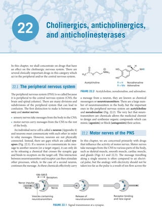
a message from a neuron, they are known as chemical
messengers or neurotransmitters. There are a large num-
ber of neurotransmitters in the body, but the important
ones in the peripheral nervous system are acetylcholine
and noradrenaline (Fig. 22.2). The very fact that neuro-
transmitters are chemicals allows the medicinal chemist
to design and synthesize organic compounds which can
mimic (agonists) or block (antagonists) their action.
22.2 Motor nerves of the PNS
In this chapter, we are concerned primarily with drugs
that influence the activity of motor nerves. Motor nerves
take messages from the CNS to various parts of the body,
such as skeletal muscle, smooth muscle, cardiac muscle,
and glands (Figs 4.1 and 22.3). The message travelling
along a single neuron is often compared to an electri-
cal pulse, but the analogy with electricity should not be
taken too far as the pulse is a result of ion flow across the
Cholinergics, anticholinergics,
and anticholinesterases
22
Receptor binding
and new signal
Release of
neurotransmitter
Vesicles containing
neurotransmitters
100–500Å
Receptors
Nerve impulse
Nerve
Nerve
FIGURE 22.1 Signal transmission at a synapse.
Acetylcholine R = H Noradrenaline
R = Me Adrenaline
H3C
C
O
O
NMe3
HO
HO
NHR
HO H
FIGURE 22.2 Acetylcholine, noradrenaline, and adrenaline.
2. membranes of neurons and not a flow of electrons (see
Appendix 4).
It should be evident that the workings of the human
body depend crucially on an effective motor nervous
system. Without it, we would not be able to operate our
muscles and we would end up as flabby blobs, unable to
move or breathe. We would not be able to eat, digest, or
excrete our food because the smooth muscle activity of
the gastrointestinal tract (GIT) and the urinary tract is
controlled by motor nerves. We would not be able to con-
trol body temperature, as the smooth muscle controlling
the diameter of our peripheral blood vessels would cease
to function. Finally, our heart would resemble a wobbly
jelly rather than a powerful pump. In short, if the motor
nerves failed to function, we would be in a mess! Let us
now look at the motor nerves in more detail.
The motor nerves of the PNS have been classified into
three subsystems: the somatic motor nervous system,
the autonomic motor nervous system, and the enteric
nervous system. These are considered in the following
sections.
22.2.1 The somatic motor nervous system
The somatic motor nerves carry messages from the CNS
to the skeletal muscles. There are no synapses en route
and the neurotransmitter at the neuromuscular junc-
tion is acetylcholine. Acetylcholine binds to cholinergic
receptors within the cell membranes of muscle cells and
the final result is contraction of skeletal muscle.
22.2.2 The autonomic motor nervous
system
The autonomic motor nerves carry messages from the
CNS to smooth muscle, cardiac muscle, and the adrenal
medulla. This system can be divided into the sympa-
thetic and parasympathetic nervous systems.
Sympathetic neurons leave the CNS and synapse
almost immediately with a second neuron using acetyl-
choline as neurotransmitter. The second neuron then
proceeds to various tissues and organs around the body.
Noradrenaline is the neurotransmitter released from the
second neuron, and this interacts with adrenergic recep-
tors present in target cells and organs. At the heart, the
action of noradrenaline leads to contraction of cardiac
muscle and an increase in heart rate. Elsewhere, it relaxes
smooth muscle and reduces the contractions of the gas-
trointestinal and urinary tracts. It also reduces salivation
and the dilatation of the peripheral blood vessels. In gen-
eral, the sympathetic nervous system promotes the ‘fight
or flight’ response by shutting down the body's house-
keeping roles (digestion, defecation, urination, etc.),
while stimulating the heart.
There are some neurons in the sympathetic nervous
system which do not synapse with a second neuron,
but go directly to a gland called the adrenal medulla.
Acetylcholine is the neurotransmitter released by these
neurons and it stimulates the adrenal medulla to release
the hormone adrenaline, which then circulates through
the blood system. Adrenaline reinforces the actions
of noradrenaline by activating adrenergic receptors
throughout the body, whether they are supplied directly
with nerves or not.
Parasympathetic neurons leave the CNS, travel
some distance, then synapse with a second neuron using
acetylcholine as neurotransmitter. The second neu-
ron then proceeds to synapse with the same target tis-
sues and organs as the sympathetic neurons. However,
acetylcholine acts as the neurotransmitter, rather than
noradrenaline, and activates cholinergic receptors on
the target cells. The resulting effects are the opposite to
those caused by activation of adrenergic receptors. For
CNS (somatic)
CNS (autonomic)
Sympathetic
Parasympathetic
Ach
Ach
NA
Skeletal muscle
(N)
Smooth muscle
Cardiac muscle
Adrenoceptors
Smooth muscle
Cardiac muscle
(M)
Ach
Ach
Ach
Adrenal
medulla
Adrenaline
(N)
(N)
FIGURE 22.3 Motor nerves of the peripheral nervous system. N = nicotinic receptor; M = musarinic receptor;
AcH = acetylcholine; NA = noradrenaline.
Motor nerves of the PNS 579
3. example, cardiac muscle is relaxed, whereas the smooth
muscle of the digestive and urinary tracts is contracted.
As the sympathetic and parasympathetic nervous
systems oppose each other in their actions, they can be
looked upon as acting like a brake and an accelerator
on the different tissues and organs around the body. The
analogy is not quite apt because both systems are always
operating and the overall result depends on which effect
is the stronger.
22.2.3 The enteric system
The third constituent of the PNS is the enteric system,
which is located in the walls of the GIT. It receives mes-
sages from sympathetic and parasympathetic nerves,
but it also responds to local effects to provide local reflex
pathways which are important in the control of GIT func-
tion. A large variety of neurotransmitters are involved
including serotonin, neuropeptides, and ATP. Nitric
oxide (NO) is also involved as a chemical messenger.
22.2.4 Defects in motor nerve
transmission
Defects in motor nerve transmission would clearly lead
to a large variety of ailments involving the heart, skel-
etal muscle, GIT, urinary tract, and many other organs.
Such defects might be the result of either a deficit or an
excess of neurotransmitter. Therefore, treatment involves
the administration of drugs which can act as agonists or
antagonists, depending on the problem. There is a dif-
ficulty with this approach, however. Usually, the prob-
lem we wish to tackle occurs at a certain location where
there might, for example, be a lack of neurotransmitter.
Application of an agonist to make up for low levels of
neurotransmitter at the heart might solve the problem
there, but would lead to problems elsewhere in the body
where the levels of neurotransmitter would be normal.
At those areas, the agonist would cause too much activ-
ity and cause unwanted side effects. Therefore, drugs
showing selectivity for different parts of the body would,
clearly, be preferred. This selectivity has been achieved
to a great extent with both cholinergic and adrenergic
agents. In this chapter, we concentrate on cholinergic
agents (adrenergic agents are covered in Chapter 23).
22.3 The cholinergic system
22.3.1 The cholinergic signalling system
Let us look first at what happens at synapses involv-
ing acetylcholine as the neurotransmitter. Figure 22.4
shows the synapse between two neurons and the events
involved when a message is transmitted from one neuron
to another. The same general process takes place when a
message is passed from a neuron to a muscle cell.
1. The first stage involves the biosynthesis of acetylcho-
line (Fig. 22.5). Acetylcholine is synthesized from
choline and acetyl coenzyme A at the end of the
presynaptic neuron. The reaction is catalysed by the
enzyme choline acetyltransferase.
2. Acetylcholine is incorporated into membrane-bound
vesicles by means of a specific transport protein.
3. The arrival of a nerve signal leads to an opening of
calcium ion channels and an increase in intracellular
calcium concentration. This induces the vesicles to
fuse with the cell membrane and release the transmit-
ter into the synaptic gap.
4. Acetylcholine crosses the synaptic gap and binds to
the cholinergic receptor, resulting in stimulation of
the second neuron.
5. Acetylcholinemovestoanenzymecalledacetylcholin-
esterase, which is situated on the postsynaptic neuron,
and which catalyses the hydrolysis of acetylcholine to
produce choline and acetic acid (ethanoic acid).
6. Choline is taken up into the presynaptic neuron by a
transport protein to continue the cycle.
The most important thing to note is that there are
several stages where it is possible to use drugs to either
promote or inhibit the overall process. The greatest suc-
cess so far has been with drugs targeted at stages 4 and 5
(i.e. the cholinergic receptor and the acetylcholinesterase
enzyme). These are considered in more detail in subse-
quent sections.
22.3.2 Presynaptic control systems
Cholinergic receptors (called autoreceptors) are pre-
sent at the terminus of the presynaptic neuron (Fig.
22.6). The purpose of these receptors is to provide a
means of local control over nerve transmission. When
acetylcholine is released from the neuron, some of it
will find its way to these autoreceptors and switch them
on. This has the effect of inhibiting further release of
acetylcholine.
The presynaptic neuron also contains receptors for
noradrenaline, which act as another control system for
acetylcholine release. Branches from the sympathetic
nervous system lead to the cholinergic synapses and when
the sympathetic nervous system is active, noradrenaline
is released and binds to these receptors. Once again, the
effect is to inhibit acetylcholine release. This indirectly
enhances the activity of noradrenaline at target organs by
lowering cholinergic activity.
580 Chapter 22 Cholinergics, anticholinergics, and anticholinesterases
4. The chemical messenger nitric oxide (NO) can also
influence acetylcholine release, but, in this case, it pro-
motes release. A large variety of other chemical mes-
sengers including co-transmitters (see below) are also
implicated in presynaptic control. The important thing
to appreciate is that presynaptic receptors offer another
possible drug target to influence the cholinergic nervous
system.
22.3.3 Co-transmitters
Co-transmitters are messenger molecules released along
with acetylcholine. The particular co-transmitter released
depends on the location and target cell of the neurons.
Each co-transmitter interacts with its own receptor on
the postsynaptic cell. Co-transmitters have a variety
of structures and include peptides, such as vasoactive
intestinal peptide (VIP), gonadotrophin-releasing
hormone (GnRH), and substance P. The roles of these
agents appear to be as follows:
• they are longer-lasting and reach more distant targets
than acetylcholine, resulting in longer-lasting effects;
• the balance of co-transmitters released varies under
different circumstances (e.g. presynaptic control) and
so can produce different effects.
ACh
NO (from endothelial cells)
ACh
Noradrenaline receptor
Cholinergic receptor
Promotes acetylcholine release
Inhibits acetylcholine release
Target
receptor
NA
FIGURE 22.6 Presynaptic control systems.
4
3
Postsynaptic
nerve
E1=Choline acetyltransferase
Ach
SCoA Vesicle
1
E1
2
6
Acetylcholine (Ach)
Choline
Acetic acid
Presynaptic
nerve
FIGURE 22.4 Synapse with acetylcholine acting as the neurotransmitter.
Choline acetyltransferase
Choline Acetylcholine
+
H3C
C
SCoA
O
HO
NMe3
H3C
C
O
O
NMe3
FIGURE 22.5 Biosynthesis of acetylcholine.
The cholinergic system 581
5. 22.4 Agonists at the cholinergic
receptor
One point might have occurred to you. If there is a lack of
acetylcholine acting at a certain part of the body, why not
just administer more acetylcholine? After all, it is easy
enough to make in the laboratory (Fig. 22.7).
There are three reasons why this is not feasible.
• Acetylcholine is easily hydrolysed in the stomach by
acid catalysis and cannot be given orally.
• Acetylcholine is easily hydrolysed in the blood by
esterase enzymes (esterases).
• There is no selectivity of action. Additional acetylcho-
line will switch on all cholinergic receptors in the body.
Therefore, we need analogues of acetylcholine that are
more stable to hydrolysis and more selective with respect to
where they act in the body. We shall look at selectivity first.
Therearetwowaysinwhichselectivitycanbeachieved.
Firstly, some drugs may be distributed more efficiently to
one part of the body than another. Secondly, there are
different types of cholinergic receptor, which vary in the
way they are distributed in tissues. It is possible to design
synthetic agents that show selectivity for these receptors
and, hence, have tissue selectivity.
This is not just a peculiarity of cholinergic receptors.
Differences have been observed for other types of recep-
tors, such as those for dopamine, noradrenaline, and ser-
otonin, and there are many types and subtypes of recep-
tor for each chemical messenger (see Chapter 4).
The first indications that different types of cholinergic
receptor existed came from the action of natural com-
pounds. It was discovered that the compounds nicotine
(present in tobacco) and muscarine (the active principle
of a poisonous mushroom) (Fig. 22.8) were both cholin-
ergic agonists, but that they had different physiological
effects.
Nicotine showed selectivity for cholinergic receptors
present on skeletal muscle or at the synapses between
different neurons, whereas muscarine showed selectiv-
ity for cholinergic receptors present on smooth muscle
and cardiac muscle. From these results, it was concluded
that there was one type of cholinergic receptor on skeletal
muscles and at nerve synapses (the nicotinic receptor),
and a different type of cholinergic receptor on smooth
muscle and cardiac muscle (the muscarinic receptor)
(Fig. 22.3).
Muscarine and nicotine were the first compounds to
indicate that receptor selectivity was possible, but they
are unsuitable as medicines because they have undesir-
able side effects resulting from their interactions with
other receptors. In the search for a good drug it is impor-
tant to gain selectivity for one class of receptor over
another (e.g. the cholinergic receptor in preference to
an adrenergic receptor) and selectivity between recep-
tor types (e.g. the muscarinic receptor in preference to
a nicotinic receptor). It is also preferable to gain selec-
tivity for particular subtypes of a receptor. For example,
not every muscarinic receptor is the same throughout the
body. At present, five subtypes of the muscarinic receptor
have been discovered (M1–M5) and ten subtypes of the
nicotinic receptor (α1−α10).
The principle of selectivity was proven with nicotine
and muscarine, and so the race was on to design novel
drugs which had the selectivity of nicotine or muscarine,
but not the side effects.
KEY POINTS
• The cholinergic nervous system involves nerves which use
the neurotransmitter acetylcholine as a chemical messen-
ger. These include the motor nerves which innervate skel-
etal muscle, nerves which synapse with other nerves in the
peripheral nervous system (PNS), and the parasympathetic
nerves innervating cardiac and smooth muscle.
• There are two types of cholinergic receptor. Muscarinic recep-
tors are present in smooth and cardiac muscle. Nicotinic
receptors are present in skeletal muscle and in synapses
between neurons.
• Acetylcholine is hydrolysed by the enzyme acetylcholinester-
ase when it departs the cholinergic receptor. The hydrolytic
product choline is taken up into presynaptic neurons and
+
O
HO
+H+ NMe3
AcO
NMe3
Ac2O
NMe3
FIGURE 22.7 Synthesis of acetylcholine
Nicotine L (+) Muscarine
N O
HO
Me
NMe3
N
Me
H
FIGURE 22.8 Nicotine and muscarine.
582 Chapter 22 Cholinergics, anticholinergics, and anticholinesterases
6. acetylated back to acetylcholine. The cholinergic receptor
and the enzyme acetylcholinesterase are useful drug targets.
• Acetylcholine cannot be used as a drug, because it is rapidly
hydrolysed by acid and enzymes. It shows no selectivity for
different types and subtypes of cholinergic receptor.
22.5 Acetylcholine: structure,
structure–activity relationships,
and receptor binding
The first stage in any drug development is to study the lead
compound and to find out which parts of the molecule are
important to activity so that they can be retained in future
analogues [i.e. structure–activity relationships (SARs)].
These results also provide information about what the
binding site of the cholinergic receptor looks like and help
decide what changes are worth making in new analogues.
In this case, the lead compound is acetylcholine itself.
The results described below are valid for both the nico-
tinic and muscarinic receptors, and were obtained by the
synthesis of a large range of analogues.
• The positively charged nitrogen atom is essential to
activity. Replacing it with a neutral carbon atom elimi-
nates activity.
• The distance from the nitrogen to the ester group is
important.
• The ester functional group is important.
• The overall size of the molecule cannot be altered
much. Bigger molecules have poorer activity.
• The ethylene bridge between the ester and the nitrogen
atom cannot be extended (Fig. 22.9).
• There must be two methyl groups on the nitrogen. A
larger, third alkyl group is tolerated, but more than one
large alkyl group leads to loss of activity.
• Bigger ester groups lead to a loss of activity.
Clearly, there is a tight fit between acetylcholine and its
binding site, which leaves little scope for variation. The
findings listed tally with a receptor binding site as shown
in Fig. 22.10.
It is proposed that important hydrogen bonding inter-
actions exist between the ester group of acetylcholine
and an asparagine residue. It is also thought that a small
hydrophobic pocket exists which can accommodate the
methyl group of the ester, but nothing larger. This inter-
action is thought to be more important in the muscarinic
receptor than the nicotinic receptor.
The evidence suggests that the NMe3
+ group is placed
in a hydrophobic pocket lined with three aromatic amino
acids. It is also thought that the pocket contains two
smaller hydrophobic pockets, which are large enough to
accommodate two of the three methyl substituents on the
NMe3
+ group. The third methyl substituent on the nitro-
gen is positioned in an open region of the binding site and
so it is possible to replace it with other groups. A strong
ionic interaction has been proposed between the charged
nitrogen atom and the anionic side group of an aspartate
residue. The existence of this ionic interaction represents
the classical view of the cholinergic receptor, but there is
an alternative suggestion which states that there may be
an induced dipole interaction between the NMe3
+ group
and the aromatic residues in the hydrophobic pocket.
There are several reasons for this. Firstly, the positive
charge on the NMe3
+ group is not localized on the nitrogen
atom, but is spread over the three methyl groups (com-
pare section 17.7.1). Such a diffuse charge is less likely to
be involved in a localized ionic interaction and it has been
shown by model studies that NMe3
+ groups can be stabi-
lized by binding to aromatic rings. It might seem strange
that a hydrophobic aromatic ring should be capable of sta-
bilizing a positively charged group, but it has to be remem-
bered that aromatic rings are electron-rich, as shown by
the fact they can undergo reaction with electrophiles. It is
thought that the diffuse positive charge on the NMe3
+ group
is capable of distorting the π electron cloud of aromatic
rings to induce a dipole moment (section 1.3.4). Induced
ion–dipole interactions between the NMe3
+ group and an
aromatic residue such as tyrosine would then account for
Acetoxy
Ethylene
bridge
Quaternary
nitrogen
Me O
NMe3
O
FIGURE 22.9 Acetylcholine.
O N
H H
Asn-617
Hydrophobic
pocket
Hydrophobic
pockets
Hydrophobic
pocket
Tyr-616 Trp-613
Trp-307 Asp-311
O O
N
CH3
CH3
CH3
CH3
CO2
FIGURE 22.10 Muscarinic receptor binding site.
Acetylcholine: structure, structure–activity relationships, and receptor binding 583
7. the binding. The fact that three aromatic amino acids are
present in the pocket adds weight to the argument.
Of course, it is possible that both types of binding inter-
actions are taking place, which will please both parties!
A large amount of effort has been expended trying to
identify the active conformation of acetylcholine, i.e. the
shape adopted by the neurotransmitter when it binds to
the cholinergic receptor. This has been no easy task, as
acetylcholine is a highly flexible molecule (Fig. 22.11)
where bond rotation along the length of its chain can lead
to many possible stable conformations (or shapes).
In the past, it was assumed that a flexible neurotrans-
mitter would adopt its most stable conformation when
binding. In the case of acetylcholine, that would be the
conformation represented by the sawhorse and Newman
projections shown in Fig. 22.12. However, there is not a
massive energy difference between alternative stable con-
formations such as the gauche conformation shown in
Figure 22.13. The stabilization energy gained from bind-
ing interactions within the binding site could more than
compensate for any energy penalties involved in adopt-
ing a slightly less stable conformation.
In order to try and establish the active conformation
of acetylcholine, rigid cyclic molecules have been stud-
ied which contain the skeleton of acetylcholine within
their structure; for example muscarine and the analogues
shown in Fig. 22.14. In these structures, the portion of
the acetylcholine skeleton which is included in a ring
is locked into a particular conformation because bonds
within rings cannot rotate freely. If such molecules bind
to the cholinergic receptor, this indicates that this par-
ticular conformation is ‘allowed’ for activity.
Test your understanding and practise your molecu-
lar modelling with Exercise 22.2.
Many such structures have been prepared, but it has
not been possible to identify one specific active confor-
mation for acetylcholine. This probably indicates that
the cholinergic receptor has a certain amount of latitude
and can recognize the acetylcholine skeleton within the
rigid analogues, even when it is not in the ideal active
conformation. Nevertheless, such studies have shown
that the separation between the ester group and the
quaternary nitrogen is important for binding, and that
this distance differs for the muscarinic and the nicotinic
receptor (Fig. 22.15).
Having identified the binding interactions and phar-
macophore of acetylcholine, we shall now look at how
acetylcholine analogues were designed with improved
stability.
Test your understanding and practise your molecu-
lar modelling with Exercises 22.1 and 22.2.
22.6 The instability of
acetylcholine
As described previously, acetylcholine is prone to
hydrolysis. This is explained by considering one of the
conformations that the molecule can adopt (Fig. 22.16).
In this conformation, the positively charged nitrogen
interacts with the carbonyl oxygen and has an electron-
withdrawing effect. To compensate, the oxygen atom
pulls electrons from the neighbouring carbon atom and
makes that carbon atom electron deficient and more
prone to nucleophilic attack. Water is a poor nucleophile,
but, because the carbonyl group is more electrophilic,
hydrolysis takes place relatively easily. This influence of
the nitrogen ion is known as neighbouring group par-
ticipation or anchimeric assistance.
We shall now look at how the problem of hydrolysis
was overcome, but it should be appreciated that we are
doing this with the benefit of hindsight. At the time the
problem was tackled the SAR studies were incomplete
and the format of the cholinergic receptor binding site
was unknown.
Me
C
O
N
O
Me
Me
Me
H
H
H
H
6
5
4
3
2
1
FIGURE 22.11 Bond rotations in acetylcholine leading
to different conformations.
Looking along
bond 4–3
Looking along
bond 5–4
NMe3
H
OAc
H
H
H
COCH3
H H
CH2NMe3
Me O
N
Me
O H
H
H
H
Me
Me
1
2
3
4 5
6
FIGURE 22.12 The sawhorse and Newman projections
of acetylcholine.
Looking along
bond 5–4
Gauche
interaction
NMe3
H H
OAc
H
H
Me O
H
O H
H
H
NMe3
1
2
3
4 5
6
FIGURE 22.13 A gauche conformation for acetylcholine.
584 Chapter 22 Cholinergics, anticholinergics, and anticholinesterases
8. 22.7 Design of acetylcholine
analogues
There are two possible approaches to tackling the inher-
ent instability of acetylcholine: steric shields and elec-
tronic stabilization.
22.7.1 Steric shields
The principle of steric shields was described in section
14.2.1 and can be demonstrated with methacholine
(Fig. 22.17). Here, an extra methyl group has been placed
on the ethylene bridge as a steric shield to protect the
carbonyl group. The shield hinders the approach of any
potential nucleophile and also hinders binding to ester-
ase enzymes, thus slowing down chemical and enzymatic
hydrolysis. As a result, methacholine is three times more
stable to hydrolysis than acetylcholine.
The obvious question now is why not put on a big-
ger alkyl group like an ethyl group or a propyl group?
Alternatively, why not put a bulky group on the acyl half
of the molecule, as this would be closer to the carbonyl
centre and have a greater shielding effect?
In fact, these approaches were tried. They certainly
increased stability but they lowered cholinergic activ-
ity. We should already know why—the fit between ace-
tylcholine and its receptor is so tight that there is little
scope for enlarging the molecule. The extra methyl group
is acceptable, but larger substituents hinder the mol-
ecule binding to the cholinergic receptor and decrease its
activity.
Introducing a methyl steric shield has another useful
effect. It was discovered that methacholine has signifi-
cant muscarinic activity, but very little nicotinic activity.
Therefore, methacholine shows good selectivity for the
muscarinic receptor. This is perhaps more important
than the gain in stability.
Selectivity for the muscarinic receptor can be
explained if we compare the proposed active conforma-
tion of methacholine with muscarine (Fig. 22.18), as
the methyl group of methacholine occupies the same
position as a methylene group in muscarine. This is
only possible for the S-enantiomer of methacholine and
when the two enantiomers of methacholine were sepa-
rated, it was found that the S-enantiomer was, indeed,
the more active enantiomer. It is not used therapeuti-
cally, however.
O
Me
HO
NMe3
O
O
Me
NMe3
Me O
NMe3
O
H
H
Muscarine
FIGURE 22.14 Rigid molecules incorporating the acetylcholine skeleton (C–C–O–C–C–N).
O
N
O
N
4.4 Å 5.9 Å
Nicotinic receptor
Muscarinic receptor
FIGURE 22.15 Pharmacophore of acetylcholine.
O
C
O
Me3N
Me
δ−
δ+
FIGURE 22.16 Neighbouring group participation.
The arrow indicates the inductive pull of oxygen
which increases the electrophilicity of the carbonyl
carbon (see Molecular modelling exercise 22.1).
Me O
NMe3
O Me
*
Asymmetric centre
Steric shield
FIGURE 22.17 Methacholine (racemic mixture).
O
HO
Me CH2NMe3
Muscarine
Me O CH2NMe3
O Me
H
H
(S)-Methacholine
Me O H
O Me
CH2NMe3
(R)-Methacholine
FIGURE 22.18 Comparison of muscarine and the R- and S-enantiomers of methacholine.
Design of acetylcholine analogues 585
9. 22.7.2 Electronic effects
The use of electronic factors to stabilize functional groups
was described in sections 14.2.2 and 14.2.3, and was used
in the design of carbachol (Fig. 22.19)—a long-acting
cholinergic agent which is resistant to hydrolysis. Here,
the acyl methyl group has been replaced by NH2 which
means that the ester has been replaced by a urethane or
carbamate group. This functional group is more resistant
to hydrolysis because the lone pair of electrons on nitro-
gen can interact with the neighbouring carbonyl group
and lower its electrophilic character (Fig. 22.20).
The tactic worked, but it was by no means a foregone
conclusion that it would. Although the NH2 group is equiv-
alent in size to the methyl group, the former is polar and the
latter is hydrophobic, and it was by no means certain that
a polar NH2 group would be accepted into a hydrophobic
pocket in the binding site. Fortunately, it is and activity is
retained, which means that the amino group acts as a bio-
isostereforthemethylgroup.Abioisostereisagroupwhich
can replace another group without affecting the pharmaco-
logical activity of interest (sections 13.3.7 and 14.2.2). Thus,
the amino group is a bioisostere for the methyl group as far
as the cholinergic receptor is concerned, but not as far as
the esterase enzymes are concerned.
The inclusion of the electron-donating amino group
greatly increases chemical and enzymatic stability.
Unfortunately, carbachol shows very little selectiv-
ity between the muscarinic and nicotinic receptors.
Nevertheless, it is used clinically for the treatment of
glaucoma where it can be applied locally, thus avoiding
the problems of receptor selectivity. Glaucoma arises
when the aqueous contents of the eye cannot be drained.
This raises the pressure on the eye and can lead to blind-
ness. Agonists cause the eye muscles to contract and
allow drainage, thus relieving the pressure.
22.7.3 Combining steric and electronic
effects
We have seen that the β-methyl group of methacho-
line increases stability and introduces receptor selectiv-
ity. Therefore, it made sense to add a β-methyl group
to carbachol. The resulting compound is bethanechol
(Fig. 22.21) which is both stable to hydrolysis and selec-
tive in its action. It is occasionally used therapeutically in
stimulating the GIT and urinary bladder after surgery.
Both these organs are ‘shut down’ with drugs during sur-
gery (section 22.9).
22.8 Clinical uses for cholinergic
agonists
22.8.1 Muscarinic agonists
A possible future use for muscarinic agonists is in the
treatment of Alzheimer’s disease. However, current clini-
cal uses include:
• treatment of glaucoma;
• ‘switching on’ the GIT and urinary tract after surgery;
• treatment of certain heart defects by decreasing heart
muscle activity and heart rate.
Pilocarpine (Fig. 22.22) is an example of a mus-
carinic agonist which is used in the treatment of glau-
coma. It is an alkaloid obtained from the leaves of shrubs
belonging to the genus Pilocarpus. Although there is no
quaternary ammonium group present in pilocarpine, it
is assumed that the drug is protonated before it inter-
acts with the muscarinic receptor. Molecular model-
ling shows that pilocarpine can adopt a conformation
having the correct pharmacophore for the muscarine
receptor; i.e. a separation between nitrogen and oxygen
of 4.4 Å.
Pilocarpine is also being considered for the treat-
ment of Alzheimer's disease, as are other muscarinic
agonists such as oxotremorine and various arecoline
analogues (Fig. 22.22). At present, anticholinesterases
are used clinically for the treatment of this disease
(section 22.15).
22.8.2 Nicotinic agonists
Nicotinic agonists are used in the treatment of myas-
thenia gravis. This is an autoimmune disease where the
body has produced antibodies against its own cholinergic
H2N O
NMe3
O
FIGURE 22.19 Carbachol.
δ+
δ−
H2N
O
H2N
O
H2N
O
FIGURE 22.20 Resonance structures of carbachol.
*
H2N O
NMe3
O Me
Asymmetric centre
*
FIGURE 22.21 Bethanechol.
586 Chapter 22 Cholinergics, anticholinergics, and anticholinesterases
10. receptors. As a result, the number of available receptors
drops and so fewer messages reach the muscle cells. In
turn, this leads to severe muscle weakness and fatigue.
Administering an agonist increases the chance of activat-
ing what few receptors remain. An example of a selective
nicotinic agonist is the first structure shown in Fig. 22.23.
This agent is very similar in structure to methacholine,
and differs only in the position of the methyl substituent.
This is sufficient, however, to completely alter receptor
selectivity. Despite that, this particular compound is not
used clinically and anticholinesterases (section 22.15.1.2)
are the preferred treatment. Varenicline is used clini-
cally, however. It is a partial agonist at nicotinic receptors
and was approved in 2006 as an aid to stop smoking.
KEY POINTS
• Acetylcholine fits snugly into the binding site of cholinergic
receptors and there is little scope for variation. Two of the
N-methyl groups and the acyl methyl group fit into hydropho-
bic pockets. The ester is involved in hydrogen bonding, and
the quaternary nitrogen is involved in ionic interactions and/or
induced dipole interactions.
• Rigid analogues of acetylcholine have been used to try and
identify the active conformation.
• Acetylcholine is unstable to acid because of neighbouring
group participation. Stable analogues have been designed
using steric shields and/or electronic effects.
22.9 Antagonists of the muscarinic
cholinergic receptor
22.9.1 Actions and uses of muscarinic
antagonists
Antagonists of the cholinergic receptor are drugs which
bind to the receptor but do not ‘switch it on’. By binding
to the receptor, an antagonist acts like a plug at the recep-
tor binding site and prevents acetylcholine from binding
(Fig. 22.24). The overall effect on the body is the same as
if there was a lack of acetylcholine. Therefore, antagonists
have the opposite clinical effect from agonists.
The antagonists described in this section act only at
the muscarinic receptor and therefore affect nerve trans-
missions to glands, the CNS, and the smooth muscle of
the GIT and urinary tract. The clinical effects and uses of
these antagonists reflect this.
The clinical effects of muscarinic antagonists are:
• reduced saliva and gastric secretions;
• reduced motility of the GIT and urinary tract by relax-
ation of smooth muscle;
• dilatation of eye pupils;
• CNS effects
O
N
N
Me
O
Me
Pilocarpine
N
N
O
Oxotremorine
N
O
O
R
Me
Arecoline (R=Me) and analogues
FIGURE 22.22 Examples of muscarinic agonists.
HN
Varenicline
N
N
*
Me O
NMe3
O
Me
* Asymmetric centre
+
FIGURE 22.23 Examples of selective nicotinic agonists.
Postsynaptic
nerve
Antagonist
Ach Ach
Ach
Postsynaptic
nerve
FIGURE 22.24 Action of an antagonist to block a receptor.
Antagonists of the muscarinic cholinergic receptor 587
11. The clinical uses are:
• shutting down the GIT and urinary tract during surgery;
• ophthalmic examinations;
• relief of peptic ulcers;
• treatment of Parkinson's disease;
• treatment of anticholinesterase poisoning;
• treatment of motion sickness;
• a potential use for M2 antagonists is in the treatment of
Alzheimer's disease.
22.9.2 Muscarinic antagonists
The first antagonists to be discovered were natural prod-
ucts—in particular alkaloids (nitrogen-containing com-
pounds derived from plants).
22.9.2.1 Atropine and hyoscine
Atropine (Fig. 22.25) is present in the roots of Atropa
belladonna (deadly nightshade) and is included in a root
extract which was once used by Italian women to dilate
their eye pupils. This was considered to enhance beauty,
hence the name belladonna. Clinically, atropine has been
used to decrease gastrointestinal motility and to counter-
act anticholinesterase poisoning.
Atropine has an asymmetric centre but exists as a
racemate. Usually, natural products exist exclusively as
one enantiomer. This is also true for atropine, which is
present in the plants of the genus Solanaceae as a single
enantiomer called hyoscyamine. As soon as the natural
product is extracted into solution, however, racemization
takes place. The asymmetric centre in atropine is easily
racemized as it is next to a carbonyl group and an aro-
matic ring. This makes the proton attached to the asym-
metric centre acidic and easily removed.
Hyoscine (or scopolamine) (Fig. 22.25) is obtained
from the thorn apple (Datura stramonium) and is very
similar in structure to atropine. It has been used in the
treatment of motion sickness.
These two compounds bind to the cholinergic recep-
tor, but, at first sight, they do not look anything like acetyl-
choline. If we look more closely though, we can see that
a basic nitrogen and an ester group are present, and if
we superimpose the acetylcholine skeleton on to the
atropine skeleton, the distance between the ester and the
nitrogen groups is similar in both molecules (Fig. 22.26).
There is, of course, the problem that the nitrogen in atro-
pine is uncharged, whereas the nitrogen in acetylcholine
has a full positive charge. This implies that the nitrogen
atom in atropine must be protonated and charged when
it binds to the cholinergic receptor.
Therefore, atropine has two important binding features
shared with acetylcholine—a charged nitrogen when pro-
tonated and an ester group. It is able to bind to the recep-
tor, but why is it unable to switch it on? Because atropine
is a larger molecule than acetylcholine, it is capable of
binding to other binding regions within the binding site
which are not used by acetylcholine itself. As a result, it
interacts differently with the receptor and does not induce
the same conformational changes (induced fit) as acetyl-
choline. This means that the receptor is not activated.
Test your understanding and practise your molecu-
lar modelling with Exercise 22.3.
As both atropine and hyoscine are tertiary amines
rather than quaternary salts, they are able to cross the
blood–brain barrier as the free base. Once they are in the
brain, they can become protonated and antagonize mus-
carinic receptors which causes CNS effects; for example
hallucinogenic activity is brought on with high doses,
and both hyoscine and atropine were used by witches
in past centuries to produce that very effect. Other CNS
effects observed in atropine poisoning are restlessness,
agitation, and hyperactivity.
Easily racemized
*
N
Me
H
O
O
CH2OH
*
N
Me
H
O
O
CH2OH
O
Atropine Hyoscine
H
H
FIGURE 22.25 Atropine and hyoscine.
N
Me
O
NMe3
O
CH3
O
O
H
H
OH
O
O
O
FIGURE 22.26 Acetylcholine skeleton superimposed
on to the atropine skeleton.
588 Chapter 22 Cholinergics, anticholinergics, and anticholinesterases
12. In recent times, the disorientating effect of scopolamine
has seen it being used as a truth drug for the interrogation
of spies and so it is no surprise to find it cropping up in
various novels. An interesting application for scopolamine
was described in Jack Higgins' novel Day of Judgement
where it was used in association with suxamethonium
(Fig 22.33) to torture one hapless victim. Suxamethonium
was applied to the conscious victim in order to create
initial convulsive muscle spasms, followed by paralysis,
inability to breathe, agonizing pain, and a living impres-
sion of death. Scopolamine was then used to erase the
memory of this horror, so that the impact would be just as
bad when the process was repeated!
22.9.2.2 Structural analogues based
on atropine
In order to reduce CNS side effects, quaternary salts of atro-
pine and atropine analogues are used clinically (Fig. 22.27).
For example, ipratropium is used as a bronchodilator in
chronic obstructive pulmonary disease. Atropine methoni-
trate acts at the intestine to relieve spasm.
A large number of different analogues of atropine were
synthesized to investigate the SAR of atropine, revealing
the importance of the aromatic ring, the ester group, and
the basic nitrogen (which is ionized).
It was further discovered that the complex ring system
was not necessary for antagonist activity, so simplifica-
tion could be carried out. For example, amprotropine
(Fig. 22.28) is active and has an ester group separated
from an amine by three carbon atoms.
Chain contraction to two carbon atoms can be car-
ried out without loss of activity, and a large variety of
active antagonists have been prepared having the general
formula shown in Fig 22.29, for example tridihexethyl
chloride and propantheline bromide.
These studies came up with the following generaliza-
tions:
• the alkyl groups (R) on nitrogen can be larger than
methyl (in contrast to agonists);
• the nitrogen can be tertiary or quaternary, whereas ago-
nists must have a quaternary nitrogen. Note, however,
that the tertiary nitrogen is probably charged when it
interacts with the receptor;
• verylargeacylgroupsareallowed(R1 andR2 = aromatic
or heteroaromatic rings). This is in contrast to agonists
where only the acetyl group is permitted.
This last point appears to be the most crucial in deter-
mining whether a compound will act as an antagonist or
not. The acyl group has to be bulky, but it also has to have
that bulk arranged in a certain manner; in other words,
there must be some sort of branching in the acyl group.
The conclusion that can be drawn from these results is
that there must be hydrophobic binding regions next to the
normal acetylcholine binding site. The overall shape of the
acetylcholine binding site plus the extra binding regions
would have to be T- or Y-shaped in order to explain the
importance of branching in antagonists (Fig. 22.30). A
structure such as propantheline, which contains the com-
plete acetylcholine skeleton, as well as the hydrophobic
acyl side chain binds more strongly to the receptor than
acetylcholine itself. The extra binding interactions mean
that the conformational changes induced in the receptor
will be different from those induced by acetylcholine and
will fail to induce the secondary biological response. As
long as the antagonist is bound, acetylcholine is unable to
bind and pass on its message.
For additional material see Web article 8: photo-
affinity labelling
A large variety of antagonists have proved to be use-
ful medicines (Fig. 22.31), with many showing selectiv-
ity for specific organs. For example, tropicamide and
cyclopentolate are used in eye drops to dilate pupils for
ophthalmic examination, while trihexyphenidyl and
benzatropine are used centrally to counteract movement
disorders caused by Parkinson's disease. Some agents act
selectively to decrease gastric secretion; others are useful
in ulcer therapy. The selectivity of action for these drugs
owes more to their distribution properties than to receptor
selectivity. In other words, the compounds can reach some
parts of the body more easily than others. Having said that,
the antagonist pirenzepine, which is used in some coun-
tries for the treatment of peptic ulcers, is a selective M1
antagonist with no activity against M2 receptors.
N
Me
R
H
O
O
CH2OH
+
X
–
R = Me, X = NO3
–; Atropine methonitrate
R = iPr, X = Br–; Ipratropium
FIGURE 22.27 Structural analogues of atropine.
O
O
CH2OH
N
Et
Et Me
Me
FIGURE 22.28 Amprotropine.
Antagonists of the muscarinic cholinergic receptor 589
13. 22.10 Antagonists of the nicotinic
cholinergic receptor
22.10.1 Applications of nicotinic
antagonists
Nicotinic receptors are present in nerve synapses at gan-
glia, as well as at the neuromuscular synapse. However,
drugs are able to show a level of selectivity between these
two sites, mainly because of the distinctive routes which
have to be taken to reach them. Antagonists of gangli-
onic nicotinic receptor sites are not therapeutically use-
ful because they cannot distinguish between the ganglia
of the sympathetic nervous system and the ganglia of
the parasympathetic nervous system (both use nicotinic
receptors) (Fig. 22.3). Consequently, they have many side
effects. However, antagonists of the neuromuscular junc-
tion are therapeutically useful and are known as neuro-
muscular blocking agents.
R1 and R2 = Aromatic
or heteroaromatic
R2N Et3N
O
R2
O
R1
OH
Cl
Tridihexethyl chloride
O
O
O
N
Me
Me2CH
Me2CH
Br
Propanthreline bromide
FIGURE 22.29 Simplified analogues of atropine.
CO2
H2N Asn
van der Waals binding
regions for antagonists
Acetylcholine binding site
Receptor surface
O
O
O
NMeR2
FIGURE 22.30 The binding of propantheline to the
muscarinic receptor.
N
H
O
Me
Tropicamide Cyclopentolate
Benzatropine
Trihexyphenidyl
Pirenzepine
Me2N
O
O
OH
O
N
N
Me
CH2OH
N
OH
N
O
N
N
Me
NH
N
O
FIGURE 22.31 Some examples of clinically useful cholinergic antagonists.
590 Chapter 22 Cholinergics, anticholinergics, and anticholinesterases
14. 22.10.2 Nicotinic antagonists
22.10.2.1 Curare and tubocurarine
Curare was first identified in the sixteenth century
when Spanish soldiers in South America found them-
selves under attack by indigenous people using poi-
soned arrows. It was discovered that the Indians
were using a crude, dried extract from a plant called
Chondrodendron tomentosum, which stopped the heart
and also caused paralysis. Curare is a mixture of com-
pounds, but the active principle is a cholinergic antag-
onist that blocks nerve transmissions from nerve to
muscle.
It might seem strange to consider such a compound
for medicinal use, but at the right dose levels and under
proper control, there are useful applications for this
sort of action. The main application is in the relaxation
of abdominal muscles in preparation for surgery. This
allows the surgeon to use lower levels of general anaes-
thetic than would otherwise be required and increase the
safety margin for operations.
As mentioned previously, curare is actually a mixture
of compounds, and it was not until 1935 that the active
principle (tubocurarine) was isolated. The determination
of the structure took even longer, and it was not estab-
lished until 1970 (Fig. 22.32). Tubocurarine was used
clinically as a neuromuscular blocker, but it had unde-
sirable side effects as it also acted as an antagonist at the
nicotinic receptors of the autonomic nervous system (Fig.
22.2). Better agents are now available.
The structure of tubocurarine presents a problem
to our theory of receptor binding. Although it has a
couple of charged nitrogen centres, there is no ester to
interact with the acetyl binding region. Studies on the
compounds discussed so far show that the positively
charged nitrogen on its own is not sufficient for good
binding, so why should tubocurarine bind to the nico-
tinic receptor?
Test your understanding and practise your molecu-
lar modelling with Exercise 22.4.
The answer lies in the fact that the molecule has two pos-
itively charged nitrogen atoms (one tertiary, which is pro-
tonated, and one quaternary). Originally, it was believed
that the distance between the two centres (1.15 nm) might
be equivalent to the distance between two separate cholin-
ergic receptors and that the tubocurarine molecule could
bridge the two binding sites, and act as a steric shield for
both. However pleasing that theory may be, the dimen-
sions of the nicotinic receptor make this impossible. The
nicotinic receptor is a protein dimer made up of two iden-
tical protein complexes separated by 9–10 nm—far too
large to be bridged by the tubocurarine molecule (Fig.
22.33 and section 22.11).
Another possibility is that the tubocurarine molecule
bridges two acetylcholine binding sites within the one
protein complex. As there are two such sites within the
complex, this appears to be an attractive theory. However,
the two sites are more than 1.15 nm apart and so this too
has to be ruled out. It has now been proposed that one of
the positively charged nitrogens on tubocurarine binds
to the anionic binding region of an acetylcholine binding
site, while the other binds to a nearby cysteine residue
0.9–1.2 nm away (Fig. 22.33).
Despite the uncertainty surrounding the binding
interactions of tubocurarine, it seems highly probable
that two ionic binding regions are involved. Such an
interaction is extremely strong and would more than
N
MeO
HO
Me
Me
O
N
H
H
OH
OMe
O
Me
H
FIGURE 22.32 Tubocurarine.
8nm
9–10nm
Protein complex
(5 subunits)
(a) Receptor dimer (b) Interaction with tubocurarine
Acetylcholine
binding site
Tubocurarine
S
N N
N N
FIGURE 22.33 Tubocurarine binding to the cholinergic receptor.
Antagonists of the nicotinic cholinergic receptor 591
15. make up for the lack of the ester binding interaction. It
is also clear that the distance between the two positively
charged nitrogen atoms is crucial to activity. Therefore,
analogues that retain this distance should also be good
antagonists. Strong evidence for this comes from the
fact that the simple molecule decamethonium is a good
antagonist (section 22.10.2.2).
22.10.2.2 Decamethonium and suxamethonium
Decamethonium (Fig. 22.34) is as simple an analogue
of tubocurarine as one could imagine. It is a flexible,
straight-chain molecule and is capable of a large num-
ber of conformations. The fully extended conformation
places the nitrogen atoms 1.4 nm apart, but there are
other more folded conformations that position the nitro-
gen centres 1.14 nm apart, which compares well with the
equivalent distance in tubocurarine (1.15 nm) (see also
Box 17.4 and Molecular modelling exercise 22.4).
The drug binds strongly to cholinergic receptors and
has proved a useful clinical agent, but it suffers from sev-
eral disadvantages. For example, when it binds initially
to nicotinic receptors, it acts as an agonist rather than an
antagonist. In other words, it switches on receptors such
that sodium ion channels open up to depolarize muscle
cell membranes and cause brief contractions of the mus-
cle. Because the drug is not rapidly hydrolysed in the
same way as acetylcholine, it remains bound to the recep-
tor leading to persistent depolarization and subsequent
desensitization of the end plate. At that stage, it can be
viewed as an antagonist as it no longer stimulates mus-
cle contraction and blocks access to acetylcholine. (A
theory of how such an effect might take place is described
in section 8.6.) Another disadvantage is that it binds too
strongly, so patients take a long time to recover from its
effects.
We now face the opposite problem from the one faced
when designing cholinergic agonists. Instead of stabiliz-
ing a molecule, we need to introduce some instability—a
sort of timer control whereby the molecule can be inac-
tivated more quickly. Success was first achieved with
suxamethonium (Fig. 22.34) where two ester groups
are incorporated into the chain in such a way that the
distance between the charged nitrogens remains the
same. The ester groups are susceptible to chemical and
enzymatic hydrolysis and, once this takes place, the
molecule can no longer bridge the two binding regions
on the receptor and is inactivated. The ester groups are
also introduced such that suxamethonium mimics two
acetylcholine molecules linked end on. Suxamethonium
has a fast onset and short duration of action (5–10 min-
utes), but suffers from various side effects. Furthermore,
about one person in every 2000 lacks the plasma cho-
linesterase enzyme which hydrolyses suxamethonium.
Nevertheless, it is still used clinically in short surgical
procedures, such as the insertion of tracheal tubes.
Both decamethonium and suxamethonium are classed
as depolarizing neuromuscular blockers and have effects
on the autonomic ganglia, which explains some of their
side effects. Decamethonium also lacks total selectivity
for the neuromuscular junction and has an effect on cho-
linergic receptors in the heart. This leads to an increased
heart rate and a fall in blood pressure.
22.10.2.3 Steroidal neuromuscular
blocking agents
The design of pancuronium, vecuronium, and rocu-
ronium (Fig. 22.35) was based on tubocurarine, but
involved a steroid nucleus acting as a spacer between
the two nitrogen groups. The distance between the qua-
ternary nitrogens is 1.09 nm compared with 1.15 nm in
tubocurarine. Acyl groups were also added to introduce
one or two acetylcholine skeletons into the molecule in
order to improve affinity for the receptor sites. These
compounds have a faster onset of action than tubocurar-
ine and do not affect blood pressure. They are not as
rapid in onset as suxamethonium and have a longer
duration of action (45 minutes). Their main advantage
is that they have fewer side effects and so they are widely
used clinically. Unlike decamethonium and suxametho-
nium, these agents have no agonist activity and act as
pure antagonists, so they have no depolarizing effect on
target muscle cells. The neuromuscular blocking activity
of rocuronium can be reversed with a cyclodextrin called
sugammadex (Box 10.3).
22.10.2.4 Atracurium and mivacurium
The design of atracurium (Fig. 22.36) was based on the
structures of tubocurarine and suxamethonium. It is
Decamethonium
O
O
O
O
NMe3
Me3N
Suxamethonium
Acetylcholine
Acetylcholine
Me3N(CH2)10NMe3
FIGURE 22.34 Decamethonium and suxamethonium.
592 Chapter 22 Cholinergics, anticholinergics, and anticholinesterases
16. superior to both as it lacks cardiac side effects and is rap-
idly broken down in blood. This rapid breakdown allows
the drug to be administered as an intravenous drip.
The rapid breakdown is due to a self-destruct mech-
anism. At the slightly alkaline pH of blood (pH = 7.4),
the molecule can undergo a Hofmann elimination (Fig.
22.37). Once this happens, the compound is inactivated
because the positive charge on the nitrogen is lost and
the molecule is split in two. It is a particularly clever
example of drug design in that the very element respon-
sible for the molecule’s biological activity promotes its
deactivation.
The important features of atracurium are:
• the spacer—a 13-atom chain connects the two quater-
nary centres;
• the blocking units—the cyclic structures at either end
of the molecule which block the binding site from
acetylcholine;
• the quaternary centres—these are essential for receptor
binding. If one is lost through Hofmann elimination,
the binding interaction is too weak and the antagonist
leaves the binding site;
• the Hofmann elimination—the ester groups within the
spacer chain are crucial to the rapid deactivation pro-
cess. Hofmann eliminations normally require strong
alkaline conditions and high temperatures—hardly
normal physiological conditions. However, if a good
electron-withdrawing group is present on the carbon
that is beta to the quaternary nitrogen centre, it allows
the reaction to proceed under the much milder alka-
line conditions present in blood (pH 7.4). The electron-
withdrawing ester group increases the acidity of the
hydrogen on the beta-carbon such that it is easily lost.
The Hofmann elimination does not occur at acid pH,
and so the drug is stable in solution at a pH of 3–4 and
can be stored safely in a refrigerator.
Because the drug acts very briefly (approximately
30 minutes), it is added intravenously for as long as it is
needed. As soon as surgery is over, the intravenous drip
is stopped and antagonism ceases almost instantaneously.
Acetylcholine
skeleton
O
O Me
N
R Me
H
H
H
H
H H
H
HO
H
Me
Me
Me
O
O
O
N N
N
Me Me
O
O
Me
Acetylcholine
skeleton
Pancuronium (R = Me)
Vecuronium (R = H)
Rocuronium
FIGURE 22.35 Steroidal neuromuscular blocking agents.
N
MeO
MeO
Me
O
OMe
(CH2)5
OMe
O
O
O
N
OMe
OMe
Me
MeO
OMe
FIGURE 22.36 Atracurium.
Active Inactive
–H
N
Me
OR
Ph
O
H
N OR
Ph
O
H
Me
FIGURE 22.37 Hofmann elimination of atracurium.
Antagonists of the nicotinic cholinergic receptor 593
17. Another major advantage is that the drug does not require
enzymes to become deactivated and so deactivation occurs
at a constant rate between patients. With previous neuro-
muscular blockers, deactivation depended on metabolic
mechanisms involving enzymic deactivation and/or excre-
tion. The efficiency of these processes varies from patient
to patient and is particularly poor for patients with kidney
failure or with low levels of plasma esterases.
Mivacurium (Fig. 22.38) is a newer drug which is sim-
ilar to atracurium and is inactivated rapidly by plasma
enzymes, as well as by the Hofmann elimination. It has
a faster onset (about 2 minutes) and shorter duration of
action(about15minutes),althoughthedurationislonger
if the patients have liver disease or enzyme deficiencies.
22.10.2.5 Other nicotinic antagonists
Local anaesthetics and barbiturates appear to prevent the
changes in ion permeability which would normally result
from the interaction of acetylcholine with the nicotinic
receptor. They do not, however, bind to the cholinergic
binding site. It is believed that they bind instead to the
part of the receptor which is on the inside of the cell
membrane, perhaps binding to the ion channel itself and
blocking it.
Certain snake toxins have been found to bind irre-
versibly to the nicotinic receptor, thus blocking cho-
linergic transmissions. These include toxins such as
α-bungarotoxin from the Indian cobra. The toxin is a
polypeptide containing 70 amino acids which cross-links
the α- and β-subunits of the cholinergic receptor (sec-
tion 22.11).
Finally, the antidepressant and antismoking drug
bupropion (section 23.12.4) has been shown to be a
nicotinic antagonist, as well as a reuptake inhibitor of
noradrenaline and dopamine. It is possible that the drug's
effectiveness as an antismoking aid may be related to its
blockage of neuronal nicotinic receptors in the brain.
KEY POINTS
• Cholinergic antagonists bind to cholinergic receptors but fail
to activate them. They block binding of acetylcholine and
have a variety of clinical uses.
• Muscarinic antagonists normally contain a tertiary or qua-
ternary nitrogen, a functional group involving oxygen, and a
branch point containing two hydrophobic ring substituents.
• Nicotinic antagonists are useful as neuromuscular blockers
in surgery.
• The pharmacophore for a nicotinic antagonist consists of two
charged nitrogen atoms separated by a spacer molecule such
that the centres are a specific distance apart.
• One of the charged nitrogens binds to the cholinergic bind-
ing site; the other interacts with a nucleophilic group neigh-
bouring the binding site.
• Neuromuscular blockers should have a fast onset of action,
minimal side effects, and a short duration of action to allow
fast recovery. The lifetime of neuromuscular blockers can be
decreased by introducing ester groups which are susceptible
to enzymatic hydrolysis.
• Neuromuscular blockers which degrade chemically by means
of the Hofmann elimination are not dependent on metabolic
reactions and are more consistent from patient to patient.
22.11 Receptor structures
The nicotinic receptor has been isolated successfully from
the electric ray (Torpedo marmorata)—a fish found in the
Atlantic and the Mediterranean—allowing the receptor
to be studied carefully. As a result, a great deal is known
about its structure and operation. It is a protein complex
made up of five subunits, two of which are the same. The
five subunits (two α, one β, one γ, and one δ) form a cylin-
drical or barrel shape which traverses the cell membrane
MeO
MeO
MeO
N O
O
O
O
N
OMe
OMe
OMe
OMe
OMe
MeO
OMe
Me
Me
H
H
FIGURE 22.38 Mivacurium.
594 Chapter 22 Cholinergics, anticholinergics, and anticholinesterases
18. (section 4.6.2). The centre of the cylinder acts as an ion
channel for sodium, and a gating or lock system is con-
trolled by the interaction of the nicotinic receptor with
acetylcholine. In the absence of acetylcholine, the gate is
shut. When acetylcholine binds, the gate is opened. The
binding site for acetylcholine is situated mainly on the
α-subunit and there are two binding sites per ion chan-
nel complex. It is usually found that nicotinic receptors
occur in pairs, linked together by a disulphide bridge
between the δ-subunits.
This is the make up of the nicotinic receptor at neuro-
muscular junctions. The nicotinic receptors at ganglia
and in the CNS are more diverse in nature involving dif-
ferent α- and β-subunits. This allows drugs to act selec-
tively on neuromuscular, rather than neuronal, receptors.
For example, decamethonium is only a weak antagonist
at autonomic ganglia, whereas epibatidine (extracted
from a South American frog) is a selective agonist for
neuronal receptors. The snake toxin α-bungarotoxin is
specific for receptors at neuromuscular junctions.
Muscarinic receptors belong to the superfamily of
G-protein-coupled receptors (section 4.7) which operate
by activation of a signal transduction process (sections 5.1–
5.3). Five subtypes of muscarinic receptors have been iden-
tified and are labelled M1–M5. These subtypes tend to be
concentrated in specific tissues. For example, M2 receptors
occur mainly in the heart, whereas M4 receptors are found
mainly in the CNS. M2 receptors are also used as the autore-
ceptors on presynaptic cholinergic neurons (section 22.3.2).
The M1, M3, and M5 receptors are associated with a
signal transduction process involving the secondary
messenger inositol triphosphate (IP3) (section 5.3). The
M2 and M4 receptors involve a process which inhibits
the production of the secondary messenger cyclic-AMP
(section 5.2). Lack of M1 activity is thought to be associ-
ated with dementia.
KEY POINTS
• The nicotinic receptor is an ion channel consisting of five pro-
tein subunits. There are two binding sites for each ion channel.
• The muscarinic receptor is a G-protein-coupled receptor.
Various subtypes of muscarinic receptor predominate in dif-
ferent tissues.
22.12 Anticholinesterases and
acetylcholinesterase
22.12.1 Effect of anticholinesterases
Anticholinesterases are inhibitors of acetylcholinester-
ase—the enzyme that hydrolyses acetylcholine (section
22.3.1). If acetylcholine is not destroyed, it can return to
reactivate the cholinergic receptor and increase choliner-
gic effects (Fig. 22.39). Therefore, an acetylcholinesterase
inhibitor will have the same biological effect as a cholin-
ergic agonist.
22.12.2 Structure of the
acetylcholinesterase enzyme
The acetylcholinesterase enzyme has a fascinating tree-
like structure (Fig. 22.40). The trunk of the tree is a col-
lagen molecule which is anchored to the cell membrane.
There are three branches with disulphide bridges that lead
off from the trunk, each of which holds the acetylcholin-
esterase enzyme above the surface of the membrane. The
enzyme itself is made up of four protein subunits, each
of which has an active site. Therefore, each enzyme tree
has 12 active sites. The trees are rooted immediately next
to the cholinergic receptors such that they efficiently
capture acetylcholine as it departs the receptor. In fact,
the acetylcholinesterase enzyme is one of the most effi-
cient enzymes known. A soluble cholinesterase enzyme
called butyrylcholinesterase is also present in various
tissues and plasma. This enzyme has a broader substrate
specificity than acetylcholinesterase and can hydrolyse a
Enzyme blocked
by inhibitor
Reactivation
of Ach receptor
Postsynaptic
nerve
Ach
Ach
FIGURE 22.39 Effect of anticholinesterases
(Ach = acetylcholine).
Enzyme
Enzyme
Enzyme
S
S
S
S
S
S
S
S
S S
S
S
S
S
S S
S
S
FIGURE 22.40 The acetylcholinesterase enzyme.
Anticholinesterases and acetylcholinesterase 595
19. variety of esters. Its physiological function is not totally
clear, but it has been found to catalyse the hydrolysis of
toxic esters, such as cocaine, and appears to have a non-
catalytic role in cell differentiation and development. It
is also more effective than acetylcholinesterase at hydro-
lysing high levels of acetylcholine when the acetylcholin-
esterase enzyme itself becomes substrate inhibited.
22.12.3 The active site of
acetylcholinesterase
The design of anticholinesterases depends on the shape
of the enzyme's active site, the binding interactions
involved with acetylcholine, and the mechanism of
hydrolysis. The active site itself is at the foot of a narrow
gorge (Fig. 22.41a) and, at the entrance to the gorge, there
is a peripheral binding site. It is believed that this site
plays a crucial role in recognizing acetylcholine as the
substrate. One of the key interactions is a weak π-cation
interaction between the heteroaromatic ring of a tryp-
tophan residue and the charged quaternary nitrogen of
acetylcholine (Fig. 22.41b). After acetylcholine has been
‘captured’ it is rapidly transferred down the gorge to
the active site (Fig. 22.41c). This process is aided by the
fact that the gorge is lined with 14 conserved aromatic
residues, which can also form π-cation interactions with
acetylcholine and thus channel the substrate down the
gorge into the active site. Once acetylcholine enters the
active site, another tryptophan residue forms yet another
π-cation interaction (Fig. 22.41d). An electrostatic gradi-
ent running down the gorge encourages the movement
of acetylcholine. The gradient is due to several negatively
charged amino acid residues in the active site, which cre-
ate a dipole that points down the gorge to serve as an
electronic steering mechanism for the positively charged
substrate. The tryptophan residues in the peripheral
binding site and the active site are 12A apart and this is
Peripheral binding site
Active site Active site
Active site
(a)
(c) (d)
(b)
G
o
r
g
e
Me
Me
Me
O
O
O
O
O
O
+
NMe3
+
NMe3
+
NMe3
Trp
Trp Trp
Trp
Trp
Trp
Trp
Trp
FIGURE 22.41 Process by which acetylcholine is recognized and bound.
596 Chapter 22 Cholinergics, anticholinergics, and anticholinesterases
20. significant when it comes to designing potential dual-
action drugs (section 22.15.2).
22.12.3.1 Crucial amino acids within the active site
The important amino acids within the active site are those
which bind acetylcholine, as well as those involved in the
mechanism of hydrolysis. As far as binding is concerned,
several amino acids are thought to be involved, but a key
interaction is the interaction between a tryptophan resi-
due and the quaternary nitrogen atom (Fig. 22.42). The
key amino acid residues involved in the catalytic mecha-
nism are serine, histidine, and glutamate.
22.12.3.2 Mechanism of hydrolysis
The histidine residue acts as an acid–base catalyst
throughout the mechanism, while serine acts as a nucleo-
phile. This is not a particularly good role for serine, as an
aliphatic alcohol is a poor nucleophile and is unable to
hydrolyse an ester, but the acid/base catalysis provided
by histidine overcomes that disadvantage. The glutamate
residue interacts with the histidine residue and serves to
orientate and activate the ring (compare chymotrypsin—
section 3.5.3). There are several stages to the mechanism
(Fig. 22.43):
1. Acetylcholine approaches and binds to the active
site. Serine acts as a nucleophile and uses a lone pair
of electrons to form a bond to the ester of acetylcho-
line. Nucleophilic addition to the ester takes place
and opens up the carbonyl group
2. The histidine residue catalyses this reaction by acting
as a base and removing a proton, thus making serine
more nucleophilic
3. Histidine now acts as an acid catalyst and protonates
the alkoxy (OR) portion of the intermediate, turning
it into a much better leaving group
4. The carbonyl group reforms and expels the alcohol
portion of the ester (i.e. choline)
5. The acyl portion of acetylcholine is now covalently
bound to the active site. Choline leaves the active site
and is replaced by water
6. Water acts as a nucleophile and uses a lone pair of
electrons on oxygen to attack the acyl group
7. Water is normally a poor nucleophile, but, histidine
aids the process again by acting as a basic catalyst
and removing a proton
8. Histidine acts as an acid catalyst by protonating the
intermediate
9. The carbonyl group is reformed and the serine resi-
due is released. Because it is now protonated, it is a
much better leaving group
10. Ethanoic acid leaves the active site and the cycle can
be repeated.
The enzymatic process is remarkably efficient owing to
the close proximity of the glutamate residue (not shown),
the serine nucleophile, and the histidine acid–base cata-
lyst. As a result, hydrolysis by acetylcholinesterase is 108
(one hundred million) times faster than in its absence.
The process is so efficient that acetylcholine is hydrolysed
within a 100 μs of reaching the enzyme.
22.13 Anticholinesterase drugs
Anticholinesterase drugs act as inhibitors of the enzyme
acetylcholinesterase.Thisinhibitioncanbeeitherreversible
or irreversible depending on how the drug interacts with
Me
O
O
N
Me
Me
M′e
Ser OH
N
NH
His
O
O
Glu
N
H
Gorge
Trp
–
+
FIGURE 22.42 Key amino acid residues within the active site.
Anticholinesterase drugs 597
21. the active site. Two main groups of acetylcholinesterases
are considered here—carbamates and organophosphorus
agents.
22.13.1 Carbamates
22.13.1.1 Physostigmine
As in so many fields of medicinal chemistry, it was a
natural product that provided the lead for the carbamate
inhibitors. The natural product was physostigmine
(Fig. 22.44) (also called eserine) which was discovered
in 1864 as a product of the poisonous calabar bean
(the ordeal bean, Physostigma venenosum) from West
Africa. Extracts of these beans were fed to criminals
to assess whether they were guilty or innocent. Death
indicated a guilty verdict. The structure was established
in 1925 and physostigmine is still used clinically to treat
glaucoma.
SAR studies of physostigmine demonstrate that:
• the carbamate group is essential to activity;
• the benzene ring is important;
• the pyrrolidine nitrogen is important and is ionized at
blood pH.
Working backwards, the positively charged pyrroli-
dine nitrogen is important because it binds to the ani-
onic binding region of the enzyme. The benzene ring
may be involved in some extra hydrophobic bonding
N
NH
O
O
R
C
O
CH3
H
C
O
CH3
O
NH
N
NH
O
O
H
C
CH3
O
H
C
OR
O
CH3
O N
H
NH
OH
NH
C OH
CH3
O N
NH
Histidine
(Acidic catalyst)
:
:
:
O H N
NH
:
:N
Histidine
:
:
:
:
:
_
:
Stage 6
N
NH
O
O
R
C
O
CH3
Histidine
H
Stage 5
Stage 7
C
OR
O
CH3
O N :N
NH
Stage 9
Stage 8
:
H
:
Serine
(Nucleophile)
Stage 1
:
:
:
:
:
C
O
OH
CH3
O N
NH
H
:
:
Stage 4
:
:
:
:
:
:
:
C OH
CH3
O N
NH
H
:
Stage 3
Histidine
(Basic catalyst)
Stage 2
_
H
:
:
Histidine
Stage 10
Me O
O
(CH2)2NMe3
Me OH
O
Histidine
(Acidic catalyst)
Histidine
(Basic catalyst)
ROH H2O
Histidine
(Base)
:
:
O
:
:
:
O
:
FIGURE 22.43 Mechanism of hydrolysis for the acetylcholinesterase enzyme (the glutamate component
of the catalytic triad is not shown).
Pyrrolidine N
Urethane or carbamate
N
N
Me
Me
Me
O
N
O
Me
H
H
FIGURE 22.44 Physostigmine.
598 Chapter 22 Cholinergics, anticholinergics, and anticholinesterases
22. with the active site. Alternatively, it may be important
in the mechanism of inhibition as it provides a good
leaving group. The carbamate group is the crucial group
responsible for physostigmine's inhibitory properties. To
understand why, we must look at what happens when
physostigmine acts as the substrate for acetylcholinest-
erase (Fig. 22.45).
The first four stages proceed as normal, with histidine
catalysing the nucleophilic attack of the serine residue on
physostigmine (stages 1 and 2). The leaving group (this
time a phenol) is expelled with the aid of acid catalysis
from histidine (stages 3 and 4) and departs the active site
to be replaced by a water molecule.
The next stage turns out to be extremely slow. Despite
the fact that histidine can still act as a basic catalyst,
water finds it difficult to attack the carbamoyl intermedi-
ate. This step becomes the rate-determining step for the
whole process and the overall rate of hydrolysis of physo-
stigmine is 40 × 106 times slower than that of acetylcho-
line. As a result, the cholinesterase active site becomes
blocked and is unable to react with acetylcholine.
The final stage is slow because of the stability of the
carbamoyl–enzyme complex. This is because the nitrogen
can feed a lone pair of electrons into the carbonyl group
and drastically reduce its electrophilic character (Fig.
22.46) (cf. section 22.9.2).
22.13.1.2 Analogues of physostigmine
Physostigmine has limited medicinal use because of seri-
ous side effects, and it has only been used in the treat-
ment of glaucoma or as an antidote for atropine poison-
ing. Simpler analogues, however, have been used in the
treatment of myasthenia gravis and as an antidote to
curare poisoning.
Miotine (Fig. 22.47) still has the necessary carba-
mate, aromatic, and tertiary aliphatic nitrogen groups. It
is active as an antagonist but it also has disadvantages:
it is susceptible to chemical hydrolysis and it can cross
the blood–brain barrier (section 11.4.5) as the free base,
resulting in side effects due to its action in the CNS.
Neostigmine and pyridostigmine (Fig. 22.47) were
designed to deal with both these problems. Firstly, a qua-
ternary nitrogen atom is present and so there is no chance
of the free base being formed. As the molecule is perma-
nently charged, it cannot cross the blood–brain barrier
O
:N
NH
H
Physostigmine
:
:
MeHN
MeHN
MeHN
O
Ar
O
OAr
C
O
MeHN
O
NH
N
:
:
H
:
:
O Ar
C
O
O
NH
N
H
:
:
:
:
:
:
:
:
O Ar
C
O
MeHN
O
NH
N
:
H
O
:N
NH
Stable carbamoyl
intermediate
O
O H
NH
:N
Hydrolysis
(Very slow)
–ArOH
Stage 3
Stage 1 Stage 2
Stage 4
+H2O
FIGURE 22.45 Mechanism of inhibition by physostigmine (Ar represents the tricyclic system of physostigmine).
Anticholinesterase drugs 599
23. and so the drug is free of CNS side effects. Increased
stability is achieved by using a dimethylcarbamate group
rather than a methylcarbamate group. Two further points
to note about neostigmine are:
• the quaternary nitrogen is 4.7 Å away from the carba-
mate group;
• the direct bonding of the quaternary centre to the aro-
matic ring reduces the number of conformations that
the molecule can adopt. This is an advantage if the
active conformation is retained because the molecule
is more likely to be in the active conformation when it
approaches the active site.
Both neostigmine and pyridostigmine are in use
today. They are given intravenously to reverse the actions
of neuromuscular blockers or used orally in the treat-
ment of myasthenia gravis. Pyridostigmine was one of
the drugs used in the chemical cocktail provided to allied
troops in Iraq during Operation Desert Shield. The
agent was present to help protect against possible expo-
sure to organophosphate nerve gases. Edrophonium is
a similar agent used to reverse neuromuscular blocking
and is also used as a treatment of myasthenia gravis.
22.13.2 Organophosphorus compounds
The potential of organophosphorus compounds as nerve
agents was first recognized by German scientists in the
1920s and 1930s, and research was carried out to inves-
tigate their potential as weapons of war. When World
War II broke out, governments in the UK, USA, Sweden,
and Russia recognized the danger of Germany perfect-
ing these weapons and started their own research efforts
during the 1940s. In the UK, this was carried out at the
Porton Down Defence Centre. Fortunately, these agents
were never used, but researchers in different countries
continued work to find suitable antidotes that would
protect troops from a possible attack. It has not been
proved whether the organophosphate nerve gases have
ever been used in combat, but many believe that they
were part of the chemical weapons arsenal that was used
against the Kurds by the Iraqi government. It has also
been proposed that sarin (Fig. 22.48) may have been
released when Iraqi chemical plants and ammunition
dumps were bombed during the period 1990–91, and
that this might be a possible cause of the mystery illness
that afflicted many of the veterans of that war—Gulf
War syndrome. Bosnians, Serbs, and Croats have also
been accused of using nerve agents during the break-
up of Yugoslavia in the 1990s. Certainly, nerve agents
have been used by terrorist groups: the most notorious
example was the release of sarin in the Tokyo subway
during 1995.
The organophosphate nerve agents are examples of
the weapons of mass destruction which several Western
countries feared might be used by Iraq on its neighbours
or supplied to extremist groups. The invasion of Iraq in
Carbonyl
group
‘deactivated’
O
MeHN O
O
+
MeHN O
FIGURE 22.46 Stabilization of the carbamoyl–enzyme
intermediate.
Ionized
at blood pH
O
MeHN
O
NMe2
NMe3
Me
O
Me2N Me2N
O
N
O
O
Me
Miotine Neostigmine Pyridostigmine
HO
Et
Me
Me
N
Edrophonium
FIGURE 22.47 Analogues of physostigmine. Miotine is a chiral molecule that has been studied as a racemate.
O
iPrO
iPrO
P
Dyflos
(Diisopropyl
fluorophosphonate)
Sarin Soman Tabun VX
F
O
iPrO
iPr
iPr
N
Me
P
F
O
O
But
Me
Me
P
F
O
EtO
Me2N
P
CN
O
S
EtO
P
Me
FIGURE 22.48 Examples of nerve agents.
600 Chapter 22 Cholinergics, anticholinergics, and anticholinesterases
24. 2003 was designed to combat this threat, but subsequent
searches failed to reveal any such weapons.
It would be wrong to give the impression that the only
use for organophosphates is as weapons of war and ter-
ror. They are also extremely important insecticides used
in agriculture and animal husbandry, and have a variety
of uses in medicine. We shall consider these aspects in
the following sections.
22.13.2.1 Nerve agents
The nerve gases dyflos and sarin (GB) (Figure 22.48)
were discovered and perfected long before their mode of
action was known. Both agents inhibit acetylcholinester-
ase by irreversibly phosphorylating the serine residue at
the active site (Fig. 22.49).
The early part of the mechanism is similar to the nor-
mal mechanism, but the phosphorylated adduct which is
formed is extremely resistant to hydrolysis. Consequently,
the enzyme is permanently inactivated. As acetylcholine
cannot be hydrolysed, the cholinergic system is continu-
ally stimulated. This results in permanent contraction of
skeletal muscle, resulting in death.
Other nerve agents include tabun (GA), soman (GD),
and VX. VX is the most toxic of the nerve agents, hav-
ing an LD50 of 10 mg through skin contact. It was dis-
covered at Porton Down in the UK in 1954 then traded
to the USA in exchange for technological information
on nuclear weapons. The USA produced several tons of
the material for its chemical warfare programme, but
decided to dispose of its stockpiles in the late 1960s—a
process that was only completed in 2008. Much of the
nerve agent now lies at the bottom of the Atlantic Ocean.
22.13.2.2 Medicines
Once the mechanism of action of nerve agents was dis-
covered, compounds such as ecothiopate (Fig. 22.50)
were designed to fit the active site more effectively by
including a quaternary amine to bind with the anionic
region. This meant that lower doses would be more effec-
tive. Ecothiopate is used medicinally in the form of eye
drops for the treatment of glaucoma and has advantages
over dyflos, which has also been used in this way. Unlike
dyflos, ecothiopate slowly hydrolyses from the enzyme
over a matter of days.
22.13.2.3 Insecticides
The insecticides parathion, malathion, and chlorpy-
rifos (Fig. 22.50) are good examples of how a detailed
knowledge of biosynthetic pathways can be useful in
drug design. These agents are relatively non-toxic com-
pared with nerve gases because the P = S double bond
prevents inhibition of the acetylcholinesterase enzymes.
In contrast, the equivalent compounds containing a
P = O double bond are highly lethal.
Fortunately, there are no metabolic pathways in mam-
mals which can convert the P = S double bond to a P = O
double bond. In insects, however, the insecticides act as
prodrugs and are metabolized by oxidative desulphuri-
zation. The resulting anticholinesterases prove lethal.
Stable
Serine
–H
P
O
F
iPrO
iPrO
iPrO
iPrO
O
H
O
P O
Serine
FIGURE 22.49 Simplified mechanism of action of dyflos at the active site of acetylcholinesterase.
NO2
CO2Et
CH2CO2Et
CH
Me3N
S
S
P
P
O
O
OEt MeO
MeO
OEt
EtO EtO
EtO
EtO
+
S
P
S
S
N
CI
CI
Chlorpyrifos
Malathion
Parathion
Ecothiopate
CI
P
O
FIGURE 22.50 Organophosphates used as medicines and insecticides.
Anticholinesterase drugs 601
25. In mammals, the same compounds are metabolized in
a different way to give inactive compounds which are
then excreted (Fig. 22.51). Despite this, organophosphate
insecticides are not totally safe and prolonged exposure
to them can cause serious side effects if they are not han-
dled with care. Parathion has high lipid solubility and
is absorbed easily through mucous membranes, and
can also be absorbed through the skin. Preparations of
malathion are used medicinally for the treatment of head
lice, crab lice, and scabies, but should not be used too
frequently or over prolonged periods.
22.14 Pralidoxime: an
organophosphate antidote
Pralidoxime (Fig. 22.52) is an antidote to organopho-
phate poisoning and represents one of the early examples
of rational drug design, Any antidote for organophos-
phate poisoning has to displace the organophosphate
moiety from serine by hydrolysing the phosphate–serine
bond. However, this is a strong bond and not easily bro-
ken. Therefore, a strong nucleophile is required.
The literature revealed that phosphates can be hydro-
lysed with hydroxylamine (Fig. 22.53). This proved too
toxic a compound to be used on humans, so the next
stage was to design an equally reactive nucleophilic group
which would specifically target the acetylcholinesterase
enzyme. If such a compound could be designed, then
there was less chance of the antidote taking part in toxic
side reactions.
The designers’ job was made easier by the knowl-
edge that the organophosphate group does not fill the
active site, and the anionic binding region is vacant.
The obvious thing to do was to find a suitable group to
bind to this anionic centre and attach a hydroxylamine
moiety to it. Once positioned in the active site, the
hydroxylamine group could react with the phosphate
ester (Fig. 22.52).
Active drug
Insect
oxidative
desulphurization
Mammals Insects
Mammalian
metabolism
Phosphorylates enzyme
Death
Inactive
(excreted)
EtO
P
S
EtO O NO2
Parathion
(inactive prodrug)
EtO
P
S
EtO OH
EtO
P
O
EtO O NO2
FIGURE 22.51 Metabolism of insecticides in mammals and insects.
Active site
(blocked)
Active site
(free)
Serine
Serine
CO2 O OH
CO2
N
Me
N
O
H
Pralidoxime
P
O
RO
RO
H
N
Me
N
O
P
OR
O
OR
FIGURE 22.52 Pralidoxime as an antidote for organophosphate poisoning.
602 Chapter 22 Cholinergics, anticholinergics, and anticholinesterases
26. Pralidoxime was the result. The positive charge is pro-
vided by a methylated pyridine ring and the nucleophilic
side group is attached to the ortho position, as it was cal-
culated that this would place the nucleophilic hydroxyl
group in exactly the correct position to react with the
phosphate ester. The results were spectacular, with pra-
lidoxime showing a potency as an antidote 106 times
greater than hydroxylamine.
Because pralidoxime has a quaternary nitrogen, it is
fully charged and cannot pass through the blood–brain
barrier into the CNS. This means that the antidote can-
not work on any enzymes that have been inhibited in the
brain. Pro-2-PAM (Fig. 22.54) is a prodrug of pralidox-
ime which avoids this problem. As a tertiary amine it can
pass through the blood–brain barrier and is oxidized to
pralidoxime once it has entered the CNS.
22.15 Anticholinesterases as
‘smart drugs’
22.15.1 Acetylcholinesterase inhibitors
Acetylcholine is an important neurotransmitter in the
CNS, as well as in the PNS. It has been proposed that the
memory loss, intellectual deterioration, and personal-
ity changes associated with Alzheimer's disease may, in
part, be due to the destruction of cholinergic nerves in the
brain. Such damage is associated with the appearance of
extracellular protein plaques and intracellular protein tan-
gles in nerve fibres. These aberrant protein structures are
neurotoxic and responsible for the destruction of neurons.
Although Alzheimer's disease is primarily a disease of
the elderly, it can strike victims as young as 30 years of
age and is the fourth leading cause of death in the devel-
oped world, affecting nearly 50% of those aged 85 years
or more. It has been predicted that there will be 70 mil-
lion sufferers worldwide by 2050, representing 1.2% of
the total population.
The destruction of cholinergic nerves results in a drop
in both cholinergic receptors and acetylcholine levels
in the brain. Therefore, research has been carried out
into the use of anticholinesterases for the treatment of
Alzheimer’s disease—the so-called smart drugs. There is
no evidence that these compounds assist general mem-
ory improvement and so they are not a student's answer
to exam cramming! The treatment does not offer a cure
for Alzheimer's disease either, but it can alleviate the
symptoms by increasing the duration of action of acetyl-
choline such that activation of the cholinergic recep-
tors remaining is prolonged. Unlike anticholinesterases
acting in the periphery, ‘smart drugs’ have to cross the
blood–brain barrier and so structures containing quater-
nary nitrogen atoms are not suitable. Tests with physo-
stigmine were carried out in 1979, but the compound
was not ideal as it does not enter the brain sufficiently
well and shows short-lived, non-selective inhibition. The
first drug to be approved for the treatment of Alzheimer's
disease was tacrine (Fig. 22.55) in 1993. However, this is
an extremely toxic drug and is only beneficial for about a
year. Other agents which have subsequently been intro-
duced include donepezil in 1997, rivastigmine in 2000,
and galantamine (obtained from daffodils or snowdrop
bulbs) in 2001. Rivastigmine (an analogue of physostig-
mine) was the first drug to be approved in all countries of
the European Union. It shows selectivity for the brain and
has beneficial effects on cognition, memory, concentra-
tion, and functional abilities, such as day-to-day tasks or
hobbies. The drug has a short half-life, reducing the risk
of accumulation or drug–drug interactions. Metrifonate
(an organophosphate) and anabaseine (from ants and
marine worms) have also been tested for the treatment of
Alzheimer's disease. Herbal medicines have been used in
the past to treat the symptoms of Alzheimer's disease and
may provide useful lead compounds for further research
(Box 22.1).
The anticholinesterase drugs have been shown to be
beneficial in the early stages of Alzheimer's disease, but
are of less benefit when the disease has become advanced.
One disadvantage with the long-term use of these agents
is the fact that they increase acetylcholine levels all
round the body and not just in the brain; this leads to
gastrointestinal side effects. Another problem is that the
increased acetylcholine levels result in an increased acti-
vation of presynaptic cholinergic receptors which act as
a feedback control to lower the amounts of acetylcholine
released. As a result, there has been research into finding
selective cholinergic agonists that could be used to treat
the symptoms of the disease.
+
+
NH2OH ROH
RO P OR
O
OR
O P OR
O
OR
H2N
FIGURE 22.53 Hydrolysis of phosphates.
N
H
H
CH3
NOH
FIGURE 22.54 ProPAM.
Anticholinesterases as ‘smart drugs’ 603
27. 22.15.2 Dual-action agents acting on the
acetylcholinesterase enzyme
Inrecentyears,ithasbeendiscoveredthattheacetylcholin-
esterase enzyme appears to do more than just catalyse the
hydrolysis of acetylcholine. Under normal conditions,
the enzyme plays a non-catalytic role in neural develop-
ment, cell adhesion, and differentiation. Protein–protein
interactions involving the interaction of the peripheral
binding site of acetylcholinesterase with other proteins
promote these processes, with the tryptophan residue
described previously (section 22.12.3) playing a crucial
role.
Unfortunately, it has also been discovered that the
enzymecanplayanactiveroleinpromotingthedepositsof
aberrant protein that are found in the brain of Alzheimer's
sufferers. Studies have shown that the peripheral bind-
ing site of the enzyme is capable of binding β-amyloid
N
NH2
MeO
MeO
O
CH2 N
H
C
H
O
C
N
O
Me
Et
Me
NMe2
N
O
MeO
Me
HO
MeO
P CCl3
O
MeO
OH
Tacrine (Cognex, Parke-Davis) Donepezil (Aricept, Eisai)
Rivastigmine (Exelon, Novartis) N
Me
N
S
N
O Me
Galantamine (Reminyl, Shire)
Metrifonate (Bayer)
H
N N
Xanomeline (Novo Nordisk) Anabaseine
Cl
FIGURE 22.55 ‘Smart drugs’.
BOX 22.1 Mosses play it smart
An extract from the club moss Huperzia serrata has been
used for centuries in Chinese herbal medicine to treat ail-
ments varying from confusion in Alzheimer's disease to
schizophrenia. The extract contains a novel alkaloid called
huperzine A, which acts as an anticholinesterase. Binding is
very specific and so the drug can be used in small doses,
thus minimizing the risk of side effects. Huperzine A has
been approved for clinical use in China and has been shown
to have memory-enhancing effects.
A synthetic route to the natural product has been worked
out which has allowed the synthesis of different analogues,
but none of these is as active as the natural product. The
tricyclic ring system seems to be necessary for good activity,
ruling out the possibility of significant simplification. All
the functional groups in the molecule are also required
for good activity.
H
N O
NH2
Me
Me
Huperzine A.
604 Chapter 22 Cholinergics, anticholinergics, and anticholinesterases
28. protein, which is normally soluble and has an antioxidant
role. However, on binding to acetylcholinesterase, the
protein undergoes a conformational change which causes
it to become insoluble, leading to the appearance of the
protein plaques and tangles associated with Alzheimer's
disease. The enzyme has been described as a pathological
chaperone for this process and becomes associated with
the protein deposits. Moreover, soluble oligomers of the
protein are also formed within cells, which disrupt mito-
chondria function and increase oxidative stress, result-
ing in cell toxicity and cell death. These, indeed, may be
more relevant to the disease than the visible extracellular
plaques.
There is an exciting possibility that drugs might be
developed which could halt the progression of the disease
by preventing the binding of β-amyloid protein to the
peripheral binding site of acetylcholinesterase. Research
is currently in progess aimed at designing dual-action
drugs that are capable of inhibiting this process, as well
as acting as acetylcholinesterase inhibitors. Donepezil
(Fig. 22.55) is one currently used inhibitor that can span
the gorge to interact with both the peripheral binding
site and the active site. It has also been shown to have an
inhibitory effect on protein aggregation. However, much
of the early work has looked at tacrine dimers. Tacrine
(Fig. 22.55) is believed to enter the active site of the
enzyme in a similar manner to acetylcholine; in other
words, it is protonated and binds initially to the periph-
eral binding site. It is then transferred down the gorge
into the active site (Fig. 22.41). A dimer was designed
where two tacrine molecules were linked by a hydro-
carbon chain of sufficient length to allow one tacrine
moiety to bind to the active site while the other inter-
acted simultaneously with the peripheral binding site.
Different lengths of linker were tried and it was found
that a seven-carbon chain was ideal—bis(7)-tacrine
(Fig. 22.56). This compound was found to be 150–1000
times more potent as an enzyme inhibitor, depending on
the source of enzyme studied. Studies have shown that
the key tryptophan residues in the active site and the
peripheral binding site can form π-cation interactions
with each of the tacrine components. The linker can
also form van der Waals interactions with the gorge and
there is an entropy gain achieved by the displacement
of water from the gorge. However, there is an entropy
penalty resulting from the restriction in flexibility of the
linker once it is constrained within the gorge. The tri-
cyclic hydrophobic nature of the tacrine moieties is also
important as there is only a small desolvation penalty
involved when the structure binds. Stronger π-cation
interactions would be possible if one of the tacrine ring
systems was replaced with a simpler amine, but the latter
would be strongly solvated and would require a higher
desolvation penalty.
Test your understanding and practise your molecu-
lar modelling with Exercises 22.5 and 22.6.
The introduction of an N-methyl group into the linker
resulted in further binding interactions and increased
potency. The N-methyl group is protonated when the
dimer binds and so it can form π-cation interactions with
the aromatic residues lining the gorge. Following on from
this work, a large number of structures were synthesized
including homodimers of galantamine and huperzine B,
as well as heterodimers containing two different acetylcho-
linesterase inhibitors. Other dual-action structures have
been prepared consisting of a standard acetylcholinesterase
inhibitor linked to a moiety designed to bind more effec-
tively with the peripheral binding site. Many of these have
been shown to inhibit both the catalytic activity of acetyl-
cholinesterase, as well as protein aggregation. Nevertheless,
none of these compounds has entered the clinic to date.
There is a good chance that a dual-action agent will
eventually reach the clinic. However, there are many fac-
tors involved in Alzheimer's disease and so drugs inter-
acting with acetylcholinesterase alone are unlikely to
provide a total cure. Attention is now turning to treat-
ments that can address more than one of the various tar-
gets implicated in Alzheimer's disease. These treatments
could involve a cocktail of different drugs acting at dif-
ferent targets. An alternative approach is to use an agent
that can interact with different targets in a predictable
way (multiple-target directed ligands) (see also section
13.3.14). For example, dual-action agents that inhibit the
acetylcholinesterase enzyme have been designed which
have one or more of the following properties;
• antioxidant activity and/or the ability to chelate
metals;
N
NH
NH
N
Bis(7)-tacrine
Ki 1.3nM
(CH2)7 Linker
N
NH
NH
(CH2)3
(CH2)3
NMe
N
Ki 0.06nM
FIGURE 22.56 Tacrine dimers as dual-action agents.
Anticholinesterases as ‘smart drugs’ 605
29. • the ability to inhibit enzymes, such as butyrylcholinest-
erase, monoamine oxidase, or BACE1;
• antagonist activity at α2-adrenoceptors, 5HT3 recep-
tors, N-methyl-d-aspartate (NMDA) receptors, mus-
carinic (M2) receptors, or H3 receptors;
• inhibition of serotonin reuptake from nerve synapses;
• the blockade of calcium ion channels.
22.15.3 Multi-targeted agents acting on
the acetylcholinesterase enzyme and the
muscarinic M2 receptor
As an example of one area of research into multi-targeted
directed ligands, we shall consider agents that have been
designed to act as dual-action agents at the acetylcholin-
esterase enzyme (AChE), as well as antagonists of the M2
receptor. The M2 receptor is an autoreceptor present on
presynaptic cholinergic neurons. Activation of the auto-
receptor inhibits the release of acetylcholine from the
presynaptic neuron (section 22.3.2 and 22.11) and so M2
antagonists will increase acetylcholine release and help
to raise acetylcholine levels. The lead compound for this
work was a polyamine structure called benextramine
(Fig. 22.57). This is an irreversible α-adrenoceptor
antagonist, but it also shows activity as an anticholinest-
erase and M2 receptor antagonist. Polyamines have been
identified as good lead compounds for multi-targeted
directed ligands as the protonated nitrogens present have
the capability of forming π-cation interactions with aro-
matic residues in virtually any protein target. Moreover,
the flexible linear structure allows the polyamine to
adopt a huge number of different conformations, making
it more likely that suitable conformations are present that
allow interaction with different targets. Such compounds
are defined as promiscuous ligands (section 12.2.7).
Studies showed that the 2-methoxybenzyl group was
important to activity, but not the disulphide bridge.
Varying the chain length led to methoctramine, which
had improved M2 activity, while retaining good acetyl-
cholinesterase (AChE) activity. It was also shown that a
diamine diamide backbone retained affinity for M2 and
so the two ‘internal’ amines were replaced with amides
in order to improve lipophilicity. This decreased affin-
ity for M2 receptors and butyrlcholinesterase (BuChE),
but increased affinity for AChE. N-Methylation further
increased affinity for AChE resulting in the discovery of
caproctamine.
Compared with benextramine, caproctamine was
found to be 42 times more active as an AChE inhibitor
and two times less active as a BuChE inhibitor, while
retaining affinity for the M2 receptor. It was also dem-
onstrated that the structure could bind simultaneously
to the tryptophan residues in the active and peripheral
binding sites, while the linker formed hydrophobic inter-
actions with aromatic residues in the gorge. However,
caproctamine showed very little ability to inhibit AChE-
induced Aβ aggregation, which demonstrated that the
ability to interact with the peripheral binding site does
not necessarily block protein aggregation.
(CH2)3
(CH2)3
OMe
OMe
OMe
OMe
OMe
N
H
N
H
N
Me
O
O
Me
N Me
N
N
Me
N
H
N
H
H
N
H
N
H
N
H
N
OMe
S
AChE plC50 5.14mM; IC50 7.24 mM
AChE plC50 5.27 mM
BuChE plC50 5.21 mM
BuChE plC50 6.01 mM
M2 pA2 - mM
AChE plC50 6.77mM; IC50 0.17 mM
AChE induced Ab aggregation <5% inhibition
BuChE plC50 4.93 mM
M2 pA2 6.39mM; Kb 0.41 mM
M2 pA2 7.92 mM
Benextramine
Methoctramine
Caproctamine
S
FIGURE 22.57 Development of caproctamine from benextramine.
606 Chapter 22 Cholinergics, anticholinergics, and anticholinesterases