Foundus fluoresecein angiography and indocyanine angiography (2)
•Download as PPTX, PDF•
4 likes•219 views

FFA AND ICG

Recommended
More Related Content
What's hot
What's hot (20)
Similar to Foundus fluoresecein angiography and indocyanine angiography (2)
Similar to Foundus fluoresecein angiography and indocyanine angiography (2) (20)
Recently uploaded
Recently uploaded (20)
Foundus fluoresecein angiography and indocyanine angiography (2)
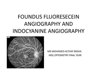
- 1. FOUNDUS FLUORESECEIN ANGIOGRAPHY AND INDOCYANINE ANGIOGRAPHY MR.MOHAMED ALTHAF BASHA MSC.OPTOMETRY FINAL YEAR
- 2. FLUORESCEIN ANGIOGRAPHY • The study and diagnosis of retinal, macular and choroidal pathologic lesions have been greatly revolutionized with the advent of fundus fluorescein angiography (FFA). • From an initial laboratory tool, it has now become a useful diagnostic tool that has aided the diagnosis and monitoring of the treatment of retinal vascular and macular diseases. • Although the retina can be readily examined by direct and indirect ophthalmoscopy and slit-lamp bio microscopy, the fluorescein angiography provides a valuable addition to these techniques.
- 3. PRINCIPLE OF FFA • Luminescence • Fluorescence • Phosphorescence • Two filters are used: • COBALT BLUE EXCITATION FILTER • YELLOW GREEN BARRIER FILTER
- 4. EXCITATION FILTER •The dye absorbs light in the blue range of the visible spectrum with absorption peaking at 465 to 490 nm. The blue flash excites the unbound fluorescein within the blood vessels or the leaked out fluorescein. • The blue filter shields out all other light and allows through only the blue excitation light. •Structures containing fluorescein within the eye emit green-yellow light. • The blue light is reflected off of the fundus structures that do not have fluorescein.
- 5. CONTRAINDICATIONS ABSOLUTE 1) Known allergy to iodine containing compounds. 2) H/O adverse reaction to FFA in the past. RELATIVE 1) Asthma 2) Hay fever 3) Renal failure 4) Hepatic failure 5) Cardiac disease – cardiac failure, Myocardial infarction 6) Previous mild reaction to dye. 7) Tonic-clonic seizures 6) Pregnancy ( especially 1st trimester)
- 7. EQUIPMENT
- 9. FFA PROVIDES THREE MAIN INFORMATION: • The flow characteristics in the blood vessels as the dye reaches and circulates through the retina and choroid. • It records fine details of the pigment epithelium and retinal circulation that may not otherwise be visible • Give a clear picture of the retinal vessels and assessment of their functional integrity
- 10. FLUORESCEIN PATHWAY • Arm-to-retina circulation time is 8-10 sec. • Normally 10-15 seconds elapse between dye injection and arrival of dye in the short ciliary arteries. • Choroidal circulation precedes retinal circulation by 1 second. • Transit of dye through the retinal circulation takes approximately 15 to 20 seconds.
- 13. CSR-SMOKE STACK
- 14. PED
- 15. NVD,NVE
- 17. NON PERFUSION AREAS VASUCLAR FILLING DEFECT
- 18. INDOCYANINE GREEN ANGIOGRAPHY • Indocyanine green (ICG) angiography (ICGA) is fast emerging as a popular and useful adjunct to the traditional fundus fluorescein angiography (FFA) in the diagnosis of macular, choroidal and outer retinal disorders. • This technique was introduced in ophthalmology in 1973 by Flower and Hochheimer. • FDA approved the ophthalmic use of ICG dye in 1975. • It remained largely unpopular owing mainly to technical difficulties. With the advent of videoangiogram recordings and the recognition of its potential in delineating occult choroidal neovascular membranes, the clinical use of ICGA has increased tremendously .
- 19. PRINCIPLES OF ICG • ICG fluorescence is only 1/25th that of fluorescein. So modern digital ICGA uses high-sensitivity videoangiographic image capture by means of an appropriately adapted camera. • Both the excitation (805 nm) and emission (835 nm) filters are set at infrared wavelengths. • Alternatively, scanning laser ophthalmoscopy (SLO) systems provide high contrast images, with less scattering of light and fast image acquisition rates facilitating high quality ICG video. • The technique is similar to that of FA, but with an increased emphasis on the acquisition of later images (up to about 45 minutes) than with FA. A dose of 25–50 mg in 1–2 ml water for injection is used
- 21. ADVERSE EFFECTS • Nausea, vomiting are uncommon. • Anaphylaxis, approximately equal incidence to FA. • Serious reactions are exceptionally rare. • ICG contains iodide and so should not be given to patients allergic to iodine or possibly shellfish. • Iodine-free preparations such as infracyanine green are available.
- 22. CONTRAINDICATIONS • ICGA is relatively contraindicated in liver disease (excretion is hepatic) • In patients with a history of a severe reaction to any allergen. • moderate or severe asthma • significant cardiac disease. • Its safety in pregnancy has not been established
- 23. PHASES OF ICGA • Early – up to 60 seconds post- injection • Early mid-phase – 1–3 minutes • Late mid-phase –3–15 minutes • Late phase – 15–45 minute
- 24. INDICATIONS OF ICGA • Polypoidal choroidal vasculopathy (PCV): ICGA is far superior to FA for the imaging of PCV. • Exudative age-related macular degeneration (AMD): Conventional FA remains the primary method of assessment, but ICGA can be a useful adjunct, particularly if PCV is suspected. • Chronic central serous chorioretinopathy often difficult to interpret areas of leakage on FA. ICGA shows choroidal leakage and the presence of dilated choroidal vessels. • Posterior uveitis. ICGA can provide useful information beyond that available from FA in relation to diagnosis and the extent of disease involvement.
- 25. ADVANTAGES OF ICGA OVER FFA • FA is an excellent method of studying the retinal circulation, it is of limited use in delineating the choroidal vasculature, due to masking by the RPE. • In contrast, the near-infrared light utilized in indocyanine green angiography (ICGA) penetrates ocular pigments such as melanin and xanthophyll, as well as exudate and thin layers of subretinal blood, making this technique eminently suitable. • ICG fluorescence can be imaged even in the presence of considerable blood, due to the phenomenon of forward scatter
- 26. • The peak absorption of ICG coincides with the emission spectrum of diode laser, which allows the selective ablation of chorioretinal lesions using ICG dye-enhanced laser photocoagulation wherein a target tissue containing ICG is exposed to the diode laser beam. • Photophobic patients tolerate ICGA better than FFA. • ICGA accurately measures the size of an occult choroidal neovascular membrane(CNVM).
- 27. LIMITATIONS OF ICGA • The choriocapillaris cannot be imaged separately with ICGA since their average cross-sectional diameter (21 μm) is much smaller than that of their feeding and draining vessels, and hence the fluorescence of the former cannot be differentiated from that arising from the latter. • The phenomenon of Mie scatter also masks the unfilled retinal vessels that cannot be visualized well in low speed angiography systems. • Bright areas do not necessarily signify dye leakage due to the phenomenon of additive fluorescence • Although superior to FFA in the imaging of occult CNVM, ICGA may underestimate the size of the CNVM.
- 29. RECENT ADVANCES IN INDOCYANINE GREEN ANGIOGRAPHY • Wide-angle angiography: This is carried out by performing ICGA with the aid of wide angle contact lenses, such as Volk SuperQuad and a traditional Topcon fundus camera. This allows real-time imaging of a wide field of the choroidal circulation up to 160 degrees of field of view. • Overlay technique: This technique allows lesion on one image to be traced on to another color or red-free image. • Digital stereo imaging: Elevated lesions such as PEDs can be better imaged in this way. • ICG as a photo sensitizer: It is considered to be a cheaper alternative to vertoporfin in photodynamic therapy of neovascularAMD& other disorders . • Digital subtraction ICGA: It uses digital subtraction of sequentially acquired ICG images along with pseudo color imaging. It shows occult CNVM in greater detail and within a shorter time than conventional ICGA.
- 30. THANK YOU