

Recommended
More Related Content
What's hot
What's hot (20)
Similar to Indocyanine Green Angiography (ICG)
Similar to Indocyanine Green Angiography (ICG) (20)
Recently uploaded
Recently uploaded (20)
Indocyanine Green Angiography (ICG)
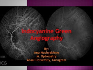
- 1. Indocyanine Green Angiography By: Anu Mushyakhwo M. Optometry Ansal University, Gurugram
- 2. Introduction Properties Principle Indications Contraindications Difference in FFA and ICG Advantages Adverse Effects Phases of ICGA Procedure Limitations
- 3. ICG dye is a water soluble tri-carbocyanine dye, primarily bound to globulins that absorbs and emits fluorescence in infra red spectrum of light Indocyanine green Angiography uses ICG dye to image choroidal and retinal circulations. Done using fundus camera with infrared filter or Laser Scanning Ophthalmoscopy (SLO)
- 4. Fluorescence: Luminescence that is maintained only by continuous excitation. Property of certain molecules to emit light energy of longer wavelength when stimulated by a shorter wavelength.
- 5. water-soluble, tricarbocyanine dye contains less than 5% sodium iodide in order to increase its solubility ICG has a half life of 150 to 180 seconds absorbs light at 805nm and emits fluorescence at 835nm 98% protein – binding ICG fluorescence is only 1/25th or 4% that of fluorescein is secreted unchanged by the liver into the bile without renal excretion there is no renal excretion and it does not cross the placenta.
- 6. the principle of fluorescence imaging used in ICG angiography (ICGA) is simple: illuminate the tissue of interest with light at the excitation wavelength (about 750 to 805 nm) while observing it at longer emission wavelengths (over 800 nm; Figure). To create a simple ICGA device, only a couple of filters are needed in addition to a proper camera and a light source, which can be quite small and suitable even for portable use . The filters are needed to prevent the mixing of the excitation (strong) and fluorescing (weak) rays to sum at the sensor. Without the filters, the weak fluorescence image cannot be seen among the strong reflection of the excitation light.
- 7. Lightsource Φ0 ΦS Sensor I0 Eb Lens R Ic Filter,Fs T Fs T Fc Filter,Fc Is It Tissue Tt Et E F,t BloodandICG Φ F ,C Hb ,C ICG Ee EF Fluorescence FIGURE 4: The principle of fluorescence imaging. The radiation from the light source is filtered by a high-pass filter, Fs, to remove the fluorescent wavelengths. The blood and ICG suspension under a tissue absorbs the excitation wavelengths and emits in fluorescent band. The emitted light is received by the sensor through a low-pass filter, Fc, to remove the excitation lightreflected fromthesource. FIGURE: The principle of fluorescence imaging. The radiation from the light source is filtered by a high-pass /excitation filter, Fs, to remove the fluorescent wavelengths. The blood and ICG suspension under a tissue absorbs the excitation wavelengths and emits in fluorescent band. The emitted light is received by the sensor through a low-pass /barrier filter, Fc, to remove the excitation light reflected from the source.
- 8. Polypoidal choroidal vasculopathy (PCV) Choroidal Neovascularization Exudative age-related macular degeneration (AMD) Posterior uveitis. Choroidal tumors RPE detachment (ICGA is superior to FFA in imaging and diagnosis of the above conditions) Chronic central serous chorio-retinopathy: often difficult to interpret areas of leakage on FA. ICGA shows choroidal leakage and the presence of dilated choroidal vessels. Breaks in Bruch membrane (When FFA is contraindicated)
- 9. contraindicated in liver disease in patients with a history of a severe reaction to any allergen moderate or severe asthma significant cardiac disease unsure for the pregnant women (safety in pregnancy has not been established)
- 11. Study of choroidal vasculature which is of limited in FFA due to RPE blockage Near-infrared light utilized penetrates melanin, xanthophylls, exudates and layers of sub-retinal blood better than the conventional fluorescein dye making this test more suitable Infrared is scattered less than visible light, thus suitable in eyes with media opacities 98% ICG molecules bound to protein, thus remaining in the blood vessels. ICG fluorescence can be imaged even in the presence of considerable blood Photophobic patients tolerate ICGA better thanFFA ICGA accurately measures the size of hidden choroidal neovascular membrane(CNVM).
- 12. mild nausea, vomiting anaphylaxis, approximately equal incidence to FA serious reactions are exceptionally rare ICG contains iodide and so should not be given to patients allergic to iodine or possibly shellfish Iodine-free preparations such as infracyanine green are available
- 13. The technique is similar to that of FA. There is an increased emphasis on the the later images (up to about 45 minutes) than with FA A dose of 25–50 mg in 1–2 ml water for injection is used.
- 14. A. Early – up to 60 seconds post-injection B. Early mid-phase – 1–3 minutes C. Late mid-phase –3–15 minutes D. Late phase – 15–45 minute Interpretation: (Same as FFA): Hypocyanescence: Blockage and filling defects Hypercyanescence: Leakage, staining, pooling and window defects
- 15. showing prominent choroidal arteries
- 16. showing greater prominence of choroidal veins as well as retinal vessels
- 17. • choroidal vessels fading but retinal vessels are still visible
- 18. showing hypofluorescent choroidal vessels and gradual fading of diffuse hyperfluorescence
- 19. FIGURE: ICG Comparison of FA and ICGA in imaging mouse retinal and choroidal vasculature.
- 20. The left picture shows the choroidal neovascularization with abnormal branching on either side but the right picture of FA shows light leakage only.
- 21. Figure: Polypoidal Choroidal Vasculopathy
- 22. the choriocapillaris cannot be imaged separately with ICGA (retinal vessels are also imaged) although superior to FFA in the imaging of occult CNVM, ICGA may underestimate the size of the CNVM
- 23. Wide-angle angiography Overlay technique Digital stereo imaging Digital subtraction ICGA
- 24. 1. Sandeep Kumar. Journal of Visualized Experiments (JoVE). February 2014 with 258 Reads. DOI: 10.3791/51061 2. Zachary Berriochoa, Alex D Jones,Yingbin Fu. Detecting Abnormalities in Choroidal Vasculature in a Mouse Model of Age Related Macular Degeneration by Time-Course Indocyanine Green. 3. Lim Jennifer I. M.D.; Flower, Robert W. Indocyanine green. International Ophthalmology Clinics: Fall 1995-Volume 35-Issue 4-ppg 59-70 4. Slakter JS , Yannuzzi LA , Guyer DR , Sorenson JA , Orlock DA. Indocyanine-green angiography. Current Opinion in Ophthalmology [01 Jun 1995, 6(3):25-32]. (PMID:10151085) 5. Kirby R. Miller. Jervey Eye Group. Greenville, S.C. Indocyanine Green Angiography. Ophthalmic Photographer’s Society 6. Jarmo T. Alander,1 Ilkka Kaartinen,2 Aki Laakso,3 Tommi Patil¨a,¨4 7. Thomas Spillmann, Valery V. Tuchin,, Maarit Venermo, and Petri Valisu0. A Review of Indocyanine Green Fluorescent Imaging in Surgery. International Journal of Biomedical Imaging. Volume 2012, Article ID 940585, 26 pages. doi:10.1155/2012/940585 8. Sarah L Owens. Indocyanine green angiography. British Journal of Ophthalmology 1996; 80: 263-266 9. Dr. Kranti Gupta, Dr.Ronel Soebam. Fluoresceince Agiography and Indo Cyanine Green. Slideshare.