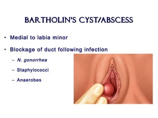
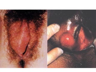
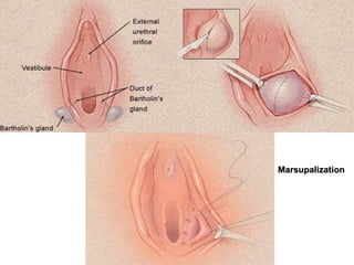
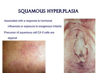
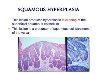
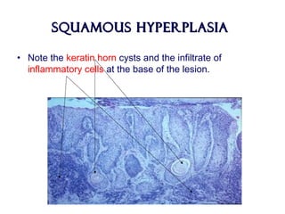
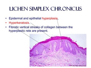
Benign diseases of theBenign diseases of the
vulva, vagina andvulva, vagina and
cervixcervix
Ahmad mukhtarAhmad mukhtar
MD.,M.B.B.Ch., M.Sc Obstetrics and GynecologyMD.,M.B.B.Ch., M.Sc Obstetrics and Gynecology
Consultant and Lecturer of Obstetrics and Gynecology,Consultant and Lecturer of Obstetrics and Gynecology,
Faculty ofFaculty of
MEDICINE, Zagazig University.MEDICINE, Zagazig University.
The vulvaThe vulva
• Is the part of the female genital tract located between the
genitocrural folds laterally, the mons pubis anteriorly, and the anus
posteriorly.
• Embryologically, it is the result of the junction of the cloacal
endoderm, urogenital ectoderm, and paramesonephric mesodermal
layers.
• This hollow structure contains
– LABIA MAJORA
– LABIA MINORA
– CLITORIS
– VESTIBULE
– URINARY MEATUS
– VAGINAL ORIFICE
– HYMEN
– BARTHOLIN GLANDS
– SKENE DUCTS.
The vulvaThe vulva
• Different epithelia, from keratinized squamous epithelium
to squamous mucosa, cover the vulva.
• The labia minora are rich with sebaceous glands but
have few sweat glands and no hair follicles.
• The epithelium of the vestibule is neither pigmented nor
keratinized and contains eccrine glands.
BENIGN LESIONS OF THE VULVABENIGN LESIONS OF THE VULVA
• According to the International Society for the Study of
Vulvar Disease (ISSVD) in 1989:
– Inflammatory diseases.
– Blistering diseases.
– Pigmentary changes.
– Benign tumors, hamartomas and cysts
– Congenital malformations.
Inflammatory diseasesInflammatory diseases
1. Lichen sclerosus
2. Squamous cell hyperplasia (+/- atypia)
3. Lichen simplex chronicus (localized
neurodermatitis)
4. Primary irritant dermatitis
5. Intertrigo
6. Allergic contact dermatitis
7. Fixed drug eruption
8. Erythema multiforme
9. Toxic epidermal necrolysis
10.Atopic dermatitis
11.Seborrheic dermatitis
12.Psoriasis
13.Reiter disease
14.Lichen planus
15.Lupus erythematosus
16.Darier disease
17.Aphthosis and Behçet
disease
18.Pyoderma gangrenosum
19.Crohn disease
20.Hidradenitis suppurativa
21.Fox-Fordyce disease
22.Plasma cell vulvitis
23.Vulvar vestibulitis syndrome
Blistering diseasesBlistering diseases
1. Familial benign chronic
pemphigus (Hailey-Hailey
disease)
2. Bullous pemphigoid
3. Cicatricial pemphigoid
4. Pemphigus vulgaris
5. Erythema multiforme
6. Epidermolysis bullosa
Pigmentary changesPigmentary changes
1. Acanthosis nigricans
2. Lentigo
3. Melanocytic nevus
4. Postinflammatory hyperpigmentation
5. Postinflammatory hypopigmentation
6. Vitiligo
Benign tumors, hamartomas,Benign tumors, hamartomas,
and cystsand cysts
1. Bartholin cysts
2. Epidermal inclusion cyst (Dermoid cyst)
3. Endometriosis
4. Hydrocele of the canal of Nuck
5. Skene duct cyst
6. Seborrheic keratosis
7. Acrochordon (fibroepithelial polyp)
8. Fibroma, fibromyoma, and dermatofibroma
8. Hidradenoma
9. Lipoma
10.Chronic Inflammatory swellings
11.Hemangioma
12.Lymphangioma
13.Angiokeratoma
14.Pyogenic granuloma
15.Sebaceous gland hyperplasia
16.Papillomatosis
BENIGN LESIONS OF THE VULVABENIGN LESIONS OF THE VULVA
• BARTHOLIN’s CYST
• ATROPHIC LICHEN (LICHEN SCLEROSUS ET
ATROPHICUS)
• SQUAMOUS HYPERPLASIA
• LICHEN SIMPLEX CHRONICUS
• HIDRADENOMA PAPILLIFERUM
Bartholin’s Cyst/AbscessBartholin’s Cyst/Abscess
• Medial to labia minor
• Blockage of duct following infection
– N. gonorrhea
– Staphylococci
– Anaerobes
• Thomas Bartholin
• Danish professor
• In 1652 he gave the first full
description of the human
lymphatic system.
MarsupalizationMarsupalization
lichenlichen
What isWhat is lichen?lichen?
A fungus, usually of the class Ascomycetes,
that grows symbiotically with algae, resulting
in a composite organism that
characteristically forms a crustlike or
branching growth on rocks or tree trunks.
In pathology….In pathology….
Any of various skin diseases characterized by
patchy eruptions of small, firm papules.
Lichen Sclerosus etLichen Sclerosus et
AtrophicusAtrophicus
• Most patients are post-menopausal women
• Stenosis of the introitus develops
Lichen Sclerosus etLichen Sclerosus et
AtrophicusAtrophicus
Note the white, parchment-like or plaque-like lesion
Lichen Sclerosus etLichen Sclerosus et
AtrophicusAtrophicus
• During early stages the patient may not have symptoms.
• Some patients develop intractable pruritus
• Burning and pain are less likely manifestations.
• Figure-of-8 or keyhole configuration.
• In late stages normal architecture may be lost
– atrophy of the labia minora, constriction of the vaginal orifice
(kraurosis), synechiae, ecchymoses, fissures.
• Squamous cell carcinoma develops in 3-6% cases
Lichen Sclerosus etLichen Sclerosus et
AtrophicusAtrophicus
• Thinning of the surface epithelium with some
hyperkeratosis.
Lichen Sclerosus etLichen Sclerosus et
AtrophicusAtrophicus
• Etiology
– Unknown. A higher prevalence of the disease in
postmenopausal women suggests hormonal factors,
but this has not been confirmed.
– Studies identifying an infection are inconclusive
– Weakly linked to autoimmune diseases and genetic
factors
– Local factors (eg, trauma, friction, chronic infection
and irritation)
– Recurrence near vulvectomy scars has been
observed.
Lichen Sclerosus etLichen Sclerosus et
AtrophicusAtrophicus
• Treatment
– Potent topical corticosteroids
– Testosterone propionate is ineffective and has
many adverse effects
– Close follow-up -----epithelial cancer.
Squamous HyperplasiaSquamous Hyperplasia
Associated with a response to hormonal
influences or exposure to exogenous irritants
Precursor of squamous cell CA if cells are
atypical
Squamous HyperplasiaSquamous Hyperplasia
• This lesion produces hyperplastic thickening of the
superficial squamous epithelium.
• This lesion is a precursor of squamous cell carcinoma
of the vulva
Squamous HyperplasiaSquamous Hyperplasia
• Note the keratin horn cysts and the infiltrate of
inflammatory cells at the base of the lesion.
Squamous HyperplasiaSquamous Hyperplasia
• ITCHING is a common symptom.
• If hyperkeratosis is not prominent, lesions may appear
as reddish plaques.
• The clitoris, labia minora, and inner aspects of the labia
majora are more commonly affected.
• Extensive lesions may result in stenosis of the vaginal
introitus.
Squamous HyperplasiaSquamous Hyperplasia
• Etiology
– Repetitive scratching or rubbing from irritants
– Treatment is aimed at halting the
itch-scratch-itch cycle.
Squamous HyperplasiaSquamous Hyperplasia
• Treatment
– The same as lichen sclerosus
– General attention to proper hygiene.
– If the skin is moist or macerated, aluminum acetate
5% solution applied 3-4 times daily for 30-60 minutes
is beneficial.
– Systemic antihistamines or tricyclic antidepressants
– Refractory lesions, intralesional injections of
triamcinolone acetonide may be an alternative.
lichen simplex chronicuslichen simplex chronicus
• Hyperkeratotic, usually ill-defined,
grayish, thickened, and sometimes
excoriated lesion.
• Usually located over the labia
majora.
• Hyperpigmentation.
• Itching is always present and may
be intense.
lichen simplex chronicuslichen simplex chronicus
• Lichen simplex chronicus of the vulva is the end stage of
the itch-scratch-itch cycle.
• The initial stimulus to itch may be:
– Underlying seborrheic dermatitis.
– Intertrigo
– Tinea.
– Psoriasis.
– In most cases, the underlying cause is not evident and may have
been transient vulvitis or vaginal discharge.
• Any itching disease of the vulva may become
secondarily lichenified.
lichen simplex chronicuslichen simplex chronicus
• Epidermal and epithelial hyperplasia,
• Hyperkeratosis.
• Fibrotic vertical streaks of collagen between the
hyperplastic rete are present.
lichen simplex chronicuslichen simplex chronicus
• Treatment
– Includes removal of irritants and/or allergens
– Topical application of mild-to-high–potency corticosteroids.
– Avoid soaps and cleansing agents other than aqueous cream.
– Discourage excessive cleaning of the genital area; use of hot
water; overheating; and wearing of synthetic, rough, and/or tight
clothing.
• Lichen simplex chronicus may be associated with
underlying diseases (eg, Paget disease, Bowen disease)
Lichen planusLichen planus
• Three types:
– Papulosquamous
– Erosive
– Hypertrophic
• Malignancy is possible in long-standing and ulcerative
lichen planus.
Lichen planusLichen planus
• The papulosquamous form:
– Occurring as part of a generalized
disease
– Is the most common and is
characterized by:
• Flat-topped
• Polyhedral,
• Violaceous, shiny, and itchy papules
located on keratinized skin of the
labia and mons pubis. Delicate and
whitish reticulated papules may be
present on the mucosa, but no
atrophy or scarring is observed.
Lichen planusLichen planus
• The erosive form:
– Involves the mucous membranes of the mouth and vulvovaginal
area and may be locally destructive, leading to atrophy and
scarring.
– Synonyms include erosive vaginal lichen planus, desquamative
inflammatory vaginitis, vulvovaginal-gingival syndrome, and
ulcerative lichen planus.
–Itching is rare, but pain, burning,
and irritation occur and may be
responsible for dyspareunia and
dysuria.
Lichen planusLichen planus
• The rare hypertrophic form:
– Resembling lichen sclerosus, manifests
with extensive white scarring of the
periclitoral area with variable degrees of
hyperkeratosis.
– It may be very itchy.
– Extensive vaginal involvement may result
in a malodorous discharge.
– Large denuded areas may become
adherent, causing stenosis of the vaginal
introitus and dyspareunia.
– Marked atrophy may develop with time.
ID/CCID/CC A 75 year old woman visits her gynecologistA 75 year old woman visits her gynecologist
for a routine checkup and is found to havefor a routine checkup and is found to have
white spots on her genitaliawhite spots on her genitalia
HPIHPI She complains of slight outerShe complains of slight outer
vaginal itching but denies anyvaginal itching but denies any
postmenopausal bleeding, vaginalpostmenopausal bleeding, vaginal
discharge, or drug intakedischarge, or drug intake
PEPE Hypochromic macules on labiaHypochromic macules on labia
majora extending to perineum andmajora extending to perineum and
inner thighs in patchy distributioninner thighs in patchy distribution
with scale formation; skin iswith scale formation; skin is
thickenedthickened
PruritPrurituus vulvas vulva
• Causes:
– General
– Local
– Psychosomatic
– Idiopathic
• General Examination
• Local examination:
– Smears
– Culture and sensitivity
– BIOPSY: KEYE’s Dermatological knife
BENIGN LESIONS OF THE VaginaBENIGN LESIONS OF THE Vagina
• CYSTIC SWELLINGS
• SOLID TUMORS
• ATROPHIC VAGINITIS
• VAGINAL ADENOSIS
Cystic swellingsCystic swellings
• Gartner’s Cyst
– Dilatation of the Gartner’s (Wollfian) duct
– Anterior and lateral vaginal walls
• Epithelial inclusion cysts
• Endometrioma
• Uretheral diverticulum
Benign diseases-of-the-vulvavagina-and - copy

Benign diseases-of-the-vulvavagina-and - copy

  • 1.
    Benign diseases oftheBenign diseases of the vulva, vagina andvulva, vagina and cervixcervix Ahmad mukhtarAhmad mukhtar MD.,M.B.B.Ch., M.Sc Obstetrics and GynecologyMD.,M.B.B.Ch., M.Sc Obstetrics and Gynecology Consultant and Lecturer of Obstetrics and Gynecology,Consultant and Lecturer of Obstetrics and Gynecology, Faculty ofFaculty of MEDICINE, Zagazig University.MEDICINE, Zagazig University.
  • 2.
    The vulvaThe vulva •Is the part of the female genital tract located between the genitocrural folds laterally, the mons pubis anteriorly, and the anus posteriorly. • Embryologically, it is the result of the junction of the cloacal endoderm, urogenital ectoderm, and paramesonephric mesodermal layers. • This hollow structure contains – LABIA MAJORA – LABIA MINORA – CLITORIS – VESTIBULE – URINARY MEATUS – VAGINAL ORIFICE – HYMEN – BARTHOLIN GLANDS – SKENE DUCTS.
  • 4.
    The vulvaThe vulva •Different epithelia, from keratinized squamous epithelium to squamous mucosa, cover the vulva. • The labia minora are rich with sebaceous glands but have few sweat glands and no hair follicles. • The epithelium of the vestibule is neither pigmented nor keratinized and contains eccrine glands.
  • 5.
    BENIGN LESIONS OFTHE VULVABENIGN LESIONS OF THE VULVA • According to the International Society for the Study of Vulvar Disease (ISSVD) in 1989: – Inflammatory diseases. – Blistering diseases. – Pigmentary changes. – Benign tumors, hamartomas and cysts – Congenital malformations.
  • 6.
    Inflammatory diseasesInflammatory diseases 1.Lichen sclerosus 2. Squamous cell hyperplasia (+/- atypia) 3. Lichen simplex chronicus (localized neurodermatitis) 4. Primary irritant dermatitis 5. Intertrigo 6. Allergic contact dermatitis 7. Fixed drug eruption 8. Erythema multiforme 9. Toxic epidermal necrolysis 10.Atopic dermatitis 11.Seborrheic dermatitis 12.Psoriasis 13.Reiter disease 14.Lichen planus 15.Lupus erythematosus 16.Darier disease 17.Aphthosis and Behçet disease 18.Pyoderma gangrenosum 19.Crohn disease 20.Hidradenitis suppurativa 21.Fox-Fordyce disease 22.Plasma cell vulvitis 23.Vulvar vestibulitis syndrome
  • 7.
    Blistering diseasesBlistering diseases 1.Familial benign chronic pemphigus (Hailey-Hailey disease) 2. Bullous pemphigoid 3. Cicatricial pemphigoid
  • 8.
    4. Pemphigus vulgaris 5.Erythema multiforme 6. Epidermolysis bullosa
  • 9.
    Pigmentary changesPigmentary changes 1.Acanthosis nigricans 2. Lentigo 3. Melanocytic nevus 4. Postinflammatory hyperpigmentation 5. Postinflammatory hypopigmentation 6. Vitiligo
  • 10.
    Benign tumors, hamartomas,Benigntumors, hamartomas, and cystsand cysts 1. Bartholin cysts 2. Epidermal inclusion cyst (Dermoid cyst) 3. Endometriosis 4. Hydrocele of the canal of Nuck 5. Skene duct cyst 6. Seborrheic keratosis 7. Acrochordon (fibroepithelial polyp) 8. Fibroma, fibromyoma, and dermatofibroma 8. Hidradenoma 9. Lipoma 10.Chronic Inflammatory swellings 11.Hemangioma 12.Lymphangioma 13.Angiokeratoma 14.Pyogenic granuloma 15.Sebaceous gland hyperplasia 16.Papillomatosis
  • 11.
    BENIGN LESIONS OFTHE VULVABENIGN LESIONS OF THE VULVA • BARTHOLIN’s CYST • ATROPHIC LICHEN (LICHEN SCLEROSUS ET ATROPHICUS) • SQUAMOUS HYPERPLASIA • LICHEN SIMPLEX CHRONICUS • HIDRADENOMA PAPILLIFERUM
  • 12.
    Bartholin’s Cyst/AbscessBartholin’s Cyst/Abscess •Medial to labia minor • Blockage of duct following infection – N. gonorrhea – Staphylococci – Anaerobes
  • 14.
    • Thomas Bartholin •Danish professor • In 1652 he gave the first full description of the human lymphatic system.
  • 15.
  • 16.
  • 17.
    What isWhat islichen?lichen? A fungus, usually of the class Ascomycetes, that grows symbiotically with algae, resulting in a composite organism that characteristically forms a crustlike or branching growth on rocks or tree trunks.
  • 18.
    In pathology….In pathology…. Anyof various skin diseases characterized by patchy eruptions of small, firm papules.
  • 19.
    Lichen Sclerosus etLichenSclerosus et AtrophicusAtrophicus • Most patients are post-menopausal women • Stenosis of the introitus develops
  • 20.
    Lichen Sclerosus etLichenSclerosus et AtrophicusAtrophicus Note the white, parchment-like or plaque-like lesion
  • 21.
    Lichen Sclerosus etLichenSclerosus et AtrophicusAtrophicus • During early stages the patient may not have symptoms. • Some patients develop intractable pruritus • Burning and pain are less likely manifestations. • Figure-of-8 or keyhole configuration. • In late stages normal architecture may be lost – atrophy of the labia minora, constriction of the vaginal orifice (kraurosis), synechiae, ecchymoses, fissures. • Squamous cell carcinoma develops in 3-6% cases
  • 22.
    Lichen Sclerosus etLichenSclerosus et AtrophicusAtrophicus • Thinning of the surface epithelium with some hyperkeratosis.
  • 23.
    Lichen Sclerosus etLichenSclerosus et AtrophicusAtrophicus • Etiology – Unknown. A higher prevalence of the disease in postmenopausal women suggests hormonal factors, but this has not been confirmed. – Studies identifying an infection are inconclusive – Weakly linked to autoimmune diseases and genetic factors – Local factors (eg, trauma, friction, chronic infection and irritation) – Recurrence near vulvectomy scars has been observed.
  • 24.
    Lichen Sclerosus etLichenSclerosus et AtrophicusAtrophicus • Treatment – Potent topical corticosteroids – Testosterone propionate is ineffective and has many adverse effects – Close follow-up -----epithelial cancer.
  • 25.
    Squamous HyperplasiaSquamous Hyperplasia Associatedwith a response to hormonal influences or exposure to exogenous irritants Precursor of squamous cell CA if cells are atypical
  • 26.
    Squamous HyperplasiaSquamous Hyperplasia •This lesion produces hyperplastic thickening of the superficial squamous epithelium. • This lesion is a precursor of squamous cell carcinoma of the vulva
  • 27.
    Squamous HyperplasiaSquamous Hyperplasia •Note the keratin horn cysts and the infiltrate of inflammatory cells at the base of the lesion.
  • 28.
    Squamous HyperplasiaSquamous Hyperplasia •ITCHING is a common symptom. • If hyperkeratosis is not prominent, lesions may appear as reddish plaques. • The clitoris, labia minora, and inner aspects of the labia majora are more commonly affected. • Extensive lesions may result in stenosis of the vaginal introitus.
  • 29.
    Squamous HyperplasiaSquamous Hyperplasia •Etiology – Repetitive scratching or rubbing from irritants – Treatment is aimed at halting the itch-scratch-itch cycle.
  • 30.
    Squamous HyperplasiaSquamous Hyperplasia •Treatment – The same as lichen sclerosus – General attention to proper hygiene. – If the skin is moist or macerated, aluminum acetate 5% solution applied 3-4 times daily for 30-60 minutes is beneficial. – Systemic antihistamines or tricyclic antidepressants – Refractory lesions, intralesional injections of triamcinolone acetonide may be an alternative.
  • 31.
    lichen simplex chronicuslichensimplex chronicus • Hyperkeratotic, usually ill-defined, grayish, thickened, and sometimes excoriated lesion. • Usually located over the labia majora. • Hyperpigmentation. • Itching is always present and may be intense.
  • 32.
    lichen simplex chronicuslichensimplex chronicus • Lichen simplex chronicus of the vulva is the end stage of the itch-scratch-itch cycle. • The initial stimulus to itch may be: – Underlying seborrheic dermatitis. – Intertrigo – Tinea. – Psoriasis. – In most cases, the underlying cause is not evident and may have been transient vulvitis or vaginal discharge. • Any itching disease of the vulva may become secondarily lichenified.
  • 33.
    lichen simplex chronicuslichensimplex chronicus • Epidermal and epithelial hyperplasia, • Hyperkeratosis. • Fibrotic vertical streaks of collagen between the hyperplastic rete are present.
  • 34.
    lichen simplex chronicuslichensimplex chronicus • Treatment – Includes removal of irritants and/or allergens – Topical application of mild-to-high–potency corticosteroids. – Avoid soaps and cleansing agents other than aqueous cream. – Discourage excessive cleaning of the genital area; use of hot water; overheating; and wearing of synthetic, rough, and/or tight clothing. • Lichen simplex chronicus may be associated with underlying diseases (eg, Paget disease, Bowen disease)
  • 35.
    Lichen planusLichen planus •Three types: – Papulosquamous – Erosive – Hypertrophic • Malignancy is possible in long-standing and ulcerative lichen planus.
  • 36.
    Lichen planusLichen planus •The papulosquamous form: – Occurring as part of a generalized disease – Is the most common and is characterized by: • Flat-topped • Polyhedral, • Violaceous, shiny, and itchy papules located on keratinized skin of the labia and mons pubis. Delicate and whitish reticulated papules may be present on the mucosa, but no atrophy or scarring is observed.
  • 37.
    Lichen planusLichen planus •The erosive form: – Involves the mucous membranes of the mouth and vulvovaginal area and may be locally destructive, leading to atrophy and scarring. – Synonyms include erosive vaginal lichen planus, desquamative inflammatory vaginitis, vulvovaginal-gingival syndrome, and ulcerative lichen planus. –Itching is rare, but pain, burning, and irritation occur and may be responsible for dyspareunia and dysuria.
  • 38.
    Lichen planusLichen planus •The rare hypertrophic form: – Resembling lichen sclerosus, manifests with extensive white scarring of the periclitoral area with variable degrees of hyperkeratosis. – It may be very itchy. – Extensive vaginal involvement may result in a malodorous discharge. – Large denuded areas may become adherent, causing stenosis of the vaginal introitus and dyspareunia. – Marked atrophy may develop with time.
  • 39.
    ID/CCID/CC A 75year old woman visits her gynecologistA 75 year old woman visits her gynecologist for a routine checkup and is found to havefor a routine checkup and is found to have white spots on her genitaliawhite spots on her genitalia HPIHPI She complains of slight outerShe complains of slight outer vaginal itching but denies anyvaginal itching but denies any postmenopausal bleeding, vaginalpostmenopausal bleeding, vaginal discharge, or drug intakedischarge, or drug intake PEPE Hypochromic macules on labiaHypochromic macules on labia majora extending to perineum andmajora extending to perineum and inner thighs in patchy distributioninner thighs in patchy distribution with scale formation; skin iswith scale formation; skin is thickenedthickened
  • 40.
    PruritPrurituus vulvas vulva •Causes: – General – Local – Psychosomatic – Idiopathic • General Examination • Local examination: – Smears – Culture and sensitivity – BIOPSY: KEYE’s Dermatological knife
  • 41.
    BENIGN LESIONS OFTHE VaginaBENIGN LESIONS OF THE Vagina • CYSTIC SWELLINGS • SOLID TUMORS • ATROPHIC VAGINITIS • VAGINAL ADENOSIS
  • 42.
    Cystic swellingsCystic swellings •Gartner’s Cyst – Dilatation of the Gartner’s (Wollfian) duct – Anterior and lateral vaginal walls • Epithelial inclusion cysts • Endometrioma • Uretheral diverticulum